Sensory - Vision Development
| Embryology - 19 Apr 2024 |
|---|
| Google Translate - select your language from the list shown below (this will open a new external page) |
|
العربية | català | 中文 | 中國傳統的 | français | Deutsche | עִברִית | हिंदी | bahasa Indonesia | italiano | 日本語 | 한국어 | မြန်မာ | Pilipino | Polskie | português | ਪੰਜਾਬੀ ਦੇ | Română | русский | Español | Swahili | Svensk | ไทย | Türkçe | اردو | ייִדיש | Tiếng Việt These external translations are automated and may not be accurate. (More? About Translations) |
Introduction

These notes introduce the development of the eye: induction and regional specification of the eye structures, maturation and formation of retina and optic tectum neuronal connections.
The adult eye has contributions from several different embryonic layers eventually forming neuronal, supportive connective tissue, optical structures, and muscular tissues.
There are additional pages shown in the vision links, covering specific topics of vision development.
| Senses Links: Introduction | placode | Hearing and Balance hearing | balance | vision | smell | taste | touch | Stage 22 | Category:Sensory |
Some Recent Findings

|
| More recent papers |
|---|
 This table allows an automated computer search of the external PubMed database using the listed "Search term" text link.
More? References | Discussion Page | Journal Searches | 2019 References | 2020 References Search term: Vision Development <pubmed limit=5>Vision Development</pubmed> Search term: Vision Embryology <pubmed limit=5>Vision Embryology</pubmed> |
Timeline
|
Embryonic Development
|

|
Carnegie Stages - Eye
The following data is from a study of human embryonic carnegie stages[6] and other sources.
|
Embryo Virtual Slides
|


Lens

The lens or crystalline lens or aquula (Latin, aquula = a little stream) has a key role in focussing light (with the cornea) upon the neural retina. The lens embryonic origin is from surface ectoderm of the sensory placodes that form in the head region (More? Week 4 - Placodes). The lens focusses by refracting light as it passes through the biconvex lens, which can be altered in shape (accommodation) by surrounding ciliary muscles. These ciliary muscles are activated (contracted) by parasympathetic innervation from the ciliary ganglion itself innervated by the oculomotor nerve (Cranial Nerve III) (More? Cranial Nerves).
surface ectoderm -> lens placode -> lens pit -> lens vesicle -> lens fibres -> lens capsule and embryonic/fetal nucleus.
- Links: Vision - Lens Development
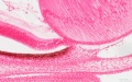
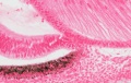
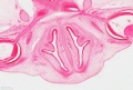
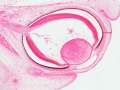
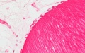
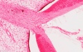
Stage 22 Eye
The images below link to virtual slides of the human developing eye at Carnegie stage 22. Click on the image to open or select specific regions from the regions of interest links.
|
|
Virtual Slide - Regions of Interest
|
Links: Embryo Virtual Slides
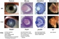
Retinotopic Map
This neuroscience term describes how the developing retina is precisely "mapped" onto the visual cortex through a series of signaling and activity dependent mechanisms. This follows from Hubel and Wiesel (1981 Nobel Prize in Physiology or Medicine) key discoveries (1959-70) of how in development system matching occurs in the visual system. The topographic map establishes an ordered neuronal connection between sensory structures and the central nervous system.
The retinotectal map (eye to brain) of birds (lower vertebrates):
- temporal (posterior) retina is connected to the rostral (anterior) part of the contralateral optic tectum
- nasal (anterior) retina to the caudal (posterior) tectum
- ventral retina to the dorsal (medial) tectum
- dorsal ventral (lateral) tectum
Retinal waves a form of coordinated spontaneous activity that occurs in the developing retina. These waves of electrical activity (action potentials) are thought to have a role in establishing the initial retinotopic map by correlating/coordinating the activity of neighbouring retinal ganglion cells.
EphA/ephrin-A molecular signaling also thought to have a role in establishing the initial retinotopic map.
Neural Crest

Mouse eye neural crest[7] |

Mouse eye TGF-beta model[7] |
- Links: Image - Mouse eye neural crest | Image - Mouse eye TGF-beta model | Vision Development | Neural Crest Development | Head Development
Schlemm's canal

Schematic showing the stages of Schlemm's canal development in the postnatal mouse by the novel process of canalogenesis.[8] (Cartoons have been drawn for clarity and are not intended to suggest that most early sprouts arise from the LVP.)
Extraocular Muscles
Extraocular muscles are required to move the eye within the orbit. Their embryonic origin requires an interaction between the cranial mesoderm and the migrating neural crest cells.
The following is from a recent paper comparing human to zebrafish muscle development.[9]
| About the Muscles | Legend | |
|---|---|---|
|
|

|
- Links: Extraocular Muscles
Additional Images
Human stage 22 developing iris region
Human stage 22 developing iris region
Human stage 22 overview of optic nerve
Human stage 22 overview of eye
Human stage 22 lens and hyaloid vessels
Human stage 22 optic nerve (stalk)
Human stage 22 retina
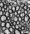
Mouse adult optic nerve axons
Pax6 eye phenotypes


Historic Images
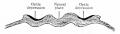
Fig. 456. Location of optic areas before the closure of the neural groove.
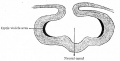
Fig. 457. Location of areas shown in Fig. 456 after the formation of the neural canal.
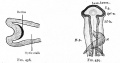
Fig. 458. Location of the optic area after the beginning of the formation of the optic cup and optic stalk. Fig. 459. Dorsal view of head of chick of 58 hours' incubation.
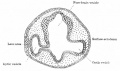
Fig. 460. Section through head of chick of two days' incubation.
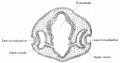
Fig. 461. Section through head of chick of three days' incubation.
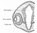
Fig. 462. Later stage in development of optic cup and lens than is shown in Fig. 461.
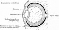
Fig. 463. Developing lens and optic cup.
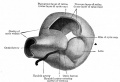
Fig. 464. Model showing lens and formation of optic cup.

Fig. 465. Stages in the development of the lens in the rabbit embryo.
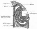
Fig. 466. Section through optic cup and lens invagination of chick of fifty-four hours' incubation.
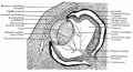
Fig. 467. Section through eye of human embryo of 13-14 weeks.
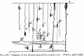
Fig. 468. Development of the retinal cells.
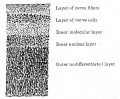
Fig. 469. Vertical section through retina of a four months' human embryo.
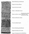
Fig. 470. Vertical section through retina of a five and one-half months' human embryo.
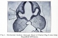
Fig. 1. Section through head of pig, 2 mm long.
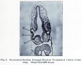
Fig. 2. Section through head of chick, 2 mm long.
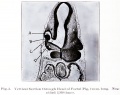
Fig. 3. Section through head of Foetal Pig, 2 mm long.
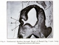
Fig. 4. Section through head of Foetal Pig, 3 mm long.
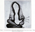
Fig. 5. Section through head of Foetal Pig, 3 mm long.
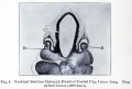
Fig. 6. Section through head of Foetal Pig, 4 mm long.
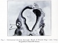
Fig. 7. Section through head of Foetal Pig, 7 mm long.
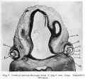
Fig. 8. Section through head of pig, 8 mm long.
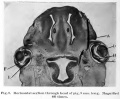
Fig. 9. Section through head of pig, 9 mm long.
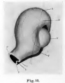
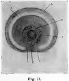
Fig. 11. colobomba of the fundus in the adult and means a lack of development.


References
- ↑ <pubmed>12186651</pubmed>| Genome Biol.
- ↑ <pubmed>22496813</pubmed>| PMC4183095 | Front Hum Neurosci.
- ↑ <pubmed>22496813</pubmed>
- ↑ <pubmed>20544023</pubmed>| PLoS ONE
- ↑ <pubmed>20459797</pubmed>
- ↑ <pubmed>7364662</pubmed>
- ↑ 7.0 7.1 <pubmed>16403239</pubmed>| J Biol.
- ↑ <pubmed>25051267</pubmed>| PLoS Biol.
- ↑ <pubmed>22132088</pubmed>| PLoS One.
Online Textbooks
- Kolb H, Fernandez E, Nelson R, editors. Webvision: The Organization of the Retina and Visual System [Internet]. Salt Lake City (UT): University of Utah Health Sciences Center; 1995-. Available from: http://www.ncbi.nlm.nih.gov/books/NBK11530/
- Developmental Biology (6th ed.) Gilbert, Scott F. Sunderland (MA): Sinauer Associates, Inc.; c2000. Evolution of the mammalian middle ear bones from the reptilian jaw | Chick embryo rhombomere neural crest cells | Some derivatives of the pharyngeal arches | Formation of the Neural Tube | Differentiation of the Neural Tube | Tissue Architecture of the Central Nervous System | Neuronal Types | Snapshot Summary: Central Nervous System and Epidermis
- Neuroscience Purves, Dale; Augustine, George J.; Fitzpatrick, David; Katz, Lawrence C.; LaMantia, Anthony-Samuel; McNamara, James O.; Williams, S. Mark. Sunderland (MA): Sinauer Associates, Inc. ; c2001 The Auditory System | The Inner Ear | The Middle Ear | The External Ear | Early Brain Development | Construction of Neural Circuits | Modification of Brain Circuits as a Result of Experience
- Molecular Biology of the Cell (4th Edn) Alberts, Bruce; Johnson, Alexander; Lewis, Julian; Raff, Martin; Roberts, Keith; Walter, Peter. New York: Garland Publishing; 2002. Neural Development | The three phases of neural development
- Clinical Methods 63. Cranial Nerves IX and X: The Glossopharyngeal and Vagus Nerves | The Tongue | 126. The Ear and Auditory System | An Overview of the Head and Neck - Ears and Hearing | Audiometry
- Health Services/Technology Assessment Text (HSTAT) Bethesda (MD): National Library of Medicine (US), 2003 Oct. Developmental Disorders Associated with Failure to Thrive
- Eurekah Bioscience Collection Cranial Neural Crest and Development of the Head Skeleton
- Webvision: The Organization of the Retina and Visual System. Kolb H, Fernandez E, Nelson R, editors. Salt Lake City (UT): University of Utah Health Sciences Center; 1995-.
Reviews
<pubmed>20855501</pubmed>| JCB
The International Journal of Developmental Biology Vol. 48 Nos. 8/9 (2004) Eye Development
Articles
<pubmed>19541779</pubmed>
Bookshelf vision development
Search Pubmed
Search Pubmed: vision development | eye development | eye embryology | retina embryology | lens embryology
Search Entrez: vision development | eye development | eye embryology | retina embryology | lens embryology
Terms
- annular tendon - (common tendinous ring, annulus of Zinn) fibrous tissue surrounding the optic nerve forming the origin for five of the six extra ocular muscles.
- canthus - (palpebral commissure) the corner of the eye where the upper and lower eyelids meet.
- extraocular muscles - six muscles that control movement of the eye (superior, Inferior, lateral and medial rectus; superior and inferior oblique).
- fovea - (fovea centralis; Latin, fovea = pit) retina region located in the center of the macula, required for sharp central vision.
- macula - (Latin, macula = spot; lutea = yellow) region near the center of the retina containing two or more layers of ganglion cells.
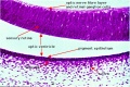
- retina - The stratified sensory structure of the eye, formed from the neural ectoderm that extends from the forebrain (diencephalon) to form initially the folded optic cup. Vertebrates have ten identifiable layers formed from nerve fibers, neurons, membranes, photoreceptors and pigmented cells. Light must pass through nearly all these layers to the photoreceptors. (1. Inner limiting membrane - Müller cell footplates; 2. Nerve fiber layer; 3. Ganglion cell layer - layer of retinal ganglion cells their axons form the nerve fiber layer and eventually the optic nerve; 4. Inner plexiform layer - another layer of neuronal processes; 5. Inner nuclear layer; 6. Outer plexiform layer; 7. Outer nuclear layer; 8. External limiting membrane - layer separating inner segment portions of photoreceptors from their cell nuclei; 9. Photoreceptor layer - rods and cones that convert light into signals; 10. Retinal pigment epithelium).
- retinal pigment epithelium - (RPE) An epethial pigmented cell layer lying outside the sensory retina, formed from the outer layer of the folded optic cup. The RPE is firmly attached to the underlying choroid and overlying retinal visual cells, for which it has a nutritional role.
- retinal waves - A form of coordinated spontaneous activity that occurs in the developing retina. These waves of electrical activity (action potentials) along with EphA/ephrin-A signaling are thought to have a role in establishing the initial retinotopic map by correlating/coordinating the activity of neighbouring retinal ganglion cells.
External Links
External Links Notice - The dynamic nature of the internet may mean that some of these listed links may no longer function. If the link no longer works search the web with the link text or name. Links to any external commercial sites are provided for information purposes only and should never be considered an endorsement. UNSW Embryology is provided as an educational resource with no clinical information or commercial affiliation.
Glossary Links
- Glossary: A | B | C | D | E | F | G | H | I | J | K | L | M | N | O | P | Q | R | S | T | U | V | W | X | Y | Z | Numbers | Symbols | Term Link
Cite this page: Hill, M.A. (2024, April 19) Embryology Sensory - Vision Development. Retrieved from https://embryology.med.unsw.edu.au/embryology/index.php/Sensory_-_Vision_Development
- © Dr Mark Hill 2024, UNSW Embryology ISBN: 978 0 7334 2609 4 - UNSW CRICOS Provider Code No. 00098G