Respiratory System - Upper Respiratory Tract
Introduction

The respiratory system does not carry out its physiological function (of gas exchange) until after birth. The respiratory tract, diaphragm and lungs do form early in embryonic development. The respiratory tract is divided anatomically into 2 main parts: 1. upper respiratory tract, consisting of the nose, nasal cavity and the pharynx; 2. lower respiratory tract consisting of the larynx, trachea, bronchi and the lungs.
In the head/neck region, the pharynx forms a major arched cavity within the phrayngeal arches.
See also sensory notes on Smell Development.
Some Recent Findings

Textbooks
- Human Embryology Larson Chapter 9 p229-260
- The Developing Human: Clinically Oriented Embryology (6th ed.) Moore and Persaud Chapter 12 p271-302
- Before We Are Born (5th ed.) Moore and Persaud Chapter 13 p255-287
- Essentials of Human Embryology Larson Chapter 9 p123-146
- Human Embryology Fitzgerald and Fitzgerald Chapter 19,20 p119-123
- Anatomy of the Human Body 1918 Henry Gray 1. The Respiratory Apparatus
Upper Respiratory Tract

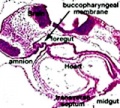
stage 11 foregut

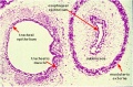
week 4 early respiratory endodermal bud

week 4 later respiratory endodermal bud
Stage 22 trachea
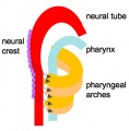
Head arches cartoon
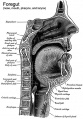
Pharynx
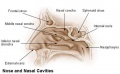
Nasal cavities
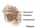
Pharynx
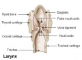
Larynx


- part of foregut development
- anatomically the nose, nasal cavity and the pharynx
- the pharynx forms a major arched cavity within the pharyngeal arches
Paranasal Sinuses
Maxillary Sinus
The data below is from a recent microscopical study of 100 human fetuses from the 9th to the 37th week (GA).[1]
- week 10 - maxillary sinus begins development.
- week 37 - the anterior-posterior diameter has a mean of 4.36 mm; ossification of the medial wall was absent, and the floor was located below the attachment of the inferior turbinate. Septa and recesses were temporarily observed.
- maxillary sinus osmium (opening) was located at the anterior third of the ethmoid infundibulum
- final dimensions were 1.96 mm in length and 0.44 mm in width.
- mean length between the ostium to the lamina papyracea and nasolacrimal duct was 1 mm.
Movies
The animations below allow a comparison of early and late embryonic lung development. Compare the size and relative position of the respiratory structures and their anatomical relationship to the developing gastrointestinal tract.
| Early embryo (stage 13)
3 dimensional reconstruction based upon a serial reconstruction from individual Carnegie stage 13 embryo slice images. | |
| Late embryo (stage 22)
3 dimensional reconstruction based upon a serial reconstruction from individual embryo slice images Carnegie stage 22, 27 mm Human embryo, approximate day 56. |
- Links: Flash Movies
References
- ↑ <pubmed>22267494</pubmed>
Reviews
<pubmed>21944636</pubmed> <pubmed>16798587</pubmed> <pubmed>15222948</pubmed>
Articles
<pubmed>21147652</pubmed> <pubmed>20966466</pubmed>
Search PubMed
Search Pubmed: Upper Respiratory Tract Development | Upper Respiratory Tract Embryology
Terms
| System Links: Introduction | Cardiovascular | Coelomic Cavity | Endocrine | Gastrointestinal Tract | Genital | Head | Immune | Integumentary | Musculoskeletal | Neural | Neural Crest | Placenta | Renal | Respiratory | Sensory | Birth |
Glossary Links
- Glossary: A | B | C | D | E | F | G | H | I | J | K | L | M | N | O | P | Q | R | S | T | U | V | W | X | Y | Z | Numbers | Symbols | Term Link
Cite this page: Hill, M.A. (2024, April 25) Embryology Respiratory System - Upper Respiratory Tract. Retrieved from https://embryology.med.unsw.edu.au/embryology/index.php/Respiratory_System_-_Upper_Respiratory_Tract
- © Dr Mark Hill 2024, UNSW Embryology ISBN: 978 0 7334 2609 4 - UNSW CRICOS Provider Code No. 00098G