Respiratory System - Histology
Introduction
This page contains information and images associated with respiratory system histology.
This can be initially divided into the 2 regions of the upper and lower respiratory tract.
- For the upper respiratory tract observe the epithelial specialisations, sensory regions and associated cartilages.
- For the lower respiratory tract observe the basic structure of the lung, alveoli and ducts, and associated cardiovascular elements.
Fetal Histology


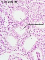
late canalicular
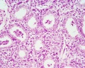
unlabeled late canalicular
Hyaline cartilage

- Fetal Respiratory: late canalicular | unlabeled late canalicular | Hyaline cartilage | Respiratory Histology
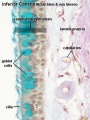
Upper Respiratory Tract - Nasal Cavity
Respiratory

|

|
| Nasal respiratory epithelium (inferior concha) | Nasal respiratory epithelium (detail) |
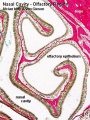
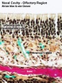
Olfactory
Nasal cavity olfactory epithelium cells
- Olfactory cells
- Sustentacular cells - located mainly in the superficial cell layer of the epithelium (difficult to distinguish from olfactory cells).
- Basal cells - identified by their location.
Epithelium
|
Lamina Propria
|

|

|
| Nasal cavity olfactory | Nasal cavity olfactory (detail) |
- Nasal Olfactory Histology: overview image | detail image | Smell Development | Histology | Histology Stains
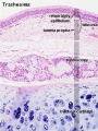
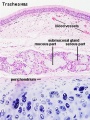
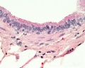
Trachea
Mucosa - formed by epithelium and underlying lamina propria.
- respiratory epithelium - (pseudostratified columnar and ciliated) ciliated cells, goblet cells, brush cells, endocrine cells, surfactant-producing cells (Clara cells), serous cells, basal cells, basement membrane.
- lamina propria - loose connective tissue, many elastic fibres.
- elastic lamina - forming the border between the mucosa and submucosa is not visible in H&E stained slides.
Submucosa - connective tissue and submucosal glands.
Submucosal Glands
(muco-serous) serous (dark) and mucous (light) parts have different staining appearance.
- Mucous secretions - "slimy" (high viscosity) mucous acini cells appear "foamy" or "frothy" and poorly stained (light). nuclei dark and smaller than serous.
- Serous secretions - "watery" (low viscosity) serous acini cell apical cytoplasm is usually well-stained (dark). nuclei round to ovoid located in cell basal cytoplasm.
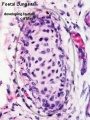
Cartilage
- perichondrium - surface of cartilage.
- tracheal cartilage - hyaline cartilage, 16 to 20 C-shaped cartilages.
- trachealis muscle - (smooth muscle) Not visible in this section, together with connective tissue fibres, join ends of the cartilages together.
Hyaline Cartilage Development
- forms from mesenchymal cells.
- precursor cells become rounded and form densely packed cellular masses, chondrification centres.
- chondroblasts - (cartilage-forming cells) begin secreting the extracellular matrix components of cartilage.
- extracellular matrix - ground substance (hyaluronan, chondroitin sulfates and keratan sulfate) and tropocollagen (polymerises into fine collagen fibres, not visible).
- Trachea Histology Links: Overview HE | Overview VG | Detail 1 HE Detail 2 HE | Respiratory Histology | Histology Stains | Histology


- Trachea Histology Links: Overview HE | Overview VG | Detail 1 HE Detail 2 HE | Respiratory Histology | Histology Stains | Histology
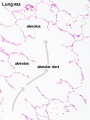
Bronchiole


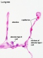
Alveolar Ducts and Alveoli
Alveolar type I cells
|
Alveolar type II cells
|


|

|

|

|
| Alveolar Duct | Alveoli | Alveoli Elastin | Lung Reticular Fibres |
Gallery
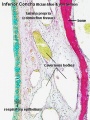
Nasal respiratory epithelium (inferior concha)
Nasal respiratory epithelium (detail)
Nasal cavity olfactory
Nasal cavity olfactory (detail)
Trachea 1
Trachea 2
Bronchiole
Respiratory bronchiole
Alveolar Duct
Alveoli

Alveoli Elastin
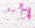
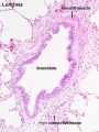
labeled lung
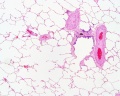
unlabeled lung
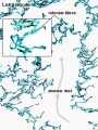
Lung reticular fibres
- Respiratory Histology: Bronchiole | Alveolar Duct | Alveoli | EM Alveoli septum | Alveoli Elastin | Trachea 1 | Trachea 2 | labeled lung | unlabeled lung | Respiratory Bronchiole | Lung Reticular Fibres | Nasal Inferior Concha | Nasal Respiratory Epithelium | Olfactory Region overview | Olfactory Region Epithelium | Histology Stains
Glossary Links
- Glossary: A | B | C | D | E | F | G | H | I | J | K | L | M | N | O | P | Q | R | S | T | U | V | W | X | Y | Z | Numbers | Symbols | Term Link
Cite this page: Hill, M.A. (2024, April 23) Embryology Respiratory System - Histology. Retrieved from https://embryology.med.unsw.edu.au/embryology/index.php/Respiratory_System_-_Histology
- © Dr Mark Hill 2024, UNSW Embryology ISBN: 978 0 7334 2609 4 - UNSW CRICOS Provider Code No. 00098G