Prize Essay on the Corpus Luteum (1851) 1
Part 1 Corpus Luteum of Menstruation
| Embryology - 24 Apr 2024 |
|---|
| Google Translate - select your language from the list shown below (this will open a new external page) |
|
العربية | català | 中文 | 中國傳統的 | français | Deutsche | עִברִית | हिंदी | bahasa Indonesia | italiano | 日本語 | 한국어 | မြန်မာ | Pilipino | Polskie | português | ਪੰਜਾਬੀ ਦੇ | Română | русский | Español | Swahili | Svensk | ไทย | Türkçe | اردو | ייִדיש | Tiếng Việt These external translations are automated and may not be accurate. (More? About Translations) |
Dalton JC. Prize essay on the corpus luteum of menstruation and pregnancy. (1851) Philadelphia: T.K. and P.G. Collins.
| Online Editor |
|---|
| This historic 1851 paper by Dalton is a very early historic description of the corpus luteum.
|
| Historic Disclaimer - information about historic embryology pages |
|---|
| Pages where the terms "Historic" (textbooks, papers, people, recommendations) appear on this site, and sections within pages where this disclaimer appears, indicate that the content and scientific understanding are specific to the time of publication. This means that while some scientific descriptions are still accurate, the terminology and interpretation of the developmental mechanisms reflect the understanding at the time of original publication and those of the preceding periods, these terms, interpretations and recommendations may not reflect our current scientific understanding. (More? Embryology History | Historic Embryology Papers) |
| Corpus Luteum 1851: Part 1 - Corpus Luteum of Menstruation | Part 2 - Corpus Luteum of Pregnancy | Part 3 - Observations on Animals | Plates |
Observation I
Appearance of the catamenia during the course of acute dysentery ; death on the following day — A Graafian vesicle in one ovary recently ruptured and filled with blood — Other Graafian vesicles in active development — Remains of preceding corpora lutea — Decidua already nearly separated from the uterine surface.
M. K., set. thirty-four, an inmate of the Insane Asylum at South Boston, was attacked with dysentery on Friday, August 9th, 1850, and died on the following Wednesday. She had been in the hospital about three months, suffering from a mild insanity, from which there were fair hopes of her recovery. The nurse stated, in general terms, that the patient's catamenia had been regular since her entrance. They made their appearance at about the regular period, on the day preceding death ; so that Dr. Stedman, the physician of the asylum, was induced to hope for a favourable change in her disease. They ceased again, however, the same night or the following morning, and were not to be seen on the day of death.
The autopsy was performed on Wednesday, and the uterus and ovaries were taken out and given to me on the Friday following. There was no menstrual fluid seen in the vagina. There was much inflammatory alteration of the mucous membrane over nearly the whole extent of the large intestine.
The uterus was three inches long, two and a quarter broad, and one and three-sixteenths thick. On cutting open the organ, it was found to contain a flattened triangular mass, corresponding by its sides and angles to the shape of the uterine cavity, which it entirely occupied. It was of a dark red colour, but assumed a brighter hue after a few moments' exposure to the air. Its texture was soft and friable, while at the same time it showed some disposition to tear in strips, like coagulable lymph. It had also a finely granulated appearance.
The greatest thickness of the mass was at its lower extremity, where it approached the internal orifice of the cervix uteri. Here it was onesixteenth of an inch thick. Superiorly, it terminated in a thin edge. The whole was very loosely adherent to the uterine parietes, from which it could be easily separated, leaving the uterine surface white, tolerably smooth, and perfectly firm. Under the microscope, it showed the following appearances : 1st. Blood-globules, rather pale, but not materially altered in size or shape. 2d. Columnar epithelium (from the oviducts). 3d. Irregularly roundish bodies, rather larger than blood-globules, and covered with very distinct granules. There were no other forms, besides these, discovered in its texture.
The right ovary was an inch and a half in length, of a yellowish white colour, firm and condensed in texture, and much puckered by cicatrices. Near its uterine extremity, and on its posterior aspect, there was a rounded prominence, about as large as the end of the little finger. This prominence, or tumour, was considerably reddened externally. It was soft and yielding, giving to the fingers quite a distinct sense of fluctuation. The ovarian coats were unnaturally thin all over the prominence, and at one spot the tunica albuginea was wanting ; and its place occupied only by a thin, smooth membrane (peritoneum), through which appeared the dark red colour of the interior. This spot was nearly semicircular in shape, and rather less than a quarter of an inch long, by a little over an eighth of an inch broad. Its edges were quite sharply defined, looking as if the tunica albuginea had been ruptured. About its centre was a very minute perforation, which allowed a little dark blood to flow out on compressing the tumour, but which was too small to be distinctly seen otherwise.
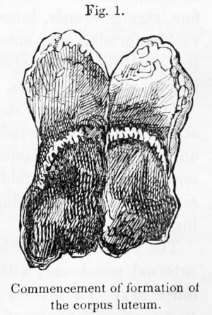
On dividing the ovary longitudinally, at the point of rupture, a cavity was opened, situated immediately beneath, of an ovoid shape, and measuring five-eighths of an inch in its long diameter. (See Plate I, Fig. 1.) It contained seven or eight drops of very dark, thickish blood, and a very dark, almost black coagulum. The coagulum lay loose in the cavity, except just at the point of rupture, where it was slightly adherent to the investing membrane. The walls of the cavity were composed of a single, firm, semi-transparent, vascular membrane, with a smooth internal surface, which could be readily stripped off entire from the cut surface of the ovary. There was no distinct membrane external to this, though irregular, vascular-looking strips of cellular tissue could still be raised by the forceps.
There was no yellow matter anywhere, and no puckering of the membrane.
There were two other well-developed Graafian vesicles in this ovary filled with clear fluid ; one of them nearly as large as the bloody cavity just described.
The right oviduct was free and pervious throughout. It contained, particularly toward its ovarian extremity, some thick whitish fluid, which under the microscope showed only nucleated columnar epithelium.
The left ovary was of about the same size as the right, and similarly seamed and puckered. It had upon its surface two Graafian vesicles, filled with clear fluid ; the larger of them about the size of a small hazelnut. It also contained one or two soft, granulous-looking, thin, compressed, yellow bodies, the remains of former menstruations. These were entirely similar, both in their gross and microscopic characters, to the other retrograde corpora lutea, hereafter to be described.
The left oviduct was pervious throughout, and contained white fluid like that in the right. There was nothing else remarkable about the uterine organs.
This case has several points of interest. In the first place, the condition of the patient, and the circumstances under which she was living, are such as almost to preclude the idea of any sexual intercourse having taken place. Probably we should never be able to meet with a case where, in the human female, the rupture of a Graafian vesicle, independent of coition, could be more certainly established than in the present instance.
It is probable that the patient's illness had not caused any very great disturbance in the periodical return of the menstrual function, since it was stated that it had heretofore been regular, and that its return at that time was only " a little" in advance of the expected period. Still, as the menstrual flow lasted only during one day, it is impossible to say whether this corresponded to the earlier or later part of its regular period of duration. The persistence of the ovarian function, notwithstanding the grave disease under which the patient was laboring ; the fact that the vesicle was burst, and filled with blood, but had not yet commenced its transformation into a corpus luteum ; and the coincidence in time between the rupture of the vesicle and the separation of a decidua from the internal surface of the uterus, are all circumstances which deserved particular notice.
Observation II
Death from cholera during menstruation — Excessive hemorrhage into a Graafian vesicle — Decidua (probably) expelled — Old corpora lutea in the opposite ovary.
J. C, a married woman, thirty-eight years of age, died at the Cholera Hospital in Boston, Sept. 3d, 1849, after an illness of twenty hours. Her general health was stated to have been good.
At the autopsy, the vagina was smeared with a dingy red secretion. The OS uteri was rather open, and purplish in colour. The cavity of the cervix contained a little adhesive secretion. The uterus, generally, was of natural appearance, and measured three inches in length, two in breadth, and one in thickness. Its internal surface was quite smooth, firm, and pale, without any appearance like the formation of a decidua ; but was smeared with a reddish fluid, like that of menstruation.
The right ovary was enlarged to six or eight times its natural size. It was ovoid in shape, and two and a quarter inches long ; the other measurements in proportion. It had the aspect and feel of a serous cyst, capable of containing one ounce of fluid ; but the red colour of its contents showed through at the thinner part of its walls. It was filled with a dark, tolerably firm, recent-looking bloody clot, intersected by one or two very delicate enclosed (false) membranes. The whole clot could be easily turned out, leaving a very red smooth lining membrane, with several spots of complete ecchymosis on its surface. The clot occupied nearly the whole of the organ ; the proper structure of the ovary being reduced to a very small proportion of the whole.
The right oviduct was somewhat convoluted. Its ovarian extremity was closed and adherent to the ovary. It could, however, be easily dissected off", without opening the cavity of either. The oviduct was distended so as to contain a drachm, or more, of thinnish, dingy-red fluid similar to that in the cavity of the uterus. Its internal surface was natural, and it was pervious throughout, except at its ovarian extremity.
The left ovary was one and three-quarters of an inch long. It was of a natural form and colour, and had several cicatrices on its surface. Internally, it contained one or two small, old-looking corpora lutea. The left oviduct was free from adhesions. Its internal surface was smeared with a small quantity of whitish fluid secretion. The amount of hemorrhage in this case was so great as perhaps almost to constitute a morbid appearance. It shows, however, the connection between the ovarian hemorrhage and menstruation, and the expulsion of the decidua ; which, a comparison with other cases will show, had probably been recently thrown off, leaving the uterine surface smooth and firm. It should also be remembered that the patient's "general health" was said to be good ; so that the deviation from a normal condition was not probably very essential.
Observation III
Sudden death in consequence of an extensive burn — A Graafian vesicle in each ovary filled with blood, and the internal surface of the uterus soft and shaggy.
M. M., a remarkably healthy-looking and well developed young woman, unmarried, twenty years of age, died at the Mass. Gen. Hospital, January 29th, 1848. Three days before, she had suffered a very extensive burn in consequence of the bursting of a camphene lamp, by which the skin was more or less completely destroyed over the greater part of her face, neck, body, and arms. She suffered but little pain after the first day, and finally died collapsed, and partially comatose. No information was obtained regarding her menstruation.
At the autopsy, it was found that the inner surface of the larynx was extensively scorched, and oedematous. There was also some pneumonia of the right lung. The uterus was of natural size : three inches long, by two broad. Its mouth was transverse, and sufficiently open to admit the end of the little finger. The cavity of the uterus was filled with an abundant reddish, very tenacious secretion. Its internal surface was soft, and somewhat shaggy. Both Fallopian tubes, easily laid open through their whole extent, contained a thick, yellowish, opake fluid.
Both ovaries were remarkably large. The left had on its surface an irregularly oval spot, three-fourths by three-eighths of an inch in size, where the investing membranes were depressed, thin, and red ; very distinct from the remainder of the surface, which had its usual thick, yellow, opake appearance. More than half the substance of the ovary was occupied by a roundish cavity, situated immediately beneath the above-mentioned spot, and filled with a firm, pretty adherent coagulum, dark red internally, more yellowish externally. The lining membrane of the cavity, immediately external to the coagulum, was of a dull red and brownish colour, and opake. It was moderately adherent, but could be raised and separated entire. External to this were cellular layers, which could only be raised in strips, the outermost of which were intimately connected with the ovarian tissue. The opposite ovary had a similar spot on its surface, and a bloody cavity underneath. The coagulum here was rather less firm than the other, and had a fresher appearance. The surrounding membrane could not be so easily traced.
The history of this, case being somewhat imperfect, it does not afford any direct proof of the connection between ovarian hemorrhage and menstruation. A comparison with other observations, however, makes it probable that menstruation had either just terminated, or was suppressed by the accident which resulted in death. The case is somewhat remarkable, like the last, for the large amount of blood effused within the Graafian vesicle, and shows, moreover, the connection of the ovarian hemorrhage with the softening of the uterine surface.
Observation IV
Piapld death from cholera — A Graafian vesicle filled with blood, and just commencing the transformation into a corpus luteum — Softening of the uterine surface.
E. D., unmarried, thirty-six years of age, died at the Cholera Hospital in Boston, Sept. 11th, 1849, after an illness of eighteen hours. No information was obtained regarding the patient's menstruation, but she was stated to have been in her " usual health" until the period of attack.
The vagina was smeared with a moderate quantity of the ordinary starchy secretion of cholera. The uterus measured three and threeeighth inches in length, one and seven-eighths in breadth, and one and a quarter in thickness. It was firm and pale anteriorly, but posteriorly had rather a tumefied appearance, and in its posterior wall it contained a fibrous tumour, a quarter of an inch in diameter, near Avliicli the uterine substance was somewhat reddened and softened. Elsewhere it was natural The os uteri was small and round, the cervix pale, firm, and empty. The internal surface of the body of the organ was much softened and reddened to the depth of threesixteenths of an inch. It was, in fact, converted for that depth into a dark purplish red, soft, shaggy coat, in which numerous slender, wavy fibres were distinctly visible, projecting toward the interior. This reddened coat was sufficiently tender to be scraped off with considerable readiness by the edge of the knife, leaving the uterine surface pale, and somewhat uneven. It was confined to the body of the organ, and did not descend at all into its neck.
The right ovary was one and seven-eighth inches in length. Nearly half of it was occupied by a loose, fluctuating tumour, having much the appearance of a serous cyst, only the red colour of its contents showed through. It was roundish and bulging, and at its most prominent part showed a small abraded spot, but nothing like a wellmarked cicatrix. Internally, it presented a cavity which was filled partly with a thin bloody fluid, and partly with a tolerably firm, adherent coagulum, from which the red colouring matter had not yet been completely absorbed. The walls of the cavity consisted apparently, at its bulging extremity, of the thinned and distended coats of the ovary, but at its deeper part of a yellowish, well-defined layer of moderately firm consistency, and irregularly folded and convoluted, like the convolutions of the brain. This yellow layer had a thickness at the deepest part of the tumour, of one-sixteenth of an inch ; but it gradually became thinner as it approached the surface, until it appeared to blend with the coats of the ovary.
The left ovary, one and a quarter inches long, contained three or or four moderately developed Graafian vesicles. There was nothing remarkable about cither oviduct.
Observation V
Corpus luteum of menstruation of about two weeks' date — Another, in the opposite ovary, in a retrograde condition — Softening of the uterine surface.
M. H., a servant girl, aged twenty-two, of a plump and healthy aspect, died at the Cholera Hospital, in Boston, Sept. 16th, 1849, after an illness of but little over thirty hours. In the recorded history of her case, it was stated that she had "menstruated" two weeks previously, but no account was obtained of the exact time at which the flow commenced or terminated.
At the autopsy, the vagina was found smeared with a starchy secretion. The OS uteri was rather open. The cavity of the cervix contained a very little glutinous secretion, and its internal surface showed one or two distended follicles beneath the lining membrane. The body of the uterus was empty and of natural appearance ; firm and whitish. Its internal surface was softened to the depth of three-sixteenths of an inch, and converted into a loose, vascular, shaggy texture, in which fine zigzag vessels, intermixed with whitish fibres, were distinctly visible, forming very much the same appearance as that described in the preceding observation. The uterus measured three inches in length, one in thickness, and one and three-quarters in breadth.
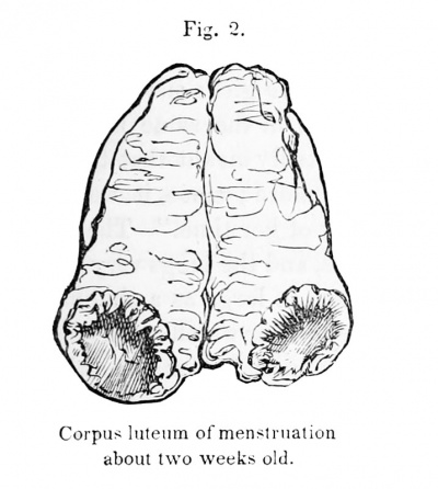
The left ovary, one and three-quarter inches in length, contained at one extremity a roundish, ovoid tumour, measuring five-eighths of an inch in its long diameter. It was composed of a light-yellow external layer, strongly folded and convoluted, and enclosing a fibrinous, semi-translucent coagulum, stained with some remains of the colouring matter.
The yellow wall of this corpus luteum was invested by a delicate external membrane, with which it could be enucleated entire from the ovary. Its texture was friable and granular. On superficial examination, it appeared to contain some bundles of fine vessels, proceeding from without inward ; but on closer inspection they were seen to exist only in the interstices between the convolutions, where they had been accidentally exposed in making the section ; in the same manner as the vessels of the pia mater may be exposed, dipping down between the convolutions of the brain. The substance of the yellow wall itself contained no vessels.
The tumour formed by this corpus luteum had just firmness sufiicient to allow its contour to be felt through the ovarian texture ; but it had very little resistance, and might easily have been crushed between the thumb and finger. Before being cut open, it even gave an indistinct and deceptive sense of fluctuation.
The following represents the ovary, of natural size, cut open longitudinally.
|
|
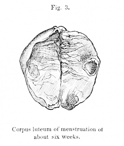
| Fig. 2. Corpus luteum of menstruation about two weeks old. | Fig. 3. Corpus luteum of menstruation of about six weeks. |
The opposite ovary was of natural size and appearance. It contained at one spot, immediately beneath its surface, a small flattened cavity, a quarter of an inch in diameter. The cavity had no bloody contents, but was surrounded by a thin wall, of a dull yellowish colour, Avhich showed indistinct appearances of being more or less convoluted. This wall was surrounded by a thin delicate membrane, with which, like the other, it could be enucleated from the substance of the ovary. A drawing of it is given above (Fig. 3).
The ovaries also contain several Graafian vesicles in active development. Both oviducts were in a natural condition, and contained only a little whitish fluid.
Observation VI
Corpus luteum of menstruation, three weeks from the termination of the menstrual period — at its maximum of development — Five others, in different stages of retrogression — Uterine surface soft, but pale.
L. E. B., a factor operative at Lowell, unmarried, twenty years of age, was attacked with hsemoptysis on Friday, June 28th, 1850, at eight P. M., while walking in the street, and died the next morning at four. The uterus and ovaries were taken out at the autopsy, and sent to me by Dr. J. 0. Dalton, Sr., by whom the following particulars, relating to menstruation, &c., were also ascertained.
The girl had been in perfectly good health for many months, and had menstruated for the last time just three weeks previous to her death. This last fact was ascertained of the patient's landlady, who had inquired particularly concerning it of the girl's sisters, immediately after her death. The sisters worked in the same mill, and boarded in the same house. It was undoubtedly meant that the girl had ceased menstruating just three weeks before ; since it was also remarked that she was then "within a week of her time." The body generally "appeared somewhat exsanguious, and the lungs very much engorged. No tubercles, but one old ulcer, as large as a shagbark, lined with false membrane, at the right apex ; near which was a strong pleuritic adhesion. Other thoracic and abdominal organs healthy."
The uterus was empty, and of natural appearance externally. Its internal surface was pale, but softened to the depth of about a quarter of an inch, exhibiting distinctly the velvety, shaggy appearance already described.
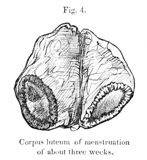
The right ovary was one inch and seven-eighths in length. Its surface was generally smooth, and pale yellowish in colour, and showed only here and there a very few fine superficial vessels. Its outer extremity presented a rounded tumour, about the size of the end of the finger, imbedded in the substance of the ovary, but projecting slightly from its posterior surface (Plate I. Fig. 2). It was globular in shape, and had but little solidity, feeling as if it might easily be crushed between the thumb and finger. The surface of the ovary, over the tumor, was quite vascular, showing many red and blue vessels. At about its most prominent part was a cicatrix ; i. e., a small oval spot, very slightly depressed, about one-twentieth of an inch in length, where the tunica albuginea was entirely wanting, and its place supplied by a thin, delicate, transparent membrane, which allowed the pale reddish colour of the contents of the tumour to show through.
On making a longitudinal section of the organ, through the cicatrix, the corpus luteum was exposed (Plate I. Fig. 3, a). It was three-quarters of an inch in its long diameter, and nearly one-half an inch in depth. It consisted of a thin, pale, reddish-yellow, irregularly convoluted wall, enclosing a solid, semi-transparent, fibrinous coagulum, which showed in various parts remains of the red colouring matter of the blood. There was no empty cavity. The wall was thickest at the deepest part of the tumour, and grew gradually thinner as it approached the surface and the situation of the external cicatrix, where finally it was entirely wanting ; showing conclusively that the rupture of the Graafian vesicle had taken place at this spot.
The relative proportions of the convoluted wall and its contents are represented in the drawing. The wall itself was not lined by any membrane, but was in immediate contact with the fibrinous coagulum. Externally, it was invested by a thin, semi-transparent, vascular membrane, with which it could be readily enucleated from the substance of the ovary, leaving a smooth and somewhat vascular surface of condensed cellular tissue. This investing membrane was intimately connected with the substance of the yellow wall, and could not readily be separated from it; so that, when raised in strips, portions of the wall adhered to it. The interstices of the convolutions were vascular, but no vessels could be detected in the substance of the yellow wall itself.
Toward the uterine extremity of the ovary, there was another yellow body (Plate I. Fig. 3, b), much smaller than the first, situated immediately beneath a minute transparent cicatrix on the surface, which was surrounded by a slight vascularity. The colour of this body was a much more decided yellow than that of the first. It appeared to consist of a wall, the opposite sides of which were nearly or quite in contact, owing to the absorption of the contained coagulum. The wall also showed some indications of a convoluted arrangement ; but the whole body was so small, and its texture so friable, that it was difiicult to establish this point distinctly.
At a short distance was situated another yellow body, similar in character to that last described, only much smaller.
The left ovary was one inch and five-eighths long. It showed on its surface several cicatrices, both shallow and depressed, and contained three very small, obsolete yellow bodies, like those described as found in the right ovary.
Both ovaries contained many Graafian vesicles, in different stages of active development, but neither showed anything else at all resembling a corpus luteum.
Both oviducts were free and natural in appearance throughout.
Observation VII
Corpus luteum of menstruation of about the same date as the preceding — Softening of the uterine surface.
E. B., a widow, thirty years of age, died at the Cholera Hospital in Boston, August 20th, 1849, after an illness of ten and a half hours. No information was obtained regarding her menstruation, but she was "as well as usual" till the period of her attack. The abdomen, externally, showed many scars of pregnancy.
The uterus was somewhat "bombde" posteriorly, and had rather a softish feel. It was empty. Its internal surface was much softened and reddened to the depth of three-sixteenths of an inch, having undergone the same change already described in previous observations. The internal surface of the cervix uteri was pale, and had but little of the adhesive secretion in its cavity ; but one or two follicles, distended with this secretion, could be seen beneath its mucous membrane. The uterus measured three and a quarter inches in length, and two and a quarter in breadth. (The subject was large and stout.)
The right ovary was one inch and three-eighths long ; generally white and natural in appearance. One end of it was occupied by an ovoid tumour, not very firm to the touch, the dull reddish colour of which showed through the integuments of the ovary. A section of the ovary showed the tumour to be three-quarters of an inch long, by nearly half an inch deep. It consisted, like the others, of a yellow convoluted wall, which enveloped a homogeneous, opaline, greenish, fibrinous-looking coagulum, which still presented, in spots, some traces of the colouring matter of the blood. The point of rupture is not seen in this drawing ; probably because, in making the section, sufficient care was not taken to cut through the cicatrix.
Observation VIII
Death during the menstrual period — Corpus lutcum of the last menstruation commencing to diminish in size — Graafian vesicle not yet burst — Decidua just separated from the uterine surface.
C. M., a married woman, thirty years of age, died at the Cholera Hospital in Boston, August 23d, 1849, after an illness of four days. She was said to have had her usual health till the time of the attack.
The uterus was rather large, particularly in proportion to the size of the patient, Avho was small and slightly built. It measured three and three-eighths inches in length, tAvo and a half in breadth, and one and a half in thickness. It was much bulging posteriorly, and had altogether a tense, stiff, swollen aspect and feel. The os uteri was widely open. The cavity of the cervix was filled with an abundance of tough, tenacious secretion, strongly coloured with blood. The vagina, also, and external organs were smeared with a bloody fluid.
The internal surface of the body of the uterus was quite pale and smooth, without any appearance of the softening elsewhere described; but its cavity contained a flattened triangular mass, like that mentioned in Observation I,, of a reddish-gray colour and loose texture, having no appearance of an organized structure, nor any attachment to tlie walls of the uterus. It had evidently taken its shape from that of the uterine cavity, its two upper angles corresponding to the orifices of the oviducts. Its longitudinal diameter was a little over one inch ; its transverse, at its widest part, something less.
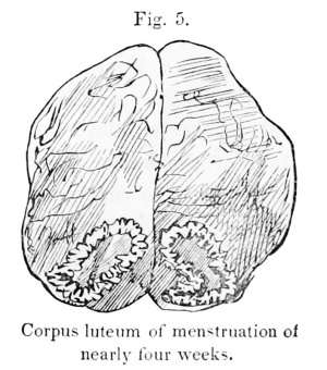
The left ovary, one and a half inches in length, had a natural colour and appearance externally. At the uterine extremity of the organ was a spot where the integuments were thinned, and the reddish colour of the corpus between showed through. The tumour was ovoid in shape, but had very little firmness, or resistance to the touch. It consisted of a thin, yellow, very irregular, convoluted wall, and an internal greenish, semi-transparent, fibrinous clot, stained with some remains of red colourmg matter. There was no strongly-marked cicatrix on the surface of the ovary. The accompanying drawing (Fig. 5) represents the ovary and corpus luteum, of the natural size. The left oviduct was empty and pale internally.
The right ovary, of about the same size as the left, presented several Graafian vesicles, moderately developed. The right oviduct contained, in its uterine half, a considerable amount of thickish, bloody fluid ; otherwise it was natural.
Observation IX
Death just at the termination of a menstrual period — A Graafian vesicle prominent, and on the point of bursting — Corpus luteum of the preceding menstruation, in a retrograde condition — Seven others, quite atrophied and obsolete — Uterine surface pale, but soft.
Mrs. a., a widow, forty-five years of age, a patient of Dr. W. H. Thayer, was attacked suddenly with apoplexy, Oct. 14th, 1850, at 5 P. M. ; and, after" lying insensible for sixteen hours, died on the following morning. Mrs. A.'s youngest child was about twelve years of age. Her husband had been dead ten years. Her maidservant, an intelligent girl, reported to Dr. Thayer that Mrs. A.'s catamenia had been regular at intervals of four weeks, from commencement to commencement ; and that they always continued a week, accompanied with much pain in the pelvic region, sometimes so great as to oblige her to keep her bed for a day. Her last catamenial period closed on Sunday the loth, the day before her attack, as Mrs. A. had herself told her. She was in general well acquainted with the course of her mistress's catamenia, as she washed her linen, and was also a kind of confidential servant.
At the autopsy, Oct. 16, twenty-five hours after death, a hemorrhage of about three ounces was found in the substance of the right hemisphere of the brain, and another, much smaller, in the right optic thalamus. The cerebral substance was much softened in the immediate neighbourhood of each coagulum. There was also considerable hypertrophy of the left ventricle of the heart, but the other organs were healthy. The stomach contained much undigested food.
The uterus was quite large, measuring three and a half inches in length, and two and a quarter in its transverse diameter. Its walls were proportionably thick, but natural in texture. It was empty. Its internal surface was rather pale, but quite soft, shaggy, and velvety in appearance. The os uteri was transverse, wide, and much fissured.
The left ovary was an inch and five-eighths long. It contained two large and somewhat prominent Graafian vesicles, with clear fluid contents. One of them was particularly distended and prominent; being covered, over a considerable part of its surface, by peritoneum alone. On the anterior surface of the organ was an irregularly oval spot, three-eighths of an inch long, where the tunica albuginea was wanting. Immediately beneath was a corpus luteum, the bright yellow colour of which showed through the peritoneal coat. There was also a black speck upon its surface, which marked the spot where the rupture of the vesicle had taken place. On making a section of the ovary, through this black spot, the corpus luteum was exposed, of a somewhat flattened, oval shape, three-eighths of an inch in length, and a little over a quarter of an inch in depth. (Plate I. Fig. 4.) It consisted, like the others, of a bright yellow, pulpy, friable wall, thickest at its deepest part, and gradually thinning off toward the situation of the rupture ; strongly folded upon itself, and destitute of vessels. It enclosed a little, very dark, moderately firm coagulum, which was easily removed, having no organic connection with the yellow wall. The Avail itself was not lined by any membrane, but vfas in immediate contact with the coagulum. When put upon the stretch, it Avas not more than one-twentieth of an inch in thickness; but, when allowed to remain folded up, appeared somewhat thicker. It could easily be enucleated entire, and externally had the same relations to the investing membrane and the substance of the ovary as in the preceding cases.
The same ovary contained two other old corpora lutea, one of them about a quarter part the size of the first, the other still smaller. They were both, in their general characters, similar to the first. The larger had also a small black spot on its surface, marking the situation of the rupture ; and a section being made at a little distance from this spot, the foldings of the wall were distinctly seen to radiate from it.
The right ovary was one and three-quarters of an inch long. Its section discovered a yellow body, about a quarter of an inch in length, much flattened, and containing a little very dark blood. There were also four others, perfectly distinguishable, but diminishing successively in size ; all superficially situated beneath shallow or moderately depressed cicatrices. The two smallest, alone, contained no noticeable remains of blood. They were all similar in structure, but the smaller were whiter, and of a less decided yellow, than the larger. There were no other remarkable appearances.
Both oviducts were quite natural. They contained only a little opake, whitish secretion, which, under the microscope, showed an abundance of ciliated epithelium.
Everything makes it probable that, in this instance, the largest corpus luteum was the result of the last menstruation but one, and that the most prominent vesicle on the left ovary was just upon the point of bursting when the patient suffered her attack of apoplexy.
So far this case, together with the preceding, would go to sustain the opinion of Pouchet and Raciborski, that the ovum is discharged in the human female, not during, but at the termination of, the menstrual period.
Observation X
Death from inflammation of the brain of several weeks' standing — Corpus luteum of menstruation thirty days after the termination of the menstrual period — Three others still more obsolete — No prominent Graafian vesicles — Uterine surface only a little softened.
M. A., a married woman, twenty-nine years of age, who had had one child four years ago, entered the Almshouse Hospital at South Boston, June 10th,1850, suffering from symptoms resembling chorea. They consisted in a capricious, semi-involuntary, twisting motion of the right arm and hand, and some disposition to a drawing up of the left leg. There was also an almost constant, slow winking motion of the right eyelid, accompanied by a sinister leering expression of the countenance, as if the patient were partially idiotic or hysterical. The intelligence was rather dull, but otherwise not much altered.
She was treated with antimonials, low diet, and blisters to the back of the neck ; afterwards by carbonate of iron and full diet.
Under this treatment she improved much, and on the 29th of June, being to all appearance well, was discharged from the Hospital and sent to the Almshouse. Two days afterward, however, she returned, suffering under a fresh attack, much more severe than the former. This time she had lost much of her muscular power ; and lay nearly prone on the floor of the ward, apparently unable to rise, and very stupid. After much urging, she managed to raise herself from the ground with the assistance of a chair, but had great difficulty in doing so. There was some paralysis of the left side, and a want of control over the muscles of the right. She was treated with purgatives and counter-irritation to the head and spine, but sunk into a deeply typhoid condition, and died on the 12th of July.
The nurse reported that the patient was menstruating at the time of her first entrance into the hospital, and continued to do so June 10th and 11th. On the 12th, the discharge returned slightly, and then finally ceased. The nurse was positive that she had had no menstruation since, as she must have known it, if such had been the case. There was found much inflammatory softening of the right hemisphere of the brain, and of the right corpus striatum ; some also of the right optic thalamus and of the left hemisphere.
The uterus, of about the normal size, was somewhat retroverted, and inclined to the left side of the pelvis, where it was confined by numerous old, organized, bridle-shaped adhesions. It was empty. Its internal surface was quite vascular and a little pulpy, but much less so than in some of the preceding observations, as the pulpy portion at its thickest part did not exceed one-sixteenth of an inch.
The right ovary was one inch in length ; white, firm, and covered with cicatrices. It contained two yellow bodies. (Plate I. Fig. 5). The larger was flattened from without inward, and contained within the yellow wall an appreciable amount of reddish fibrin which could be easily separated from its cavity. This body was a little over a quarter of an inch in its long diameter. It was situated immediately beneath a strongly depressed cicatrix on the surface of the ovary. On very minute examination traces of the convoluted arrangement of its walls were visible.
The second body was irregularly globular in shape, and much smaller than the first. Its centre was occupied by a radiated cicatrix, which no longer contained any noticeable amount of coagulum. The right ovary also contained one more very minute yellow body, too small to show any definite structure. There was also one more in the left ovary, but nothing else in either that presented any appearance like that of a corpus luteum.
There were but very few Graafian vesicles to be seen, and these were small and of an inactive appearance.
The right oviduct was much convoluted, and confined by adhesions. The fimbriated extremity was entirely adherent to the ovary, enclosing an oval space on the surface of the organ five-eighths of an inch long. The oviduct was somewhat distended, and contained about half a drachm of thickish, brownish-red fluid, approaching to bistre in its colour. The internal surface was vascular, and stained by the brownish fluid ; otherwise natural. The left oviduct was adherent like the right, but empty and collapsed. It was impervious in two or three spots, toward its uterine extremity. But the right was pervious throughout.
- cicatrix - A scar left by the formation of new connective tissue over a healing sore or wound.
Observation XI
Absence of catamenia for over two months — Corpus luteum of last period very small — Four others, quite obsolete — No softening of uterine surface.
M. S., unmarried, twenty-three years of age, died at the Massachusetts General Hospital, Aug. 26th, 1850, of tubercular meningitis. She entered the house Aug. 18th, at which time her intelligence was so much afiected, that she was unable to give any account of herself. Her sister, however, reported that she had had no catamenia for two months, and none appeared while she was in the hospital ; making, in all, a period of two months and eight days that the catamenia had been absent.
At the autopsy, the substance of the brain was found somewhat softened, with a rather copious eff"usion of serum into its cavities, and an abundance of minute, whitish, opake (tuberculous) grains, scattered through the meshes of the pia mater between the convolutions ; more particularly at the base of the brain. The pleurae were also sprinkled with tuberculous granulations, without the existence of any considerable collection in the pulmonary substance. The liver and spleen also showed an abundance of similar tubercles in their interior.
The uterus was rather small and firm. Its neck was conical, and its mouth small and perfectly circular. The cavity of the organ contained a moderate quantity of transparent mucus, slightly tinged with red. A part of its internal surface was rather vascular, but it was everywhere smooth and firm, and of its natural consistency.
The right ovary was one and a half inches in length, and of the ordinary yellowish-Tfhite colour. Its surface showed several shallow, bluish cicatrices, none of which had a very recent appearance. The ovary contained four old corpora lutca, the largest of which was three-sixteenths of an inch in diameter, and entirely similar to that described in the preceding observation, except for being smaller. (Plate II. Fig. 1.) It consisted of a delicate, flattened, thin, yellowish-white wall, destitute of vessels, and enclosing a thin fibrinous coagulum, which was still somewhat coloured with blood. Traces of a convoluted arrangement of the yellow wall were quite evident on close inspection. It was neither lined nor surrounded by any membrane that could be demonstrated, but was immediately in contact with the fibrinous clot internally, and externally appeared closely connected with the ovarian tissue.
The remaining yellow bodies in the ovaries were smaller, but otherwise similar. The left ovary also contained one, very small. Both ovaries contained many Graafian vesicles, varying in size from a quarter of an inch or less to three-eighths of an inch in diameter.
Both oviducts were free and pervious throughout. They contained a moderate quantity of thick, whitish, opake fluid, like that met with in Observation I. There was nothing else remarkable.
From the foregoing observations we can deduce a more or less complete history of the corpus luteum. In the first place, it is certain that, in connection with the appearance of the menstrual period, a Graafian vesicle arrives at maturity, protrudes from the surface of the ovary, ruptures, and becomes filled with blood. Sometimes this process takes place in each ovary at the same period. It will even hereafter be seen that two vesicles may become ruptured simultaneously in the same ovary. During the final enlargement of the vesicle, and its protrusion from the ovarian surface, it strongly distends the albugineous coat, the fibres of which become separated and pushed aside to such a degree that this tunic is at last wanting over a spot of considerable size ; and the vesicle is covered, at its most prominent part, by peritoneum alone ; so that, at the time of the rupture, it is only the peritoneal coat which becomes lacerated.
The precise period, during the catamenial flow, at which this rupture takes place, does not seem, as yet, to be definitely ascertained. The inherent difficulty of making these observations in the human subject, and the rarity of opportunities for examining the ovaries of healthy women, who have died by violence or after a rapid illness, oppose great obstacles to our arriving at a correct conclusion on this subject. The difficulty, too, of estimating the influence which even a short illness may have had on the exact period of rupture is so great, that many further observations will be necessary, in order to determine with certainty so nice a point. Perhaps this period is not, even in the ordinary course of nature, invariable ; and if we are to rely on such information as we already possess, this would seem to be the most probable supposition. Bischoff,* Pouchet,t and Raciborski| all agree that the ovum is discharged regularly, only at the termination of the menstrual period, and this opinion is certainly sustained by Observations VIII. and IX. of the present paper. Dr. Robert Lee§ also relates the case of a woman who died " while menstruating." The uterus and oviducts were lined with menstrual fluid, and on one ovary was a large, projecting, Graafian vesicle, having over it an abrasion of the peritoneum. On the other hand, there are not wanting abundant observations of Graafian vesicles ruptured during the menstrual flow. Such an observation by Cruikshank, recorded in the Philosophical Transactions, 1797, has already been alluded to. Dr. Lee|| gives an instance of a Graafian vesicle filled with blood, death having taken place during menstruation, and cites other similar ones from Gendrin. Bischoff^ states that he has opened the bodies of four females, dead suddenly during menstruation. Three of them presented, in one of the ovaries, a vesicle burst and filled with blood ; in the fourth, there was remarked only a vesicle not yet ruptured. It would therefore appear that the rupture takes place sometimes earlier and sometimes later, but that it always has an intimate relation with the appearance of the catamenia.
Another remarkable variation in the appearance of the vesicle at this period depends upon its larger or smaller size at the time of rupture, and the greater or smaller quantity of blood effused. In Observation I., this quantity was very moderate, as may be seen by reference to the drawing. In Observation III., it Avas much larger; and in Observation II., it was so excessive as to be estimated at about one ounce. This variation may probably be explained by the difference in the age of the subjects, and in the condition of the vascular system generally, whether of full health, of plethora, or of deficiency of vascular action. It also depends, without doubt, to a certain extent, on the greater or less activity of the uterine organs in particular. Whatever may be its cause, it is, perhaps, not more remarkable than the differences which are observed in the size of the ovaries themselves, or of the other internal organs. It will hereafter be seen, in another division of this paper, that the size of the vesicles, even in the same ovary, is not always the same at the period of their full development, those which are matured and ruptured not always being the largest.
• Op. cit., p. G2. t IljiJ- P- 244. { Ibul. p. 414.
§ London Med. Gazette, Nov. 5th, 1S4-2. || Ibid.
TT Op. cit., p. 50.
Another point which still remains somewhat doubtful relates to the period at which the hemorrhage takes place ; whether it occurs before or after the time of rupture. From some of the accompanying observations, it would seem probable that it succeeds to, and is a consequence of, the bursting of the vesicle ; since we have seen the vesicle, at a late period, very large and prominent, with its contents still clear and unmixed with blood. Pouchet,[1] however, states that the ovum is pushed from the bottom of the vesicle toward the surface by a gradual hemorrhage beneath its inner membrane (membrana granulosa) ; and that the vesicle is, in this way, already filled With blood, before the final rupture takes place. This matter must be considered as still uncertain. Pouchet's opinion may, no doubt, receive support from some observations in which the vesicle has been found filled with blood without presenting any lesion of its peritoneal coat. It must, however, be remarked that, though the albugineous tunic remains for a long time deficient at the spot where the protrusion of the vesicle took place, yet the peritoneal coat is very rapidly reproduced ; consequently, no perforation might be visible in this membrane, only a very short time after the occurrence of the rupture.
The rupture of a Graafian vesicle, with hemorrhagic effusion into its cavity, as accompaniments of menstruation, have been recognized by some writers, who are, nevertheless, unwilling to acknowledge that an ovum is at the same time discharged. Dr. Robert Leef considers it probable that " all the phenomena of menstruation depend on, or are connected with, some peculiar changes in the Graafian vesicles, in consequence of which an opening is formed in their peritoneal and proper coats." Notwithstanding this, he denies explicitly that any ovum passes from the ovarium during menstruation, but appears to consider this process, when it does occur, as excited by the stimulus of the seminal fluid. Dr. Patterson[2] entertains a similar opinion, except that he does not think the rupture of the vesicle at the time of menstruation a constant, but only an occasional occurrence. This, indeed, is a matter in regard to which we must be content, at present, to rely on other evidences than direct observation ; for I am not aware that any one has yet seen, in the human female, an unimpregnated ovum in the oviduct, and recently discharged from a vesicle. It appears, however, contrary to all known physiological analogies to suppose that the vesicles should be ruptured for any other purpose than that of discharging their contents. It is conceded on all hands that the human ovum, as well as that of the lower animals, exists in the vesicle previously to impregnation. Thus in a young girl, ten years of age, dead at the Female Orphan Asylum, January 9th, 1851, of perforation of the stomach, I was able to discover three ova in one ovary, and one in the other. No doubt many more might have been found, but that the vesicles were very friable, and many became ruptured in the process of separating them from the ovary. The vesicles, containing the ova above mentioned, were all in an inactive condition, not at all prominent, but situated entirely beneath the albugineous tunic.
The regular tumefaction, also, and rupture of the vesicle take place in both the human species and the lower animals in a manner entirely analogous ; and since we know from direct observation that, in the latter, this process terminates by the discharge of an ovum, it is certainly unreasonable to suppose that it should have any other object in the former.
Shortly after the rupture of the vesicle, the formation of the corpus luteum commences. In order, however, to understand distinctly the manner in which this body is produced, it will be necessary to recall the anatomical relations of the vesicle and its neighbouring tissues. The Graafian vesicle, in its ordinary quiescent condition, consists of a simple globular sac, filled with a transparent, albuminous fluid. It is not very closely connected with the ovarian substance, and by a little manipulation Can be removed entire ; when it appears as a smooth, diaphanous cyst, usually with several distinct blood-vessels ramifying over its surface. It is said to be lined by an inner membrane, the memhrana granulosa, composed of minute, granular, cell-like bodies, similar to those Avhich, collected in a more compact mass round the ovum, constitute the proligerous disc. These bodies are of a flattened form, and nearly circular in outline Avhen isolated, but when crowded together they are compressed into more or less angular forms. They have altogether the appearance of cell-nuclei rather than of cells, and frequently exhibit a very distinct nucleolus. They are undoubtedly to be regarded as a kind of epithelium, lining the serous surface of the Graafian vesicle.
The membrana granulosa, however, is so exceedingly delicate that it is very easily broken up, and detached from the internal surface of the vesicle by the necessary manipulations ; so that, in ordinary dissections, it is seen only as a collection of soft, whitish, membranous flocculi, which give to the fluid of the vesicle an appearance of turbidity. External to the vesicle are a number of irregular vascular laminae of cellular substance, which can only be raised in strips and fragments, varying in size and thickness. These cellular layers have sometimes been described as a third tunic of the Graafian vesicle, membrana cellulosa (membrane celluleuse, Pouchet) ; but they appear rather to belong to the tissue of the ovary, and to form a sort of nidus, or bed for the reception of the vesicle.
When the ovum is expelled, the proligerous disc, and probably a considerable portion of the membrana granulosa, are expelled with it. The proper membrane of the vesicle then begins to become hypertrophied ; the hypertrophy consisting in a peculiar growth of soft, yellowish, friable substance, which takes place most abundantly at the deepest part of the vesicle, and becomes thinner and thinner as we approach the vicinity of the cicatrix. As this new growth proceeds, the membrane of the vesicle becomes, at the same time, folded upon itself in all directions, so as to present innumerable convolutions and anfractuosities, which give a radiated appearance to its cut surface. This peculiar arrangement seems to be a necessary consequence of the increased nutrition of the membrane, which is, at the same time, confined nearly within its original limits. We see the same thing take place in other tissues of the body under similar circumstances, as in varicose veins, enlarged ureters, &c. The gray substance of the brain, during the first five months of foetal life, presents a smooth surface, without any trace of convolutions. As it afterward increases rapidly in extent, the cranial cavity, at the same time, not enlarging In proportion, it becomes necessarily folded upon itself, in order to accommodate its increased bulk to a proportionally smaller cavity. The valvulse conniventes of the small intestine are another example of the same thing. These are not present during the greater part of foetal life, the mucous membrane being then of the same length as the muscular coat. But, as it afterward grows more rapidly than the latter, it is thrown into folds which project into the cavity of the intestine. It is the same process which gives rise to the formation of the corpus luteum. At the same time that the hypertrophy of the vesicular membrane proceeds, the blood eifused within its cavity also undergoes an alteration. Its fluid part is absorbed, and afterward the coagulum loses by degrees its colouring matter, becoming paler and paler, in proportion to the time which has elapsed since the rupture of the vesicle. The vascularity of the neighbouring parts also increases, or, at least, does not diminish, and at the end of about three weeks from the termination of the menstrual period, the corpus luteum has attained its maximum of development. Its situation is then marked by a concentration of vessels on the surface of the ovary, and by a globular or ovoid tumour, about the size of the end of the finger, which causes a slight prominence externally. The cicatrix is not always strongly marked at this period, and may consist only of a small, bluish, transparent spot, indicating a separation of the ruptured peritoneum. The tumour, though well defined, is soft and yielding, and does not give to the touch the impression of a very highly organized body. It does not now contain any cavity, properly speaking; but its centre is occupied by the solid, semi-transparent, gray, or greenish coagu. lum, more or less mottled with red. The coagulum is in immediate contact with the inner surface of the yellow wall, but can be separated from it by a little manipulation, as the two have no organic connection. Thewall itself still forms the smallest part of the tumour, as it is very thin ; not more than one-eighth of an inch thick at its deepest part, when allowed to remain folded up ; but if the convolutions are unfolded, it is seen to be, in reality, much thinner.
The point of rupture of the vesicle is still very well marked on a section of the corpus luteum, provided the incision be made accurately through the external cicatrix ; for at that spot the yellow wall terminates, and the central coagulum is in immediate relation with the peritoneal coat. If a horizontal section be made, and the coagulum removed from over the point of rupture, the sinuosities of the yellow wall may also be seen radiating from it as a centre.
The new growth or deposit of yellow matter, when examined under the microscope, is seen to consist of an abundance of peculiar, irregularly-shaped, granular cells, varying in size, and sometimes enclosing minute, opaline, yellowish globules, like oil. The contour of the cellwall can, in most cases, be completely traced throughout ; but, in some instances, it is indistinct at certain parts of the circumference. There are also to be seen, in the field of the microscope, a few oilglobules, similar to those in the interior of the cells; but for the most part of larger size. The cells are rendered paler by acetic acid, and their (oily) granules and globules become more distinct, but no proper nucleus is visible.
Dr. Frank Reuaud[3] has given an excellent description of these cells. He is disposed, however, to regard the oil-globules as the " nuclei" of the cells, whereas they do not have at all the appearance characteristic of ordinary nuclei, but seem to be rather a production of the cell itself. Indeed, the oil-globules evidently increase in quantity in proportion as the cells become atrophied and disappear.
According to the present observations, the yellow matter is essentially a growth of the proper membrane of the Graafian vesicle. Much difference of opinion, however, has prevailed among writers with regard to this point, some asserting, as above, that the new growth consists in a hypertrophy of the outer membrane (Knox, Pouchet), others that it is a development of the inner membrane (De Baer), others that it is deposited between the two (Montgomery), and others that it is external to both (Lee). A part of this discrepancy, no doubt, arises from confusion and misunderstanding as to what membranes are designated by the terms "inner" and "outer." Dr. Montgomery, for instance, f could never have spoken of the inner coat of the Graafian vesicle as a " strong white cyst," if, under the name of the "inner coat, he had intended to designate the membrana granulosa. Some writers, indeed, recognize the cellular laminae, already spoken of, lying external to the outer membrane, as a proper tunic of the vesicle. In reality, they cannot be so considered ; and when we take into view the exceedingly friable nature of the "membrana granulosa," it appears most correct to describe, with Raciborski, only one proper membrane of the Graafian vesicle, the "ovisac" of Barry, the '■^ vesicule ovulifere" of Pouchet.
This is the membrane, which, by its development and consequent folding up, forms the corpus luteum. That the yellow wall is not lined by any second membrane, I have made certain by frequent observations. At the same time, it must be remarked that, after a longer period of time, the enclosed coagulum, flattened and compressed, and deprived by absorption of all its colouring matter, may present a deceptive resemblance to a true membrane ; particularly as at advanced periods, and in the corpus luteum of pregnancy, it becomes more closely adherent to the yellow wall. Dr. Robert Lee, in his Lectures on Midwifery, published in the London Medical G-azette, even gives two drawings of corpora lutea (of pregnancy), in which, in accordance with his ideas, there are demonstrated two distinct membranes within the yellow wall. These must certainly have been formed accidentally, by an artificial separation of the central coagulum into laminae. In the corpus luteum of menstruation, and also in that of pregnancy, if of recent date, it is easy to demonstrate the entire absence of any real lining membrane.
Externally the corpus luteum is invested with an extremely thin, transparent, vascular membrane, to which it is intimately connected, and which sends prolongations between the convolutions of the yellow matter. This layer, indeed, seems to form rather a memhra^ious surface than a distinct tunic, and cannot be stripped off without bringing with it torn fragments of the yellow matter. It is, in fact, the outer surface, merely of the membrane of the vesicle, the new growth having sprouted from its internal aspect, toward the cavity of the vesicle. External to this, are the vascular layers of cellular tissue, which have already been described as surrounding the Graafian vesicle, and forming a nidus for it in the ovarian tissue, previous to the time of its complete development.
The substance of the corpus luteum is not vascular, though minute red vessels may often bo seen, apparently running into the yellow matter, from without inward. A close examination, however, shows these vessels to exist only in the interstices of the convolutions, where they have penetrated from the vascular network on the external surface of the body.
It lias been already shown that the development of the yellow matter commences soon after the rupture of the vesicle has taken place. The strange theory regarding these bodies, entertained by Sir Everard Home, and afterwards adopted by N^grier, viz., that the yellow matter is deposited before the rupture, that it is intended to aflford nutrition to the ovum, and that, consequently, corpora lutea are only one stage of the ascending development of the Graafian vesicle, must certainly be regarded as entirely erroneous. It is disproved in the most positive manner by several of the accompanying observations. We see, for instance, in one case the Graafian vesicle large, prominent on the surface of the ovary, distended with serosity, and apparently on the point of rupture, while the vesicular membrane has, as yet, undergone no change.
Secondly, we have Graafian vesicles, freshly ruptured and filled with blood, with the vesicular membrane equally unaltered as before. Thirdly, beyond this period we have corpora lutea in all stages of retrogression ; but wherever the deposit of yellow matter has taken place, the vesicle contains blood, and has evidently been ruptured. The only exception to this rule is in the case of some very small corpora lutea, which have already been long subjected to the process of absorption, and in which it is therefore difficult to recognize the remains of the coagulum.
It is not, however, necessary to rely entirely on the accompanying observations, to disprove the theory above alluded to. N^grier himself refers to the cases " communicated to him by Dr. Ollivier D'Angers," of two women who committed suicide during a menstrual period. In each case, one ovary presented a bloody ruptured vesicle (" rupture vesiculaire saignante"), containing a soft and recent coagulum. But nothing is mentioned, in either case, about any alteration of the inner surface of the vesicle. In the three cases by Bischoff, already referred to, of recently ruptured vesicles, there is no mention made of any yellow deposit being visible. There is also another exceedingly interesting case of the rupture of a vesicle during menstruation reported by Dr. Myddleton Michel in the Charleston, S. 0. 31edical Journal (quoted in the Amerhcan Journal of 3Iedical Sciences, July 1848). It was that of a woman who was executed for murder. She had been imprisoned several weeks previously to the execution, and had undoubtedly been deprived of sexual intercourse. At the examination, the OS uteri was found "open, tumefied, and dripping with blood," and its inner surface coated with blood, and " congested" at its upper part. The right ovary contained a number of vesicles in process of development, containing clear fluid. The left liad a ruptured vesicle on its anterior surface, with its orifice partially closed by a bloody coagulum. The membrana granulosa (examined by the microscope) also protruded through the opening. The cavity of the Graafian vesicle was filled with coagulated blood. Its tunic was very vascular, and " had not yet hegim to fold, to form the corpus luteum." The precise period during menstruation at which death took place in this instance was not ascertained with certainty.
Ndgrier, however,[4] gives one observation on this point which is not very easily explained. He describes very accurately a recent corpus luteum of menstruation found by him in an ovary which, as he says, did not present on its surface " any trace of rupture." With regard to this, I can only say that I have not met with a similar case, and that it is contrary to the experience of most other observers.
After the end of the third week from the close of menstruation, the corpus luteum passes into a retrograde condition. It diminishes in size ; and the fibrinous coagulum becomes absorbed, and loses still farther its colouring matter. The diminution of the size of the body takes place more particularly in a certain direction ; so that, from having a globular or ovoid form, it becomes collapsed and flattened, either laterally or from within outward. In consequence of this flattening, the corpus luteum at this period may appear to vary considerably in size, according as the section has been made in the plane of its compressed surfaces or perpendicularly to them. Thus, in Observations IX. and X., both of which give specimens of corpora lutea four weeks after menstruation, the apparent size of these bodies is very diff"erent. But, if we consider that one has evidently been cut across, while the other was probably divided in the plane of its compression, the diff'erence will not appear so striking. Still, it is undoubtedly the case that these bodies do not, in all instances, become atrophied with the same rapidity, and that more or less variation is apparent in this, as in most other physiological operations of nature. It is certain, too, that the effused coagulum does not always lose its colour with the same readiness ; since, in some of the drawings which represent corpora lutea of four weeks or over, the clot is even redder than in Observation VI, where it is represented at the termination of the third week.
Fig. 8.
The colour of the yellow wall, however, during the early part of the retrograde stage, instead of fading like that of the fibrinous clot, becomes more strongly marked. From the dull yellowisli-red hue, similar to that of recent lymph, -which it had at first, it assumes a brighter and more decided yellow, and contrasts, therefore, more strongly with the neighbouring tissues. This yellow hue of the corpus luteum has been attributed by various writers to an alteration in the colouring matter of the efi'used blood, similar to that which takes place in the neighbourhood of old apoplectic clots. Pouchet* so considers it, and Raciborskif entertains the same opinion. A microscopic examination of the corpus luteum at this period, however, demonstrates that this colour depends principally on the presence of oil, which is seen in larger or smaller globules, and having the same yellowish tinge which is presented by the whole body when viewed by the naked eye. The oil-globules, in fact, which, at an earlier period, were seen in small numbers and of minute size, and for the most part enclosed within the irregular granulated cells, have now become large and abundant, so as to occupy nearly the whole field, while the cells themselves have disappeared, or are with difficulty distinguishable. Everything leads us to believe that the cells are a formation destined to produce the oil, as a gland its secretion ; and that, when they have accomplished this purpose, they disappear and are replaced by the ordinary fibrous tissue of the ovarian stroma.
As the corpus luteum diminishes in size, the yellow matter becomes softer and more friable, and shows less distinctly the markings of its convolutions. At the same time, it is more intimately connected with the neighbouring tissues, so that it can be less readily enucleated from the ovary. The central coagulum is no longer anything more than a faint, whitish, stellate cicatrix ; while the yellow matter itself often also assumes a stellated, triangular, or other similar form, and pursues constantly a retrograde course, which terminates finally in its obliteration. When the corpus luteum, however, has reached this advanced stage of retrogression, it appears to become comparatively stationary, and its alterations proceed less rapidly than at an earlier period ; so that it may remain for some months afterward as a minute, faint, obsolete, yellow spot, imbedded in the ovarian substance. In the case of Mrs. A., Observation IX., seven of these obsolete yellow bodies were distinguishable, besides that resulting from the last rupture of a vesicle, the oldest of which probably dated from a period of eight months. In general terms, then, it may be stated that a period of six to eight weeks is sufficient to reduce the corpus luteum of menstruation to an insignificant, soft, thin, compressed sac, measuring less than a quarter of an inch in its longest diameter, and with its opposite walls nearly or quite in contact ; but that after this time it remains more quiescent, and that a period of eight months sometimes elapses before its final and complete destruction.
Oil-globules from the smaller corpus luteum in PI. I. Fig. 3 (same scale).
• Page 14G.
t Page 437.
There is good reason to believe that, in connection with the processes of menstruation, a peculiar change takes place in the internal surface of the uterus, which results in the formation and separation of a decidual membrane. Pouchet,[5] indeed, considers the regular monthly formation and discharge of a decidua as satisfactorily established. Dr. Robert Leef states that the uterine decidua is a thing of frequent occurrence in unmarried females, and does not consider its presence as the slightest evidence of the existence of pregnancy. He cites also Drs. Baillie and Blundell in support of the same views. The last-mentioned writers, however, are not disposed to consider the formation of a decidua, unconnected with pregnancy, as a regular physiological phenomenon, but rather as an occasional appearance, dependent perhaps on difficulty of menstruation. In reality, however, it is very probably a process which recurs spontaneously somewhere about each menstrual period.
The mucous membrane of the uterus, in its ordinary inactive condition, presents a smooth, pale surface, of considerable firmness. It does not have the appearance of a distinct membrane, but rather of a membranous surface, owing to the softness of its structure, and its intimate connection with the subjacent uterine tissue. It is only occasionally that we can succeed in raising with the forceps very thin, delicate strips, which in reality appear to be rather portions of epithelium than of the mucous membrane itself. At this time, it is difficult for the eye to distinguish any peculiarities of structure, but when, at certain periods, the membrane has become swollen and hypertrophied, numerous, very minute, whitish, wavy or zigzag lines may be seen running from the inner part of the substance of the uterine parietes, and terminating on its free surface. In the ewe, these lines are much more distinctly seen, particularly at those spots where the cotyledons are afterward to be developed. These appearances, together with the remaining parts of the uterine mucous membrane, are described at length by C. B. Reichert in the first number of Muller's Archiv for 1848.[6] According to him, these whitish lines arc the glandular ducts or tubuli of the uterine mucous membrane ; which open upon its free surface, and are lined by a continuation of its epithelium. After conception, this portion of the membrane becomes much hypertrophied ; the tubuli are lengthened, and their orifices, destined afterwards to receive the villi of the chorion, become visible on the surface. The membrane increases also in vascularity, and becomes soft and spongy, and finishes by being transformed into the decidua vera, and at last expelled from the uterus. This change has also been recognized by other writers as taking place after conception, and Dr. Carpenter[7] even states that it "occurs whether the ovum reach the uterus or not, it being probably invariable in cases of extra-uterine pregnancy."
The preceding observations, however, make it probable that this formation accompanies the regular processes of menstruation, whether the ovum be fecundated or not. It will be noticed that, in all the foregoing cases except four, viz., Obs. I., II., VIII., and XI., there existed more or less softening and sponginess of the uterine mucous membrane. In two of these four cases, Obs. I. and VIII., a decidual membrane had just been separated, or was nearly so, and was found in the cavity of the uterus. In Obs. II., it had very probably been expelled from the organ, and in Obs. XI. all the functions of the generative system had been suspended for more than two months ; a circumstance which accounts satisfactorily for the absence of any alteration in the mucous surface of the uterus. In Obs. X., in which the softening existed, but only to a slight degree, these functions had been arrested for a period of about four weeks.
The separation and expulsion of the decidual membrane seem to take place about the time of the rupture of the Graafian vesicle, that is to say, toward the termination of the menstrual period. After this separation, the internal surface of the uterus is left firm, pale, and smooth. It soon recommences, however, to become hypertrophied, and in the course of a short time it presents a soft, velvety, shaggy alteration, extending to the depth of three-sixteenths or a quarter of an inch, and showing very distinctly the whitish, wavy, or zigzag tubuli, intermixed with numerous fine blood-vessels, extending toward the free surface of the membrane. The decidual membrane is, in this way, in process of preparation during the descent of the ovum through the tubes, and while it is yet unprepared to make any vascular connection with the walls of the uterus.
References
- ↑ Page 138. f Cyclop. Pract. Med. iii. 4 J 4.
- ↑ J Edinburgh Med. and Surg. Journal. Jan. 1840.
- ↑ London and Edinburgh Monthly Journal of Medical Science, August, 1845. f Op. cit. p. 226.
- ↑ Op. cit. page 22.
- ↑ Page 249. f London Med. Gazette, vol. xxxi. p. 412.
- ↑ Ueber die Biklung der liinfalligen Hilute der Gebiir mutter, und deren VerhiLltniss zur placenta uterina.
- ↑ Principles of Human Physiology, p. 579.
| Embryology - 24 Apr 2024 |
|---|
| Google Translate - select your language from the list shown below (this will open a new external page) |
|
العربية | català | 中文 | 中國傳統的 | français | Deutsche | עִברִית | हिंदी | bahasa Indonesia | italiano | 日本語 | 한국어 | မြန်မာ | Pilipino | Polskie | português | ਪੰਜਾਬੀ ਦੇ | Română | русский | Español | Swahili | Svensk | ไทย | Türkçe | اردو | ייִדיש | Tiếng Việt These external translations are automated and may not be accurate. (More? About Translations) |
Dalton JC. Prize essay on the corpus luteum of menstruation and pregnancy. (1851) Philadelphia: T.K. and P.G. Collins.
| Online Editor | ||||
|---|---|---|---|---|
| This historic 1851 paper by Dalton is a very early historic description of the corpus luteum.
|
| Historic Disclaimer - information about historic embryology pages |
|---|
| Pages where the terms "Historic" (textbooks, papers, people, recommendations) appear on this site, and sections within pages where this disclaimer appears, indicate that the content and scientific understanding are specific to the time of publication. This means that while some scientific descriptions are still accurate, the terminology and interpretation of the developmental mechanisms reflect the understanding at the time of original publication and those of the preceding periods, these terms, interpretations and recommendations may not reflect our current scientific understanding. (More? Embryology History | Historic Embryology Papers) |
| Corpus Luteum 1851: Part 1 - Corpus Luteum of Menstruation | Part 2 - Corpus Luteum of Pregnancy | Part 3 - Observations on Animals | Plates |


