Primordial Germ Cell Development
Introduction
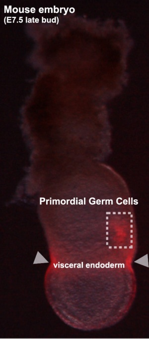
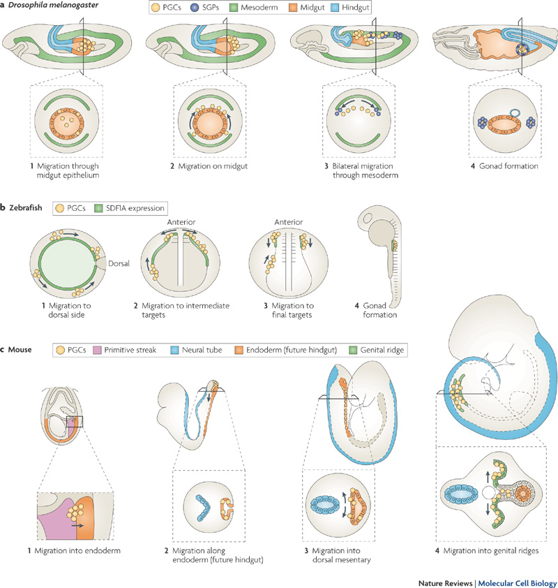
Early in development at the time of gastrulation a small group of cells are "put aside" to later form oocytes and spermatozoa. This population of cells is described as the primordial germ cells (PGCs). These cells also migrate initially into the posterior endoderm that forms the hindgut and from there into the genital ridge that will be the site of the developing gonad. The maintenance of pluripotency within this cell population may arise through epigenetic modifications that suppress somatic differentiation programs.
This population of cells when transformed is also thought to give rise to testicular germ cell tumours.
Some Recent Findings
|
(More? Recent References)
Textbooks
- Human Embryology (2nd ed.) Larson Chapter 10 p261-306
- The Developing Human: Clinically Oriented Embryology (6th ed.) Moore and Persaud Chapter 13 p303-346
- Before We Are Born (5th ed.) Moore and Persaud Chapter 14 p289-326
- Essentials of Human Embryology, Larson Chapter 10 p173-205
- Human Embryology, Fitzgerald and Fitzgerald Chapter 21-22 p134-152
- Developmental Biology (6th ed.) Gilbert Chapter 14 Intermediate Mesoderm
Primordial Germ Cell Migration
Species Comparison of Migration
Stages of primordial germ cell migration[6]
Mouse Migration Movies
The movies below show labeled primordial germ cells (green) migrating within the mouse embryo between the periods of E9.0 to E10.5 into the genital ridge region that will later form the gonad.
| E9.0 | E9.5 | E10.5 |
| Quicktime version | Quicktime version | Quicktime version |
| Flash version | Flash version | Flash version |
Cell Structure
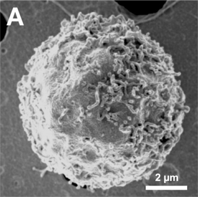
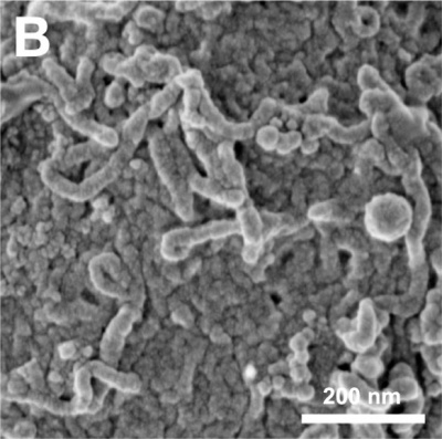
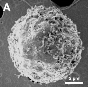
The images below are scanning electron micrographs of the surface of a chicken primordial germ cell that has been grown in culture.[7]
The first image shows the whole cell and the second image shows detail of the cell surface showing extensions.
DNA Methylation
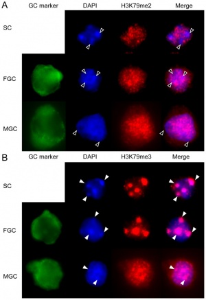
| Mouse primordial germ cell DNA methylation[8]
Demethylation
|
|
X-linked Gene Expression
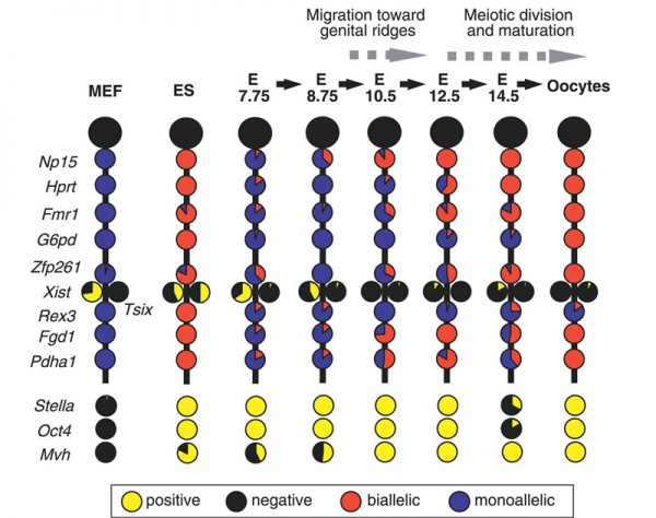
Mouse- X-linked gene expression during primordial germ cell development.[9]
Each circle graph indicates the ratio of cells that are positive (yellow) and negative (black) for each gene, and biallelically (red) and monoallelically (blue) expressed in cells positive for each gene.
- Links: X Inactivation | Mouse Development
Molecular
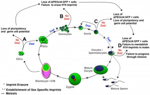
Study has recently identified 11 genes that are specifically expressed in male and female fetal germ cells, both in vivo and in vitro, but are not expressed in embryonic stem cells.[11]
PGC Markers: alkaline phosphatase-positive, Oct4 (POU5F1), Fragilis (IFITM1)[12], Stella (DPPA3), Dazl, and Vasa (DDX4).
- Steel factor - (KITLG) a ligand for the KIT tyrosine kinase receptor.
- DAZL
- dead end - coding an RNA binding protein mainly expressed in the germ cells of vertebrates.
- Blimp1 - B-Lymphocyte induced maturation protein-1 (PRDM1)
- Prmt5 - protein arginine methyltransferase-5
- Nanog - knockdown induces apoptotic cell death in mouse migrating primordial germ cells.[13]
- AID - Activation-Induced cytidine Deaminase enzyme required for demethylation (removal of CpG methylation). Within the genome, DNA methylation is associated with epigenetic mechanisms and occurs at cytosine residues that are followed by guanines.[14]
Abnormalities
Teratomas
Common group of fetal tumors occuring along the body midline, anywhere from the coccyx to the pineal gland, reflecting the developmental PGC migration pathway (for review see [15]).
- Histologically classified as either mature or immature.
- Immature elements consisting principally of primitive neuroglial tissue and neuroepithelial rosettes and have have a generally favorable prognosis.
- Sacrococcygeal teratomas - most common site (70%–80% of all teratomas).
- classified into four types based on the amount of mass present externally versus internally.
Testicular germ cell tumours (seminoma)
References
- ↑ <pubmed>20886037</pubmed>| PLoS One.
- ↑ <pubmed>19997484</pubmed>| PMC2777314 | PLoS Genet.
- ↑ <pubmed>20845430</pubmed>
- ↑ <pubmed>19468308 </pubmed>
- ↑ <pubmed>19279135</pubmed>
- ↑ <pubmed> 20027186</pubmed>| Nature Reviews Molecular Cell Biology
- ↑ <pubmed>20886037</pubmed>| PLoS One.
- ↑ <pubmed>21886830</pubmed>| PLoS One.
- ↑ <pubmed>17676999</pubmed>| PMC1950944 | PLoS Genet.
- ↑ <pubmed>19468308</pubmed>| PLoS One.
- ↑ <pubmed>20940145</pubmed>
- ↑ <pubmed>12659663</pubmed>
- ↑ <pubmed>19906868</pubmed>
- ↑ <pubmed>20236475</pubmed>
- ↑ <pubmed>15653597</pubmed>
Reviews
<pubmed>20371640</pubmed>| Reproduction <pubmed>20027186</pubmed> <pubmed>19875497</pubmed> <pubmed>19442193</pubmed> <pubmed>17446386</pubmed> <pubmed>15666347</pubmed> <pubmed>11565804</pubmed> <pubmed>11061420</pubmed>
Articles
<pubmed>19578360</pubmed> <pubmed>18953407</pubmed>
Search PubMed
Search Pubmed: Primordial Germ Cell Migration | Primordial Germ Cell | Testicular germ cell tumours
External Links
External Links Notice - The dynamic nature of the internet may mean that some of these listed links may no longer function. If the link no longer works search the web with the link text or name. Links to any external commercial sites are provided for information purposes only and should never be considered an endorsement. UNSW Embryology is provided as an educational resource with no clinical information or commercial affiliation.
Glossary Links
- Glossary: A | B | C | D | E | F | G | H | I | J | K | L | M | N | O | P | Q | R | S | T | U | V | W | X | Y | Z | Numbers | Symbols | Term Link
Cite this page: Hill, M.A. (2024, April 24) Embryology Primordial Germ Cell Development. Retrieved from https://embryology.med.unsw.edu.au/embryology/index.php/Primordial_Germ_Cell_Development
- © Dr Mark Hill 2024, UNSW Embryology ISBN: 978 0 7334 2609 4 - UNSW CRICOS Provider Code No. 00098G

