Placenta - Cord: Difference between revisions
| Line 22: | Line 22: | ||
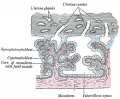
==Hofbauer Cells== | ==Hofbauer Cells== | ||
* human villous macrophages | |||
* located the core of placental villi | * located the core of placental villi | ||
* macrophages with micropinocytotic activity and phagocytosis ability | * macrophages with micropinocytotic activity and phagocytosis ability | ||
Revision as of 09:39, 17 November 2012
Introduction

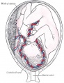
The placenta (Greek, plakuos = flat cake) named on the basis of this organs appearance. The placental cord (umbilical cord) is the connecting region between the functional placenta and the embryo/fetal umbilical region. The human cord varies greatly in overall length increasing to about 60 to 70 cm at term. This extraembryonic structure contains the placental blood vessels and allantois.
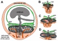
There are essentially 3 separate aortic/venous circulatory systems: umbilical, systemic and vitelline. The umbilical system is lost at birth, the vitelline contributes to the portal system and the systemic (embryonic) is extensively remodelled to fom the the cardiovascular system.
| System Links: Introduction | Cardiovascular | Coelomic Cavity | Endocrine | Gastrointestinal Tract | Genital | Head | Immune | Integumentary | Musculoskeletal | Neural | Neural Crest | Placenta | Renal | Respiratory | Sensory | Birth |
Some Recent Findings
|
Hofbauer Cells
- human villous macrophages
- located the core of placental villi
- macrophages with micropinocytotic activity and phagocytosis ability
- possible paracrine role for early stages of placental vasculogenesis
- express angiogenic growth factors (VEGF)
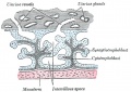
Wharton's Jelly
- placental cord connective tissue (substantia gelatinea funiculi umbilicalis)
- amorphous substance containing glycosaminoglycans, proteoglycans and hyaluronic acid.
- cells similar to smooth muscle that allows a contractile function.
- network of collagen that form canaliculi and perivascular spaces.
- maintain blood flow to the fetus during placental cord compression during pregnancy or delivery.
First described and named after Thomas Wharton (1614–1673) an English physician and anatomist.
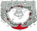
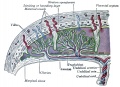
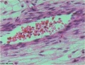
Placental Cord Histology

Placental cord cross-section

Placental vein

Placental artery

Placental allantois


Persistent Right Umbilical Vein
A fairly rare anomaly, a study of 15,237 obstetric ultrasound examinations performed after 15 weeks' gestation identified only 33 cases of persistent right umbilical vein.[3] Some studies have identified associated fetal anomalies with this condition.[4]
Cord Length
The following are lengths and classifications at term.
- Normal range - 50 to 60 cm.
- Short cord - less than 35 cm.
- Long cords - over 70 cm can be associated with wrapping around the fetus and other abnormalities.[5]
Cord Coiling
A recent review of the published literature on cord coiling.[6]
- Hypocoiling - associated with increased incidence of fetal demise, intrapartum fetal heart rate decelerations, operative delivery for fetal distress, anatomic-karyotypic abnormalities and chorio-amnionitis.
- Hypercoiling - associated with increased incidence of fetal growth restriction, intrapartum fetal heart rate decelerations, vascular thrombosis and cord stenosis.
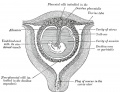
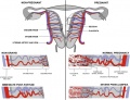
Placental Cord Ultrasound

Ultrasound image of transverse scan through the cord show the method of estimation of the cross-sectional area.

Cord Abnormalities
Cord Vessel Number

Cord Knotting

There are few abnormalities associated with umbilical cord development, other that abnormally short or long cords, which in most cases do not cause difficulties.
In some cases though, long cords can wrap around limbs or the fetus neck, which can then restrict blood flow or lead to tissue or nerve damage, and therefore effect develoment.
Cord knotting can also occur (1%) in most cases these knots have no effect, in some cases of severe knotting this can prevents the passage of placental blood.
Umbilical cord torsion
Rare umbilical cord torsion, even without knot formation can also affect placental blood flow, even leading to fetal demise.[7]
Cord Length
References
Reviews
Articles
Search PubMed
May 2010 search "Placental Cord Development]" All (650) Review (91) Free Full Text (119)
Search Pubmed: Placental Cord | Umbilical Cord | Placental Cord Development | Umbilical Cord Development | Hofbauer cells
Additional Images
see all online Placental materials

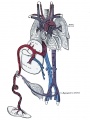
Fetal circulation overview


Glossary Links
- Glossary: A | B | C | D | E | F | G | H | I | J | K | L | M | N | O | P | Q | R | S | T | U | V | W | X | Y | Z | Numbers | Symbols | Term Link
Cite this page: Hill, M.A. (2024, April 18) Embryology Placenta - Cord. Retrieved from https://embryology.med.unsw.edu.au/embryology/index.php/Placenta_-_Cord
- © Dr Mark Hill 2024, UNSW Embryology ISBN: 978 0 7334 2609 4 - UNSW CRICOS Provider Code No. 00098G