Pig Development

Introduction
Pig (Sus scrofa) developmental model is studied extensively due to the commercial applications of pigs for meat production and for health issues such as obesity, cardiovascular disease, and organ transplantation (xenotransplantation).
Historically, there is an excellent description of the pig reproductive estrous cycle and the cyclic changes that occur within the ovary.[1]
Some Recent Findings
- How pig sperm prepares to fertilize[2]"We propose that this capacitation driven membrane docking and stability thereof is a preparative step prior to the multipoint membrane fusions characteristic for the acrosome reaction induced by sperm-zona binding."
- Axial differentiation and early gastrulation stages of the pig embryo[3] "Differentiation of the principal body axes in the early vertebrate embryo is based on a specific blueprint of gene expression and a series of transient axial structures such as Hensen's node and the notochord of the late gastrulation phase. ... Intriguingly, the round shape and gradual posterior displacement of the APD in the pig appear to be species-specific (differing from all other mammals studied in detail to date) but correlate with ensuing specific primitive streak and extraembryonic mesoderm development. APD and, hence, the earliest axial structure presently known in the mammalian embryo may thus be functionally involved in shaping extraembryonic membranes and, possibly, the specific adult body form."
Taxon
Taxonomy ID: 9823
Genbank common name: pig
Inherited blast name: even-toed ungulates
Rank: species
Genetic code: Translation table 1 (Standard)
Mitochondrial genetic code: Translation table 2 (Vertebrate Mitochondrial)
Other names: wild boar, swine, pigs
Lineage (full): cellular organisms; Eukaryota; Fungi/Metazoa group; Metazoa; Eumetazoa; Bilateria; Coelomata; Deuterostomia; Chordata; Craniata; Vertebrata; Gnathostomata; Teleostomi; Euteleostomi; Sarcopterygii; Tetrapoda; Amniota; Mammalia; Theria; Eutheria; Laurasiatheria; Cetartiodactyla; Suina; Suidae; Sus
Uterus and Ovary

Diagram showing form and dimensions of the uterus and Fallopian tubes of the sow.[4] Drawn from an average specimen taken from a young mature animal.
Estrous Cycle
Non-Pregnant

Events of the average cycle of 21 days in the non-pregnant sow.[4]
Diagram showing relationship between oestrua, ovulation, corpus luteum development, and the progress of the ova in the sow.
Pregnant

Events of the first weeks of pregnancy.[4]
Diagram showing relationship between oestrua, ovulation, corpus luteum development, and the progress of the ova in the sow.
Pig Development

- The gestation period of a pig is 112 to 114 days.
- Female pigs can become pregnant at around 8 to 18 months of age.
- The pig has an estrus cycle occurring every 21 days if not bred.
- Male pigs become sexually active at 8 to 10 months of age.
- Embryos begin to attach to the uterus on days 13–14 of pregnancy.
- A litter of piglets is between 6 and 12 piglets.
Data For Carnegie Stages Comparison Graph (Species/Days)
| Species | Stage | |||||||||||||||
| Human | Days | 20 | 22 | 24 | 28 | 30 | 33 | 36 | 40 | 42 | 44 | 48 | 52 | 54 | 55 | 58 |
| Pig | Days | 14 | 15 | 16 | 17 | 18 | 19 | 20.5 | 21.5 | 23 | 24 | 25.5 | 27.5 | 29 | 30.5 | 32.5 |
- Links: Carnegie Stage Comparison
Neural Development
The data below is summarised from an excellent study of early neural development in the pig.[6] The same authors have studied neural development in the rabbit.
- 7 somite embryo - first apposition of the neural folds occurs at somite levels 5-7. (corresponds to closure site I in mouse).
- next stage - rostral and caudal parts of the rhombencephalic folds appose, leaving an opening in between.
- at this stage four neuropores can be distinguished, of which the anterior and posterior ones will remain open longest. (two rhombencephalic closure sites have no counterpart in the mouse, but do have some resemblance to those of the rabbit)
anterior neuropore
- closes in three phases
- dorsal folds slowly align and then close instantaneously, the slow progression being likely due to a counteracting effect of the mesencephalic flexure
- dorso-lateral folds close in a zipper-like fashion in caudo-rostral direction
- final round aperture is likely to close by circumferential growth.
22 somite embryo - anterior neuropore is completely closed. (closure sites for the anterior neuropore in mouse embryo, none of these were detected in the pig embryo)
posterior neuropore
- closes initially very fast in the somitic region, but this process almost stops thereafter.
- stage 20-22 somites the posterior neuropore suddenly reduces in size but thereafter a small neuropore remains for 5 somite stages.
- closure of the posterior neuropore is completed at the stage of 28 somites.
8-20 somite embryos - the width of the posterior neuropore does not change, while the rate of closure gradually increases.
Additional Images
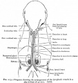
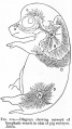
Diagram showing the arrangement of the lymphatic vessels in a pig embryo of 40 mm.


References
- ↑ Corner, G.W., Cyclic changes in the ovaries and uterus of swine, and their relations to the mechanism of implantation. Contributions to Embryology Carnegie Institution, 1922, No.64 117-146.
- ↑ <pubmed>20585455</pubmed>
- ↑ <pubmed>19683851</pubmed>
- ↑ 4.0 4.1 4.2 Corner, G.W., Cyclic changes in the ovaries and uterus of swine, and their relations to the mechanism of implantation. Carnegie Institution - Contributions to Embryology No.64 (1922) 117-146.
- ↑ <pubmed>20640155</pubmed>| PMC2904919
- ↑ <pubmed>10985427</pubmed>
Reviews
Articles
- Corner, G.W., Cyclic changes in the ovaries and uterus of swine, and their relations to the mechanism of implantation. Contributions to Embryology Carnegie Institution, 1922, No.64 117-146.
Search PubMed
- pig development - All (22158) Review (1443) Free Full Text (6175)
- pig embryo - All (7265) Review (518) Free Full Text (1792)
Search Pubmed: pig development | pig embryo | Sus scrofa development
External Links
- NCBI - Pig Genome
- USA - PigBase a computer database that includes information on papers published about gene mapping in the pig.
- NSW Agriculture - Pig breeds and breeding
- AGBU - Pig Genetics
Animal Development
| Animal Development: axolotl | bat | cat | chicken | cow | dog | dolphin | echidna | fly | frog | goat | grasshopper | guinea pig | hamster | horse | kangaroo | koala | lizard | medaka | mouse | opossum | pig | platypus | rabbit | rat | salamander | sea squirt | sea urchin | sheep | worm | zebrafish | life cycles | development timetable | development models | K12 |
Glossary Links
- Glossary: A | B | C | D | E | F | G | H | I | J | K | L | M | N | O | P | Q | R | S | T | U | V | W | X | Y | Z | Numbers | Symbols | Term Link
Cite this page: Hill, M.A. (2024, April 18) Embryology Pig Development. Retrieved from https://embryology.med.unsw.edu.au/embryology/index.php/Pig_Development
- © Dr Mark Hill 2024, UNSW Embryology ISBN: 978 0 7334 2609 4 - UNSW CRICOS Provider Code No. 00098G