Pharyngeal arches: Difference between revisions
| Line 5: | Line 5: | ||
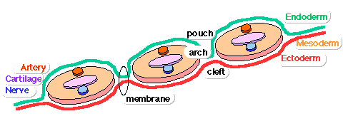
Each arch has initially identical structures: an internal endodermal pouch, a mesenchymal ([[M#mesoderm|mesoderm]] and [[N#neural crest|neural crest]]) core, a membrane ([[E#endoderm|endoderm]] and [[E#ectoderm|ectoderm]]) and external cleft ([[E#ectoderm|ectoderm]]). Each arch mesenchymal core also contains similar components: blood vessel, nerve, muscular, cartilage. | Each arch has initially identical structures: an internal endodermal pouch, a mesenchymal ([[M#mesoderm|mesoderm]] and [[N#neural crest|neural crest]]) core, a membrane ([[E#endoderm|endoderm]] and [[E#ectoderm|ectoderm]]) and external cleft ([[E#ectoderm|ectoderm]]). Each arch mesenchymal core also contains similar components: blood vessel, nerve, muscular, cartilage. | ||
:'''Links:''' [[Head Development]] | [[Neural Crest Development]] | [[ | :'''Links:''' [[Head Development]] | [[Neural Crest Development]] | [[Endocrine System Development]] | ||
==Pharyngeal Arch Development== | ==Pharyngeal Arch Development== | ||
Revision as of 00:31, 5 August 2010
Introduction
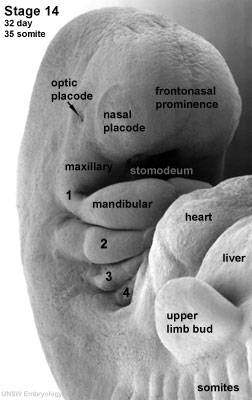
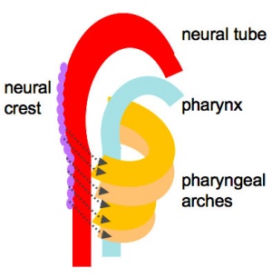
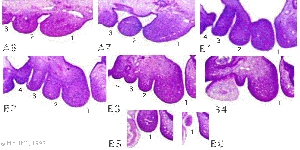
The pharyngeal arches (branchial arch, Greek, branchial = gill) are a series of externally visible anterior tissue bands lying under the early brain that give rise to the structures of the head and neck. Each arch though initially formed from similar components will differentiate to form different head and neck structures. In humans, five arches form (1,2,3,4 and 6) but only four are externally visible on the embryo.
Each arch has initially identical structures: an internal endodermal pouch, a mesenchymal (mesoderm and neural crest) core, a membrane (endoderm and ectoderm) and external cleft (ectoderm). Each arch mesenchymal core also contains similar components: blood vessel, nerve, muscular, cartilage.
Pharyngeal Arch Development
- branchial arch (Gk. branchia= gill)
- arch consists of all 3 trilaminar embryo layers
- ectoderm- outside
- mesoderm- core of mesenchyme
- endoderm- inside
Neural Crest
- Mesenchyme invaded by neural crest generating connective tissue components
- cartilage, bone, ligaments
- arises from midbrain and hindbrain region
Arch Features
Each arch contains: artery, cartilage, nerve, muscular component
Arches and Phanynx Form the face, tongue, lips, jaws, palate, pharynx and neck cranial nerves, sense organ components, glands
- Humans have 5 arches - 1, 2, 3, 4, 6 (Arch 5 does not form or regresses rapidly)
- from in rostro-caudal sequence, Arch 1 to 6 from week 4 onwards
- arch 1 and 2 appear at time of closure of cranial neuropore
- Face - mainly arch 1 and 2
- Neck components - arch 3 and 4 (arch 4 and 6 fuse)
Arch Features
- arch
- groove
- externally separates each arch
- also called a cleft
- only first pair persist as external auditory meatus
- externally separates each arch
- pouch
- internally separates each arch
- pockets from the pharynx
- membrane
- ectoderm and endoderm contact regions
- only first pair persist as tympanic membrane
- Pharyngeal Arch 1 (Mandibular Arch) has 2 prominances
- smaller upper- maxillary forms maxilla, zygomatic bone and squamous part of temporal
- larger lower- mandibular, forms mandible
- Pharyngeal Arch 2 (Hyoid Arch)
- forms most of hyoid bone
- Arch 3 and 4
- neck structures
Embryo Week: Week 1 | Week 2 | Week 3 | Week 4 | Week 5 | Week 6 | Week 7 | Week 8 | Week 9
- Carnegie Stages: 1 | 2 | 3 | 4 | 5 | 6 | 7 | 8 | 9 | 10 | 11 | 12 | 13 | 14 | 15 | 16 | 17 | 18 | 19 | 20 | 21 | 22 | 23 | About Stages | Timeline
Glossary Links
- Glossary: A | B | C | D | E | F | G | H | I | J | K | L | M | N | O | P | Q | R | S | T | U | V | W | X | Y | Z | Numbers | Symbols | Term Link
Cite this page: Hill, M.A. (2024, April 18) Embryology Pharyngeal arches. Retrieved from https://embryology.med.unsw.edu.au/embryology/index.php/Pharyngeal_arches
- © Dr Mark Hill 2024, UNSW Embryology ISBN: 978 0 7334 2609 4 - UNSW CRICOS Provider Code No. 00098G


