Pharyngeal arches: Difference between revisions
mNo edit summary |
mNo edit summary |
||
| Line 28: | Line 28: | ||
{{Pharyngeal Arch table}} | {{Pharyngeal Arch table}} | ||
== Some Recent Findings == | |||
{| | |||
|-bgcolor="F5FAFF" | |||
| | |||
|} | |||
{| class="wikitable mw-collapsible mw-collapsed" | |||
! More recent papers | |||
|- | |||
| [[File:Mark_Hill.jpg|90px|left]] {{Most_Recent_Refs}} | |||
Search term: [http://www.ncbi.nlm.nih.gov/pubmed/?term=Pharyngeal+Arch ''Pharyngeal Arch''] | |||
<pubmed limit=5>Pharyngeal Arch</pubmed> | |||
|} | |||
===Neural Crest === | ===Neural Crest === | ||
| Line 35: | Line 51: | ||
Links: [[Neural Crest Development]] | Links: [[Neural Crest Development]] | ||
===Arch Features=== | ===Arch Features=== | ||
Revision as of 09:28, 30 January 2017
| Embryology - 18 Apr 2024 |
|---|
| Google Translate - select your language from the list shown below (this will open a new external page) |
|
العربية | català | 中文 | 中國傳統的 | français | Deutsche | עִברִית | हिंदी | bahasa Indonesia | italiano | 日本語 | 한국어 | မြန်မာ | Pilipino | Polskie | português | ਪੰਜਾਬੀ ਦੇ | Română | русский | Español | Swahili | Svensk | ไทย | Türkçe | اردو | ייִדיש | Tiếng Việt These external translations are automated and may not be accurate. (More? About Translations) |
Introduction
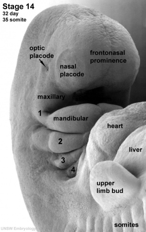
The pharyngeal arches (branchial arch, Greek, branchial = gill) are a series of externally visible anterior tissue bands lying under the early brain that give rise to the structures of the head and neck. Each arch though initially formed from similar components will differentiate to form different head and neck structures. In humans, five arches form (1, 2, 3, 4 and 6) but only four are externally visible on the embryo.
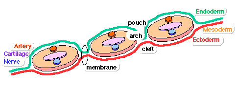
Each arch has initially identical structures: an internal endodermal pouch, a mesenchymal (mesoderm and neural crest) core, a membrane (endoderm and ectoderm) and external cleft (ectoderm). Each arch mesenchymal core also contains similar components: blood vessel, nerve, muscular, cartilage.
Note is a draft page and this topic is currently covered in more detail on the Head Development page.
Pharyngeal Arch Development
- branchial arch (Gk. branchia= gill)
- arch consists of all 3 trilaminar embryo layers
- ectoderm- outside
- mesoderm- core of mesenchyme
- endoderm- inside
Pharyngeal Arch Tables
This table gives an overview of what each arch will contribute to the embryo.
| Pharyngeal Arch | Nerve | Artery | Neural Crest (Skeletal Structures) |
Muscles | Ligaments |
|---|---|---|---|---|---|
| 1 (maxillary/mandibular) |
trigeminal (CN V) | maxillary artery (terminal branches) | mandible, maxilla, malleus, incus | muscles of mastication, mylohyoid, tensor tympanic, ant. belly digastric | ant lig of malleus, sphenomandibular ligament |
| 2 (hyoid) |
facial (CN VII) | stapedial (embryonic) corticotympanic (adult) |
stapes, styloid process, lesser cornu of hyoid, upper part of body of hyoid bone | muscles of facial expression, stapedius, stylohyoid, post. belly digastric | stylohyoid ligament |
| 3 | glossopharyngeal (CN IX) | common carotid, internal carotid arteries | greater cornu of hyoid, lower part of body of hyoid bone | stylopharyngeus | |
| 4 | vagus (CN X) superior laryngeal branch | part of aortic arch (left), part right subclavian artery (right) | thyroid, cricoid, arytenoid, corniculate and cuneform cartilages | crycothyroid, soft palate levator veli palatini (not tensor veli palatini) | |
| 6 | vagus (CN X) recurrent laryngeal branch | part of left pulmonary artery (left), part of right pulmonary artery (right) | thyroid, cricoid, arytenoid, corniculate and cuneform cartilages | larynx intrinsic muscles (not cricothyroid muscle) |
Some Recent Findings
| More recent papers |
|---|
|
This table allows an automated computer search of the external PubMed database using the listed "Search term" text link.
More? References | Discussion Page | Journal Searches | 2019 References | 2020 References Search term: Pharyngeal Arch <pubmed limit=5>Pharyngeal Arch</pubmed> |
Neural Crest
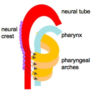
- Mesenchyme invaded by neural crest generating connective tissue components
- cartilage, bone, ligaments
- arises from midbrain and hindbrain region
Links: Neural Crest Development
Arch Features
Each arch contains: artery, cartilage, nerve, muscular component
Arches and Phanynx Form the face, tongue, lips, jaws, palate, pharynx and neck cranial nerves, sense organ components, glands
- Humans have 5 arches - 1, 2, 3, 4, 6 (Arch 5 does not form or regresses rapidly)
- from in rostro-caudal sequence, Arch 1 to 6 from week 4 onwards
- arch 1 and 2 appear at time of closure of cranial neuropore
- Face - mainly arch 1 and 2
- Neck components - arch 3 and 4 (arch 4 and 6 fuse)
Arch Features
- arch
- groove
- externally separates each arch
- also called a cleft
- only first pair persist as external auditory meatus
- externally separates each arch
- pouch
- internally separates each arch
- pockets from the pharynx
- membrane
- ectoderm and endoderm contact regions
- only first pair persist as tympanic membrane
- Pharyngeal Arch 1 (Mandibular Arch) has 2 prominances
- smaller upper- maxillary forms maxilla, zygomatic bone and squamous part of temporal
- larger lower- mandibular, forms mandible
- Pharyngeal Arch 2 (Hyoid Arch)
- forms most of hyoid bone
- Arch 3 and 4
- neck structures
Embryo Week: Week 1 | Week 2 | Week 3 | Week 4 | Week 5 | Week 6 | Week 7 | Week 8 | Week 9
- Carnegie Stages: 1 | 2 | 3 | 4 | 5 | 6 | 7 | 8 | 9 | 10 | 11 | 12 | 13 | 14 | 15 | 16 | 17 | 18 | 19 | 20 | 21 | 22 | 23 | About Stages | Timeline
Pharyngeal Arch 1
Pharyngeal Arch 2
Pharyngeal Arch 3
Pharyngeal Arch 4
Pharyngeal Arch 6
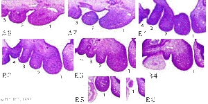
Additional Images
Historic Images
| Historic Disclaimer - information about historic embryology pages |
|---|
| Pages where the terms "Historic" (textbooks, papers, people, recommendations) appear on this site, and sections within pages where this disclaimer appears, indicate that the content and scientific understanding are specific to the time of publication. This means that while some scientific descriptions are still accurate, the terminology and interpretation of the developmental mechanisms reflect the understanding at the time of original publication and those of the preceding periods, these terms, interpretations and recommendations may not reflect our current scientific understanding. (More? Embryology History | Historic Embryology Papers) |
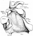
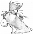
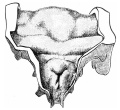
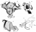
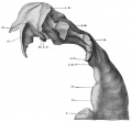
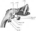
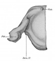
Frazer JE. The second visceral arch and groove in the tubo-tympanic region. (1914) J Anat Physiol. 48(4): 391-408. PMID 17233005
- Second Pharyngeal Arch
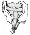
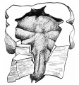
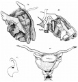
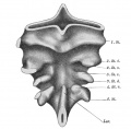
Frazer JE. Development of the larynx. (1910) J Anat. 44: 156-191. PMID 17232839
- Embryonic Larynx Development
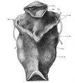
Keibel F. and Mall FP. Manual of Human Embryology II. (1912) J. B. Lippincott Company, Philadelphia.
- Pharynx
Glossary Links
- Glossary: A | B | C | D | E | F | G | H | I | J | K | L | M | N | O | P | Q | R | S | T | U | V | W | X | Y | Z | Numbers | Symbols | Term Link
Cite this page: Hill, M.A. (2024, April 18) Embryology Pharyngeal arches. Retrieved from https://embryology.med.unsw.edu.au/embryology/index.php/Pharyngeal_arches
- © Dr Mark Hill 2024, UNSW Embryology ISBN: 978 0 7334 2609 4 - UNSW CRICOS Provider Code No. 00098G
