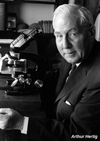
Paper - the primary human oocyte - some observations on the fine structure of Balbiani's vitelline body and the origin of the annulate lamellae
| Embryology - 19 Apr 2024 |
|---|
| Google Translate - select your language from the list shown below (this will open a new external page) |
|
العربية | català | 中文 | 中國傳統的 | français | Deutsche | עִברִית | हिंदी | bahasa Indonesia | italiano | 日本語 | 한국어 | မြန်မာ | Pilipino | Polskie | português | ਪੰਜਾਬੀ ਦੇ | Română | русский | Español | Swahili | Svensk | ไทย | Türkçe | اردو | ייִדיש | Tiếng Việt These external translations are automated and may not be accurate. (More? About Translations) |
Hertig AT. The primary human oocyte: some observations on the fine structure of Balbiani's vitelline body and the origin of the annulate lamellae. (1968) Amer. J Anat. 122: 107-37. PMID 5654499
| Online Editor |
|---|
| This historic 1968 paper describes early human oocyte structure. Draft only.
Hertig AT. and Rock J. Two human ova of the pre-villous stage, having a developmental age of about seven and nine days respectively. (1945) Contrib. Embryol., Carnegie Inst. Wash. Publ. 557, 31: 65-84. Hertig AT. and Rock J. On a normal human ovum not over 7.5 days of age. (1945) Anat. Rec. 91: 281. Hertig AT. and Rock J. On a normal ovum of approximately 9 to 10 days of age. (1945) Anat. Rec. 91: 281.
|
| Historic Disclaimer - information about historic embryology pages |
|---|
| Pages where the terms "Historic" (textbooks, papers, people, recommendations) appear on this site, and sections within pages where this disclaimer appears, indicate that the content and scientific understanding are specific to the time of publication. This means that while some scientific descriptions are still accurate, the terminology and interpretation of the developmental mechanisms reflect the understanding at the time of original publication and those of the preceding periods, these terms, interpretations and recommendations may not reflect our current scientific understanding. (More? Embryology History | Historic Embryology Papers) |
The primary human oocyte - Some observations on the fine structure of Balbiani's vitelline body and the origin of the annulate lamellae
Department of Pathology, Harvard Medical School, Boston, Massachusetts
1 The present communication forms the historical and scientific basis of the addressgiven by the author after the banquet of the American Association of Anatomists in Kansas City, Mo., April 6, 1967 entitled: “Human Oocytes, Past Present and Future; a Resurrection of Balbianfs Body.”
2 This work was supported U.S.P.H.S. grant I-ID-00137.
Abstract
The ultrastructural details of human oocytes from four primordial
follicles and one early primary follicle are presented. A fifth primordial follicle is represented by a paraflin section stained by hematoxylin and eosin. The paranuclear Balbiani vitelline body, contsisting of a centrosome surrounded by endoplasmic reticulum, Golgi complexes, compound aggregates, annulate lamellae, and mitochondria is described. The annulate larnellae arise as an evagination from the outer leaflet of the nuclear envelope and interdigitate with folds of the endoplasmic reticulum which also is continuous with the outer leaflet of the nuclear envelope. Structural aspects of annulate lamellae are discussed in relationship to current ideas of nuclear membrane ultrastructure and to their possible role in nucleo-cytoplasmic transfer.
A biographical note on the life of Edouard Gérard Balbiani is presented.
The conspicuous paranuclear mitochondrial mass surrounding the centrosome of vertebrate and invertebrate oocytes has been studied by light microscopists for many years. According to Henneguy (1887) it was described in the spider oocyte by von Wittich in 1845 and was named the “Dotterkern” by Came in 1848. Balbiani described it in detafl in oocytes of spiders and myriapods (1864a, b). He summarized his extensive observations in 1893. This structure was designated by Henneguy - Balbianifs student - as “Balbiani’s vesicle” in 1887 and as the “yolk body of Balbiani” in 1893. Van der Stricht in his classic monograph of 1923 on the developmental stages of oocytes in mammals distinguishes clearly between the central vitelline or Balbiani body and the surrounding vitellogenic or mitochondrial bed. Aykroyd (’38), following the example of Brambell, Whose 1925 paper she quotes, designated these areas as the “pallial layer” and “couche vitellogéne” respectively. These authors believed that in the past Balbiani’s name has been applied indiscriminately to either or both the components of this paranuclear complex. Beams and Sheehan (’41) designate it as the “yolk nucleus complex” whereas Raven (’61) believes that it should be called Balbiani’s vitelline body. We agree with this latter designation and apply that term to the entire paranuclear complex of apparently interrelated organelles in human oocytes within primordial and early transitional primary follicles. Four of the oocytes from primordial follicles illustrated here were included in a larger group previously reported in detafl (Hertig and Adams, 67). One previously unpublished primordial oocyte is illustrated and, in addition, one oocyte from an early transitional primary follicle (H35-2) will be described (table 1).
During the preparation of this paper and the address based upon it, the author became interested in the life and Works of Edouard Gérard Balbiani, the cytologist, comparative embryologist and supplier of medical eponyms. A biographical summary of this remarkable man is included here.
Edouard Gérard Balbiani (fig. 17) came from an Italian family of ancient lineage. During the reign of Francis the first his ancestors left Italy to reside in various European countries. Edouard descended from the German branch; his banker father having emigrated to Haiti. Balbiani was born, apparently" in Port-auPrince, about 1823. His classical schooling took place in Frankfurt-am-Main and in Paris. There he went on to study law, natural science and medicine; obtaining his M.D. in 1854 (Henneguy, ’00; Boyer, ’48; Bibliographic Anatomique, 1899; Nature, 1899).
Balbiani immediately embarked upon a scientific career. He was a patient, skillful dissector and manipulator of living cells and unicellular organisms which he observed for hours; recording the effect upon them of various reagents. He developed techniques of microdissection and coined the term “merotomy.” During his first study of the ciliated infusoria he observed their fission and conjugation. Believing that such animals Were complete and that the macronucleus was an ovary and the micronucleus a “testicule,” he totally misunderstood the process of mitosis. According to Hughes, (plate X, fig. G., ’59) the original plates of Balbiani’s 1861 paper, however, show several phases of mitotic division; the spindle fibers and chromosomes unfortunately being interpreted as a bundle of spermatozoa! Thus, although Balbiani was the first to observe and record the vital process of mitosis, it remained for Biitschli in 1876 to interpret correctly the role of the micronucleus in the sexual reproduction of ciliates. Ba.lbiani’s skills brought him to the attention of Claude Bernard who appointed him in 1867 as Director of Histological Studies of the Laboratory of Physiology at the Museum of Natural Sciences. He held this post until 1873 when he succeeded Coste in the Professorship of Comparative Embryology at the College de France.
The complex, subsequently to be called the “yolk body of Balbiani” by Henneguy in 1893, was first studied by Balbiani in spiders and myriapods ( 1864a, b). His interpretation of these structures was obscured by his belief in the hermaphroditism which he thought he had established for the ciliates. In applying such concepts to the germ cells of metazoa he attributed a role in the formation of the “germ” to the yolk nucleus. Later (1893) he was to regard the “yolk nucleus” (Dotterkern) as homologous to the Nebenkern (centrosome of Platner) of germinal cells and of the centrosome of somatic cells. He believed that the yolk nucleus originated from the nucleus of the immature oocyte and that during its formation, a more or less thick layer of yolk condensed around it. (It is now clear that what he was observing was the centrosome with its surrounding mass of organelles, undoubtedly Golgi bodies and mitochondria. Please refer to figure 1, an intact oocyte from the myriapod, Geophilus longicornis drawn by Balbiani himself and figure 18, the microscope with which he presumably studied such oocytes.)
As an applied scientist he identified the causative sporozoan of the silk-worm disease, pebrme, whose cure added to Pasteur’s growing fame. Balbiani also Worked out the life cycle of the root louse, Phylloxem vastatrix, with its parthenogenetic generations, which was infesting the French vineyards (Henneguy, ’O0; Boyer, ’-48). Although Balbiani recommended decortication (“décorticage des ceps”) of the vine stocks, and application of insecticides to the roots to control the overwintering louse eggs, the more practical solution to this pressing problem lay elsewhere. Resistant root stocks of Vitis rupestris were imported from American vineyards and upon these Were grafted the native French Vitis vinifera (Amerine, ’66).
Perhaps Balbianfs greatest cytological achievement lay in his discovery and description of the giant chromosome with its surrounding puffs or rings in the larval salivary glands of the midge, Chironomus plumosus (fig. 19). He named this structure a “cordon” (Balbiani, 1881a, b) and it is later referred to as a “filament nucléin— ien” (Bibliographic Anatomique, 1899). It is not clear whether it was accepted as a chromosome or not, even though Balbiani himself (1876) explicitly identified it as being homologous with the filaments which he had earlier observed in the nuclei of dividing cells in the ovary of the grasshopper, Stenobrothus pmtorum. He also related the “cordon” to the nuclear filaments of cells in karyokinesis as described in the salamander by Pfitzner ( 1881). [The term “mitosis” was introduced in 1882 by Flemming and the term “chromosome” by Waldeyer in 1888. It was of historic interest that at this time Miescher (1871) and Altmann (1889) were doing their pioneer work on the nucleic acids.
Balbiani described at magnifications of 100-150, the irregularly convoluted or folded cord of 15 u in diameter with its dark cross striations and discoid pale expansions. The relationship of the “cordon” to the nucleoli is seen in all three of his drawings (fig. 19). The cross striations were stained by methyl green although the intervening areas and rings were unstained. Carmine and hematoxylin, however, left the bands only slightly stained but stained the rings and the nucleoli intensely. It is ironic that Balbiani refused to speculate (1881b) on the function of the “cordon” and never returned to it in any of his subsequent publications. Nevertheless, he was the first to describe the morphologic counterpart of the gene concept as formulated by the geneticists.
It is of interest that both Mendel’s and Balbiani’s observations lay unnoticed for many years. By 1915 the location of 50 genes for the four single chromosomes of Drosophila had been mapped (Morgan et al., ’15, as cited by Hughes, ’59). It was not until 1933, however, that Heitz and Bauer realized that the giant chromosomes of dipterous larvae, described by Balbiani in 188-1 under the light microscope, were equivalent to the interphase chromosomes of ordinary cells. This led Painter and others to compare the band patterns of giant chromosomes to established genetic maps in larval Drosophila. There also emerged the chemical nature of Balbiani’s rings or puffs (Beermann, "52; Beermann and Bahr, ’54; Clever and Karlson, ’60).

