Minot CS. The veins of the Wolffian body in the pig. (1898) Proc. Bost. Soc. of Nat. Hist, 28: 265-274.
| Online Editor
|
| This historic 1889 paper by Minot studies the veins of the Wolffian body (mesonephros).
National Library of Medicine holding
See the review below of the article Science 7, No. 164 p. 229
Dr. Minot had studied especially the condition in pig-embryos of 12.0 mm. The cardinal vein ends abruptly at the cephalic end of the Wolffian body ; the vena cava inferior is also well developed and communicates widely with the middle of each mesonephros. Between the Wolffian tubules there are no capillaries, but only ‘large sinuses, the endothelium of which lies close against the epithelium of the tubules. The sinuses communicate freely with both the cardinal and cava veins. Along the dorsal side of the Wolfiian body there is no continuous cardinal vein,’but there are still two channels of reduced size, representing the lower parts of the cardinal which have become united with the cava inferior.
The author described: (1) the mesothelial villi of the allantois in the pig; (2) the development of the hypophysis and infundibular gland in the pig, Amia,Bat1'achus, Ameiurus and Necturus, confirming and extending the results of Béla Haller; (3) observations upon various vertebrate types, tending to show that the zones of His have a constant morphological value ; (4) the fore-brain‘ of Ameiurus Embryos, clearly similar to that of other types of vertebrates as concerns the hemispheres and foramen of Monro; if this observation is confirmed by further study it will show that neither the theory of Burkhardt nor that of Studniscka in regard to homologies of the Teleostean afore-brain is correct.
Modern Notes: mesonephros | vein | pig
|
| Historic Disclaimer - information about historic embryology pages
|
| Pages where the terms "Historic" (textbooks, papers, people, recommendations) appear on this site, and sections within pages where this disclaimer appears, indicate that the content and scientific understanding are specific to the time of publication. This means that while some scientific descriptions are still accurate, the terminology and interpretation of the developmental mechanisms reflect the understanding at the time of original publication and those of the preceding periods, these terms, interpretations and recommendations may not reflect our current scientific understanding. (More? Embryology History | Historic Embryology Papers)
|
The Veins of the Wolffian Body in the Pig
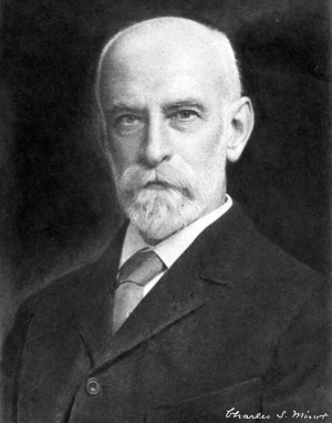
Charles Sedgwick Minot (1852–1914)
Cite this page: Hill, M.A. (2024, April 19) Embryology Paper - The veins of the Wolffian body in the pig. Retrieved from https://embryology.med.unsw.edu.au/embryology/index.php/Paper_-_The_veins_of_the_Wolffian_body_in_the_pig
- What Links Here?
- © Dr Mark Hill 2024, UNSW Embryology ISBN: 978 0 7334 2609 4 - UNSW CRICOS Provider Code No. 00098G
