Paper - The second visceral arch and groove in the tubo-tympanic region: Difference between revisions
mNo edit summary |
mNo edit summary |
||
| (14 intermediate revisions by the same user not shown) | |||
| Line 4: | Line 4: | ||
! Online Editor | ! Online Editor | ||
|- | |- | ||
| [[File:Mark_Hill.jpg|90px|left]] This 1914 paper describes human second pharyngeal arch development. | | [[File:Mark_Hill.jpg|90px|left]] This 1914 paper describes human second pharyngeal arch development. John Ernest Frazer (1870-1946) also wrote on several other embryology topics, as well as published an embryology textbook. | ||
{{Ref-Frazer1940}} | |||
<br> | <br> | ||
See pages [[Pharyngeal arches]] and [[Head Development]] | |||
|} | |} | ||
{{Historic Disclaimer}} | {{Historic Disclaimer}} | ||
=The Second Visceral Arch and Groove in the Tubo-Tympanic Region= | |||
[[File:John Ernest Frazer.jpg|thumb|alt=File:John Ernest Frazer.jpg|link=Embryology History - Ernest Frazer|J. Ernest Frazer (1870-1946)]] | |||
By J. Ernest Frazer, | |||
Lecturer on Anatomy in the Medical School of St Mary’s Hospital. | |||
In 1910 I brought the results of an investigation into the development of the naso-pharynx before the Anatomical Section of the British Medical Association, calling attention at the same time to the fact that the tubotympanic recess has in its floor the outer ends of the first two arches and grooves, and that consequently its posterior margin or wall is formed by tissues owning a third arch value: further, I showed that the first and second lateral pouches persisted and were recognisable throughout the second and third months, with the outer ends of the arches, whereas the inner portion of the second arch disappeared in the tubal region, the wall of the cavity in its neighbourhood undergoing what I called—in default of a better term—a process of atrophy. The description which I published at that time (B.M..T., 15th October) was rather of the nature of a sideissue in the work on which I was engaged, and no subsequent reference was made by me to it; but in the present paper I hope to give a more complete account of the modifications which occur in this region and affect the second arch in particular, and to indicate the rearrangement of the parts that leads to the ultimate formation of the tubo-tympanic cavity. Before entering on such a description, it is perhaps advisable to lay a little stress on the general characters and relations of the tubo-tympanic recess. The early pharynx is a cavity that is very wide from side to side in front, and narrows rapidly and considerably as it is traced back towards the stomach; it is flattened dorso-ventrally, so that the floor and roof are almost in contact, perhaps quite in contact in the living embryo. It has no proper side wall, the lateral limits of the cavity being made by the junctions of floor and roof, and the visceral arches and grooves are only found in the floor; the grooves terminate in lateral pouches (whose subdivisions do not affect the general arrangement), and these lie therefore in the region of the lateral parts of the cavity, where they form depressions deeper than the remainders of their corresponding grooves. Towards the end of the first month the narrowing of the cavity from side to side is seen to take place less gradually than in the earlier stages, and the region of the third arch is the level at which this more sudden decrcaselin width is placed, so that in front of this arch there remains a wide area, and the part of the cavity that extends out in front of the arch, beyond the general lateral limits of the cavity, constitutes the tubo-tympanic recess that will subsequently be moulded into the cavity of the middle ear and Eustachian tube. | |||
Fig. 1 gives in outline the shape of the pharyngeal cavity in a 12-mm. embryo, and illustrates this description, showing how each broad and open recess is floored by the outer parts of the first two arches, how their grooves lie behind them and end externally in deeper lateral pouches, and how the third arch, lying between the second and third pouches, necessarily forms the short posterior boundary of the recess. | |||
recess | |||
the | |||
[[File:Frazer1914 fig01.jpg|500px]] | |||
'''Fig. 1.''' From models to show (A) floor of 12-mm. pharynx, (B) View from above of left tubo-tympanic recess in 16-mm. embryo. Somewhat diagrammatic. M., R., condensations forming Meckel's and Reichert's bars respectively ; man., position of manubrial extension from first arch. This causes depression of the neighbouring wall, with a. secondary projection, 2:, behind it. | |||
Later, as the recess elongates obliquely and develops, it is rotated on its longitudinal axis so that its original floor becomes its front and outer wall, and the periotic capsule that primarily lies on its roof assumes an inner and posterior relationship to it. | |||
It follows from the foregoing statements that an account of the arches utilised in the formation of the recess must concern itself mainly with the floor, and that, as development proceeds, the arch-structures which persist will be found in association with what is described as the outer wall in the anatomy of the adult. The second drawing in fig. 1 is an outline of the -cavity seen from above in the 16-mm. embryo, and the projections of the first and second lateral pouches are easily recognised and compared with those in the 12-mm. specimen. In the older one, however, the wall of the cavity has been pushed in immediately behind the first pouch by the extension of a mass of condensed mesenchyme from the upper part of the first arch, concerned in forming the handle of the malleus, etc. This incursion is associated with temporary separation of the pouch from the ectoderm (see figure), and also with the formation of a secondary projection in the margin of the cavity just behind the portion pushed in by the manubria] condensation. | |||
[[File:Frazer1914 fig02.jpg|500px]] | |||
'''Fig. 2.''' Floor of left recess, roof removed. From model (magn. 100 X 5/6). A portion of the margin of the tongue is seen below. 16 mm. | |||
in | On removing the roof of the recess and thus exposing the floor, the disposition of the arches becomes apparent. fig. 2 is a drawing of the floor of the left recess in an embryo of 16 mm. exposed in this way, and is of interest because the key to subsequent changes is to be found in the conditions present at this stage. | ||
The front part of the field is formed by structures of the first arch, among them Meckel’s bar passing downwards and inwards, and behind this a deep but narrow sulcus marks the situation of the first visceral groove ending externally in the first lateral pouch, just above which the chorda tympani runs forward. The second lateral pouch is apparent behind the styloid bar (Reichert’s bar), between this and the glossopharyngeal, which is closely applied to the wall of the recess here: this is the definite position of the second pouch, shown also in fig. 1 in the 12-mm. specimen, and is the position maintained by it until the growth of the cartilaginous auditory capsule effects a separation between it and the nerve. | |||
The area of the second arch lies between the first and second pouches, and is plainly subdivided into two districts, an anterior one showing the prominence caused by the underlying manubrial condensation, and a posterior one exhibiting a convexity which is evidently caused by the styloid bar against which this part of the floor rests; the sulcus between these two districts corresponds, of course, with the secondary projection seen externally in fig. 1. | |||
But, if we follow the complete second arch area inwards, it becomes clear that there is already a difference apparent between the arrangement of structures in the floor here and that in the 12-mm. embryo, and this difference is due to growth of the third arch tissues. The region of the third arch, where it forms the hinder boundary of the recess, has become more prominent, and an extension forward from it has taken place, spreading across the inner part of the floor towards the first arch, and, in doing so, covering over the tissues of the second arch in this situation, so that this latter is not represented in this part of the floor. The extension of the third arch is evident in the figure, and it is seen to be separated from the first arch region by a shallow gutter towards which the limiting sulci of the second arch are directed: they reach a broader hollow from which thevfirst-mentioned shallow gutter is directed forwards and inwards. The floor of the gutter and broader hollow, as seen in the model and the drawing, is composed of much—thickened epithelium, and a reconstruction of the regions with the epithelium removed allows the surface appearance of the second arch to be followed a little lower than in the complete condition; but the general result is the same—the outer part of the arch is concerned in the formation of the floor of the cavity and is divisible there into its two districts, but the inner part has dropped out of the floor, and is separated from the cavity by a forward growth of the arch immediately succeeding it. | |||
It may be pointed out here that the floor of the recess shows a general and well-marked concavity from before backwards as well as from without inwards, a condition that is not brought out in the drawing owing to the necessity for a clear presentation of the arches, etc. In other words, the growth of the first arch has turned up the related floor, the same effect has been produced on the manubrial district of the second arch by the mesenchymal extension underlying it, the styloid bar is directed downwards as well as inwards, so that the posterior district of the arch is correspondingly disposed, and the third arch is raising the hinder part of the floor as a result of its increasing size: the part that remains unaffected by these several factors is the broad hollow toward which the other surfaces slope, and the narrower gutter leading away from it which marks the place where the first and third arches have not yet met, but where such meeting is about to occur. | |||
the | |||
The further development of the region up to the third month is a simple progression of the changes seen in their early stages in fig. 2. The first arch area stands up more, the manubrial district of the second arch is more prominent and defined, and the second pouch is recognisable between the styloid bar and the glosso-pharyngeal nerve. But a distinct change is visible when attention is directed to the third arch region; the arch, presumably as" a result of its growth, is encroaching on the lumen of the inner portion of the cavity from behind, so that already some indication of a division into tubal and tympanic parts is foreshadowed. A drawing of one of the models showing this stage is seen in fig. 3, and it is interesting to compare it with the earlier condition: recognition of the first and second arch region is easy, and the districts into which the latter is subdivided are evident and unmistakable. | |||
But equally evident and unmistakable is the change that is apparent in the back and inner boundary of the recess. The third arch has grown and has carried this part of the hinder wall in a forward direction, tending to lessen the size of the opening of the recess from behind forwards: this can be graphically demonstrated and appreciated by superimposing a tracing of the later upon the earlier stage, when the first and second arch areas will be found practically to correspond and occupy their proper positions, whereas the increased obliquity forward of the hinder wall shows at once how much this part has extended in a forward direction relative to the other parts. Such growth, moreover, is most marked at the inner or pharyngeal end of the hinder wall, so that the corresponding portion of the recess exhibits some narrowing compared with the remainder. | |||
this | |||
the | |||
The chief growth appears to be in the prominent part of the arch from which (fig. 2) the forward extension takes place, and in this extension itself. As a result of growth in the first-mentioned region, the depth and abruptness of the postero-internal wall of the broad hollow in the floor is increased and the hollow thus accentuated,.while the growth of the forward extension has enabled this to reach the mandibular arch and to obliterate in doing so the gutter that separated the two in the earlier stage. The whole process is a. continuation of that seen in its early stages at 16 mm., and indicates the results of growth forward of the third toward the first arch across those tissues of the second arch which should be in the inner part of the floor of the recess, but still leaving the outer parts" of the second arch exposed in the corresponding portion of the floor. The two districts into which the outer part of the second arch is divided are more definite at this stage, principally because of the greater prominence and definition of the manubrial swelling, which not only stands out into the cavity, but has a deeper sulcus between it and the styloid district; at one place this sulcus shows an extension deeply round the back of the manubrium, which is apparently an early indication of the posterior recess of Tr‘o‘ltsch. The styloid district is if anything relatively smaller, depending on the thickness of the bar on which it is moulded. The second pouch is seen behind, and a little internal to, the styloid district, between the styloid bar and the glosso-pharyngeal nerve. | |||
of | |||
of the | |||
which | |||
The | |||
[[File:Frazer1914 fig03.jpg|500px]] | |||
'''Fig. 3.''' Left recess, from a model. 27 mm. Observe that the forward extension, A, from third arch along the floor is much more marked and prominent than in the last figure, and has reached the first arch. | |||
The first arch region, like the manubrial mass, stands up more than in the early stages; in other words, the front and outer part of the floor is being lifted up by underlying growth, and thus being brought more into the position of an outer and front Wall, as mentioned in the beginning of this paper. It follows as a result that the growth of these parts assists in accentuating the hollow that lies at the bottom of their declivities. | |||
these | |||
[[File:Frazer1914 fig04.jpg|500px]] | |||
'''Fig. 4.''' Left recess, simplified, from model 35 mm. '''a''', bottom of tympanum practically corresponding with lower extremity mental plate and tympanic ring; '''b, c''', prominences made by back and front edges of mental plate growing in round the manubrium ; th., tympano-hyal area. | |||
It is about this time that the process begins, the effects of which are so strikingly seen in specimens of about 35 mm. length: apparently the mass of the third arch advances toward the area of the first arch with greater rapidity, along the floor, so that one is heaped up, so to speak, against the other, with an_ epithelial septum lying between them composed of the cells which at the time cover their meeting surfaces. | |||
and | Fig. 4 is from a model of a 35-mm. embryo, and shows how greatly the third arch has extended forward; comparison with the earlier stages makes this evident. The recess as a whole is slightly longer, and the growth of the third arch particularly affects the inner part and narrows it, thus definitely indicating the distinction between tube and tympanum. But it is equally plain that this narrowing has not been obtained by a raising up of the epithelial floor as a result of growth under it, but by a fusion of the surfaces of the first and third arches where they have come into contact; for an epithelial lamina is present below the newly made “ floor" in this region which is continuous with the epithelial lining of the cavity along its length from the hinder boundary of the pharyngeal opening to the hollow situated at the foot of the anterior district of the second arch in the tympanic region. Such a lamina can be nothing but the covering of the prominent arches caught between them as they meet. The lamina, as shown in the model, consists only of a solid and continuous epithelial septum; but under the microscope broken layers and masses of cells can be found which are not suitable for modelling, but indicate that the fusion. is more extensive behind and below than is suggested by the model. What is probably the beginning of this process is found in the 29—mm. embryo, in which an epithelial mass can be seen lying above the front part of the forward extension of the third arch, between the thicker posterior part of this extension and the first arch eminence. | ||
part of the floor of | |||
arch | |||
The | |||
are | |||
and | |||
the | |||
The lower part of the tympanic region is now (fig. 4) more definitely limited in front, by the fusion that narrows the cavity, and it becomes necessary to see how far back the fusion would extend if it were marked out on the earlier recess shown in fig. 3. At that stage there is a large and well-marked hollow to be seen; it lies along the lower margin of the first arch area and extends back to the foot of the manubrial swelling and forward to the forward—growing process of the third arch, which separates it from the opening of the recess. In the stage shown in fig. 2 its narrower inner part forms the gutter which is interrupted by the process of the third arch, so that the hollow seen in fig. 3 includes the commencement of the gutter. This hollow, as seen in fig. 3, is obliterated by the adhesion and fusion of its postero-internal with its antero-external wall, with the exception of the part which lies at the foot of the ma/nubrial swelling; this part is seen at a, in fig. 4:, the remainder of the hollow in front of this being obliterated and its adherent walls forming the epithelial lamina, which increases in depth as the area of adhesion increases. The convexity of the first arch which forms the antero—external wall of the hollow is preserved in the adherent state, and can be appreciated in the 35-mm. model, in which the epithelial lamina is concave forward and continuous with the curve of what is left of the first arch region above the area of fusion. | |||
The | The growth and fusion which goes on at the inner part of the recess does not affect the outer portion of the second arch directly, but leaves it exposed in the wider part of the cavity where no fusion has occurred. | ||
the | |||
the | |||
Remembering that the floor of the recess becomes the outer wall of the ultimate tympanum, it is easy to follow the changes which are apparent in the surface view of the arch that is obtained here: the changes are evident in the figures of the various stages. | |||
In this connexion it is necessary to inquire first into the nature of the structures which are in relation with the deep or outer side of the arch seen in the floor of the recess. In the 12-mm. stage the first and second lateral pouches are in contact relation with the ectoderm of the corresponding external grooves, but after this they are separated by the increasing thickness of the mesoderm which interposes itself between the outer and inner grooves. In the case of the second pouch the separation causes at first an elongation of the epithelial connexion, drawing it out into a narrow ductus branchialis connected with the lining of the precervical sinus; this soon atrophies and disappears, and no trace of the structure can be found subsequently. With the first pouch, however, the case is different so far as the sequel is concerned. Possibly the mesoderm which effects the separation is the condensed mass which extends back from the upper part of the first arch to the outer side of the chorda tympani, and constitutes what I have termed the “manubrial mass” or extension in this paper. This forms a thick mass which is continuous in the sixth week with the condensation of the anlage of Meckel’s bar, the continuity lying above and outside the first pouch. The mass formed in this way has therefore extended from the first arch into the second, pushes in the anterior part of this last arch to form the anterior district visible in the 16-mm. stage, and is placed between this part of the second arch in the floor of the recess and the epitheliumlined recess or bay of the external groove which represents the early state of the external meatus at this stage. | |||
The position of the developing meatus and the shape it assumes appear to be factors of some importance in the production of modifications -in the aspect of that part of the wall of the tympanum which is under consideration, and, without entering into the whole question of the formation of the meatus, it may be of some little use to give a brief account of the structures as they concern the tympanum. | |||
a, bottom of | The external meatus, at the stage shown in fig. 2, is represented by a broad groove directed downwards and inwards, and ending below in a pit which is placed below the level of the outer part of the tubo-tympanic recess: the structure as a whole can be described as situated obliquely below and outside the recess, and separated from it by the “manubrial mass” of cells. In fig. 2 the upper and outer part of the groove is seen out through at the top of the drawing, and from this the floor of the groove can be followed down and in, separated from the floor of the recess by the manubrial extension, until it is lost to sight under the floor of the recess. The upper part of the manubrial mass is thick, but it thins away below, so that the pit in which the meatal groove terminates below is nearer the floor of the tubo-tympanic recess than is the upper part of the groove, and the part of the floor with which the pit is in nearest relation is near the lower end of the anterior district of the second arch. In later stages the relative length of the open groove has decreased, while that of the pit has much increased,_ so that this latter is now in relation with the floor of the recess —the manubrial mass intervening—so far in as almost to reach the level of the bottom of the hollow in the floor seen in fig. 3; in other words, the inner part of the pit-like meatal extension is now applied to the outer wall of the hollow in the recess and to the lower part of the anterior district of the arch above this, and here is separated from them only by the attenuated lower part of the “ manubrial ” condensation, and is consequently plate-like in form, whereas the outer and upper part of the mental growth is further away, separated from the recess by the greater thickness of the condensation where the handle of the malleus is developing, and the edges of the meatal extension are growing out toward the recess on each side of the handle, thus limiting the condensation and fitting in between the handle and the styloid bar behind and Meckel’s bar in front. | ||
and | |||
growing in | |||
[[File:Frazer1914 fig05.jpg|500px]] | |||
'''Fig. 5.''' 0n the left, two schemes to show the relations between the recess and the mental extension (mental plate). Represented in transverse section. In the upper figure the plate is separated from the recess by thick condensation, but in lower figure the mental extension is longer and more nearly in contact with the recess, with thinned out mesenchyme intervening. Note the correspondence between the extremity of plate and bottom of tympanum. The right-hand figure represents the separated meatal plate. a, concavity receiving manubrlum; b, edges passing round manubrium to reach wall of recess; c, flat lower part applied to wall of recess below level of manubrium. | |||
the | |||
Fig. 5 shows schematically the application of the meatal extension to the floor of the recess, and in the same figure is given a sketch of the extension to exhibit its general shape. Its inner part is a solid plate of epithelial cells—the meatal plate—and is practically flat at its extremity, but higher up it is concave Where it receives the manubrial mass which separates it from the floor of the recess; it is at the edges of this concave portion that the growth inwards takes place towards the floor. | |||
We can now appreciate the appearance of the floor seen in the ninth Week (fig. 4). The lower end of the meatal plate is applied to the floor just above the point a—with, of course, a thin mesenchymal layer interveningand above this it is applied to the floor as far up as the definite prominence caused by the handle of the malleus. Here the plate becomes separated from the floor by the thicker mass of structures, but its edges have grown forward on each side of these and have produced a decided bulging of the floor on each side of the manubrial swelling, marked in the figure as b and c. Of these, the anterior one, c, is evidently produced in the first arch region in front of the remains of the first groove, while the posterior one, b, is situated behind the secondary post-manubrial sulcus (posterior recess of Troltsch), and therefore in the posterior district of the arch. A glance at fig. 2 will show that this posterior district is in its early state practically altogether in relation with the styloid bar, but in fig. 3 the bar is proportionately smaller; it has not kept pace in its growth with the. general enlargement of the recess, and thus is only occupying a part of the field of the posterior district. It is the unoccupied area antero-external to the bar which is reached by the posterior edge of the meatal plate and is pushed in by it, and thus the part of the floor which covers the styloid bar is again and more definitely mapped out, and can now be termed the “tympano-hyal ” area (th, fig. 4). At this stage, therefore, we can recognise the situation of many definite structures in relation with the outer wall of the tympanum. The area over which the meatal plate comes into relation with the floor of the recess determines the extent of the tympanic membrane, and this area is apparent in fig. 4:; the definite swellings produced by the edges of the plate at b and c are continuous below with the indefinite convexity which covers the flatter lower end of the plate, and the whole forms a horseshoeshaped area round the manubrial swelling that marks the position of the tympanic membrane. This area has the bony tympanic ring round its periphery; the ring does not produce any visible impression on the floor, but its lowest part comes down practically to the level of the point a. The chorda tympani is not shown in the drawing, but would be as in the other figures; and we can thus see that the outer wall of the tympanum, nearly if not quite up to the level of the chorda, is formed from the floor of the recess, and has both first and second arch elements in it, the share taken by the first arch being limited to the part in front of the handle of the malleus. | |||
structures | |||
in the | |||
the | |||
of the | |||
of the | |||
The | The area of the second arch includes the outer wall behind this, and turns on to the back wall to take in the tympano-hyal region. | ||
The second pouch is evident between the styloid bar and the ninth nerve, which still approaches the wall of the recess here, although the growing auditory capsule is carrying it as a whole further away. Observe the position of the pouch: it is no longer behind the styloid bar as in fig. 1, but is now definitely internal to it, a position which has been gradually assumed as the recess has developed; that is to say, that the original relation, which was postero-internal, has now lost its posterior character and remains only internal. The relationship is still, of course, morphologically posterior in the sense that if we follow the side-limits of the pharynx in an aboral direction we come to the pouch after passing the arch and bar. | |||
the | |||
The outer wall of the tympanum, so far as it has been seen, appears to be fixed in its position as a result of its relations and connexions with the structures lying deep to it, and it would seem probable that, if this is so, that part of the outer wall which lies above the chorda tympani must be formed as a secondary derivative of the roof of the recess. | |||
The limitation of the area of fusion as shown in fig. 4 strongly suggests that there is some connexion between the persistence of the non-fused tympanic region and the close relation of the ectodermal meatal plate, but what this connexion may be, if it exists directly at all, there appears to be no evidence to show. I hope to get some light on the question by examination of other forms, but in the meantime can only say that the advance and fusion with anterior structures of the third arch does not take place where the meatal plate is in relation with the floor of the recess, that the sizeof the cavity is consequently greater in this area, and that here the second arch remains in the floor and is not covered in, as in the more internal parts, by the forward advance of the arch-structures which lie behind it in a morphological sense. | |||
The growth of the third arch tissues and the consequent covering over of those of the second arch where it occurs is a phenomenon which has its parallel in the external growth of the second arch that hides the third and other arches and even extends forward over the mandibular derivatives. It is interesting to observe that the second arch begins to drop out of the floor of the recess about the time that it commences to extend its muscle cells more externally away from its own special region. Before this it lies (see fig. 1) as a complete visceral bar across the pharyngeal floor, passing to the region of the future back part of the tongue ; but after it has commenced its external growth the central portion apparently ceases to grow proportionately and seems to be only responsible for the formation of the fibres of the stylo—hyoid and digastric. This comparative atrophy of the greater part of the original arch leaves it poorly represented in the general floor, and it is in fact seemingly completely covered by the increased internal growth of the third arch ,' the only part of the arch persisting in the floor is that outer portion which I have shown in this paper to remain as part of the floor of the recess or outer wall of the tympanum, and the stapedius is developed here from the muscle cells of the original arch-tissue. While examining sections I have found some reason to think that the attenuated second arch can be followed in its proper situation— though covered by tl.JI'd arch tissues—during the second month, and that the true nerve of the arch is to be found in the remnant: this must form the subject of a future investigation, but it may be mentioned here that the nerve in question appears to be the connexion running from the facial to the glosso-pharyngeal, which, in the embryo, makes its junction deeply in the basal portion of the tongue. | |||
of the | |||
the | |||
in | |||
The increase in the pharyngeal aspect and extent of the third arch fills the deficiency resulting from the comparative atrophy of the second arch in the pharyngeal floor. This growth of the tissues of the third arch in the neighbourhood of the opening of the tubo-tympanic recess is associated with the formation of the palate. When investigating the development of the naso-pharynx and nose it was necessary to work out the development of the palate, and in 1910 I called attention to the fact that a forward growth takes place from the region of the third arch to join the (maxillary) palate-fold, and is, in fact, the basis of the pharyngeal extension of the fold. I also pointed out that the maxillary fold is really an outgrowth of the floor of the pharynx, and it can be understood, without entering further into the formation of the palate, that if the fold extends back on the floor to the hinder limit of the first arch, at the junction of the recess with the pharynx, the forward growth of the third arch will reach it when it extends across the buried remnants of the second arch. So far as I am aware, this observation and suggestion of the formation of the palate from two distinct elements was a novel one, and it interested me to find that Michio Inouyé (Anat. Hefte, 1912) described and figured the palate in moles and mice as developing in separated parts which coalesce later: in the human embryo, however, the two parts have always met before they produce any appreciable “backward extension” that can be seen on the pharyngeal wall. | |||
The | The relative position of these extensions of the third arch can be understood by referring to the stage shown in fig. 2: the palate fold will be placed along the outer side of the sulcus beside the tongue, just internal to the forward extension from the third arch, and the palatal growth from this arch will meet it in this line. Thus, when the compound ridge becomes apparent on the surface, it stands out as a fold between the opening of the recess and the sulcus beside the tongue, dividing the original floor of the pharynx in this situation into an inner and outer portion; the former is depressed with the tongue and makes the posterior part of the wall of the mouth, but the latter is raised up, supported on the mass of mandibular mesenchyme, and becomes the antero-external wall of the tube and tympanum. We have seen that the tissues of the third arch by their extension make the “floor” of the tubal opening, and this extension is continuous with the palatal growth, so that structures developed between the opening and the palate are also formed in this extension, which covers the more central parts of the second arch ; in fact, it may be shortly stated that all the structures which lie in the plane of the wall of the pharynx between the floor of the Eustachian tube and the stylo-hyoid level are derived from the secondary growth of the tissues of the third arch. | ||
on | |||
Internal to (i.e. below) the palatal growth the mcsenchyme of the third arch extends on to the tongue, covering over the remains of the second arch and forming a new posterior part for that organ. | |||
the arch and | |||
At the end of the second and early part of the third month there appears to be a rapid increase of the third arch growths, so that we find about this time that the inner part of the tubo-tympanic recess is narrowed by growth of the tissues there, the palate fold becomes more prominent preliminary to closure, and the tongue extension forms a prominent mass in sital which makes with its fellow an angular depression that marks the future foramen caacum. In this way the partial fusion which brings the tubal division of the recess into evidence can be said to be associated with the later stages of formation of the palate, not as cause or effect, but as a result of the same factor, the rapid growth of the tissues of the extension forward from the third arch which has buried under it those of the inner and larger part of the second arch. | |||
that | |||
tympanic | |||
fusion | |||
In this account of the formation of the tubo-tympanic region I have, inter alia, dealt rather fully with the question of the closure of part of the recess to produce the tubal portion of the cavity, because my later and more detailed investigations have led to results which call for a certain amount of modification as well as extension of the views outlined in my previous paper. In that account I termed the process an “atrophy,” and in one sense the term is a true one, for there is, without doubt, an atrophy of the layer immediately lining the cavity, and a failure of development of the true second arch where it lies below this part of the cavity; but, although I had made certain observations on the palate growth, I had failed to appreciate the wide extent of the forward movement of the third arch, and was thus at a loss to account for the occurrence of the narrowing, even though I described the appearances as probably indicating that it had been preceded by epithelial adhesion between the walls. It was therefore suggested that the second arch atrophies, leaving it to be understood that when it disappears the third and first arches must of necessity come together. The present investigation, aided by a series of reconstructions of the floor of the cavity, show that that suggestion rather placed the cart before the horse—that the disappearance of the second is a result and not the cause of the changes affecting the third arch, depending, like the fusion with the first arch, on the growth of the tissues of the third arch. | |||
of the | |||
to | |||
though | |||
This correction of a suggestion by a truer conception of the conditions involves as a corollary the modification of another and smaller point in the original description. This concerns the presence of -a shallow groove emerging from the opening of the recess and situated in front of what is evidently part of the third arch: such a groove, without a right conception of the extent of growth of the third arch, suggests itself at once as a portion of the second groove, and falls into line thus with the idea of a partial atrophy of the arch in the tubal region and its reappearance further in. But although it is in front of third arch structures it is really only a groove secondarily made between these and other tissues of the same arch, part of the forward growth, and cannot therefore be properly described as a portion of the second groove. | |||
The | The second groove, however, may be said to reappear below the main mass of the growth of the third arch. This mass has been described as showing three principal processes or parts—an upper forward extension in the tubal region, a palatine part, and an extension to the tongue : the last of these is separated from the pharyngo—epiglottic fold, which is the proper third arch in the floor of the pharynx, by the deep groove in which the tonsil develops and which is part of the second Visceral groove. It is true that the second arch structures in front of it are covered by those of the third arch, but the groove itself appears to remain unaltered from the 12-mm. stage, and really marks the position of the second groove. The groove first described, at the opening of the recess, may possibly mark the place where the third arch growth crosses the original situation of the second groove, but as it is on a third arch basis it ought not to be classed with the second groove, which would properly be described as reappearing below (and not above) the level of the palate-fold. | ||
of the | |||
arch | |||
The second pouch remains in its position between the upper part of the styloid bar and the glosso-pharyngeal nerve up to the stage to which we have so far followed the development, but the fate of the second groove between the pouch and the lower part of the tympanum is not so clear. Examination of the earlier stages shows that the lower portion of the groove in this fraction of its extent is filled by proliferated epithelium, and the condition in later stages suggests that the lower portion may be obliterated: probably it would be safe to say that only the upper or outer extremity of the groove remains, along the postero-internal edge of the tympano-hyal area (fig. 4«), while the lower part, extending to the bottom of the tympanum, only shows the partly obliterated remnants of the groove remaining in their proper position between structures of the second and third arches. | |||
The second pouch is affected in the direction it’assumes by the relationship of the auditory capsule to the developing recess. The capsule grows very rapidly, and, being situated above the roof of the recess, produces a concavity on the upper aspect of the roof. The pouch is at the outer and posterior part of the roof and concavity, and thus is on the whole turned somewhat upwards with reference to the general level of the roof. The condition may perhaps be rendered clearer by fig. 6, from a model of the parts in a foetus of 50 mm.: the roof of the tubo-tympanic cavity is seen from above and behind, exhibiting the depression caused by the cartilaginous (cochlear) capsule with the second pouch turned slightly up at its outer and posterior part. The cartilaginous structure is shown separated in part and reversed, so that the hollow into which the pouch fits is seen on it: this is the fossula rotunda. At this time there is no part that could be termed the tympanic sinus, the locality in which one would expect a priori to find the pouch, and if the sinus is related to the pouch it is probably as a secondary extension: on this matter, however, I cannot make any positive statement, and must be content with the account already given—that it corresponds with the fossula rotunda. in the later thirdmonth stages. Both the fossula and the sinus are internal to the tympano—hyal, so that one or other—or both—might mark the situation of the remains of the pouch. | |||
the | |||
in one | |||
[[File:Frazer1914 fig06.jpg|500px]] | |||
'''Fig. 6.''' From model of left middle ear region in 50-mm. specimen. Somewhat simplified. Lower figure shows superficial view of recess seen from above and behind. Note position of 2nd pouch and its relation to the concavity, visible on the roof, which lodges the auditory capsule. The upper figure shows the cochlear portion of capsule cut away from the rest and turned round to exhibit the fossula rotunda. which lies over the pouch. Above this the facial nerve and stapes are seen cut, their other parts remaining on the other portion of the model. The thickness of the walls, of course, makes the pouch appear more prominent than it would be if the region were exposed as in the previous figures. | |||
the | The secondary sulci in relation with the outer wall of the tympanum developed from the floor of the recess may be briefly mentioned here. The posterior recess of Troltsch has already been noticed forming immediately behind the developing manubrium; it deepens somewhat rapidly, and by the third month shows a tendency to extend up to the outer side of the chorda tympani. In this rate of extension it differs from the sulcus in front of the handle which marks apparently the remnant of the first groove ; this is deep in the 16-mm. embryo, and later becomes partly filled up and shallower, so that its presence in the third month appears to owe its evidence as much to the prominences at each side of it as to any inherent depth it may possess. Probably the anterior recess of Troltsch is secondarily derived from it, but even in the fourth month there is only a very slight increase in the relative depth of the groove. The different powers of extension which are exhibited by the two sulci may have something to do with the fact that the posterior recess is said to communicate with the pouch (Prussak’s) above it, but that this never occurs with the anterior recess. | ||
of the | |||
The | The consideration of the nerve supply of the walls of the tympanic cavity would seem to belong more appropriately to the account of the formation of the roof of the tubo-tympanic recess and its subsequent development, but, as the second arch is represented in the floor or outer wall, one might expect to find an area over which the nerve of the second arch distributes filaments. Such an area is quite unknown—at least, I am not aware of a description of a sensory supply of this sort ever having been given, and I have failed to find any indication of its existence in sections at the fourth month. If such an area did exist, one would expect it to be very small, limited to the mucous membrane over the tympano-hyal, for the remainder of the second arch has been invaded by extension from the first arch, and this has carried its nerves and vessels with it. | ||
the | |||
of | |||
second | |||
The | The condensation of this manubrial extension is placed between the meatus and the floor of the recess, and is much thicker above than below. The thick upper and outer part can again be divided into an inner part in which the handle is formed, and an outer which is continued into the thinner layers of the lower and inner portion: these two by their subsequent thinning out make the membrane, which is therefore on the outer side of the handle of the malleus. Thus the manubrial mass is not only concerned with the manubrium but also with the membrane, this portion of it being thinned out apparently by the pressure of the meatal plate growing toward the cavity: perhaps the thicker and looser and more vascular tissues of the membrane of Shrapnell may represent the upper and less compressed part of the condensation. The nerves of the condensation are carried backwards and downwards with it. They thus belong to the auriculatemporal group of the mandibular, and one of them (the superior meatal of auriculo-temporal) runs back over the top of the meatal plate and turns down in the condensation with the handle of the malleusg it lies outside and behind the handle, running parallel to it, and gives off filaments which pass radially to the whole periphery of the membrane, round which they turn to be distributed to the region of the meatus. I cannot help thinking that these branches are also supplied to the tympanic aspect of the structure. Todd describes connexions between this nerve and the chorda tympani: I have not been able to find these certainly, nor any indication of other nerve supply. It seems to me that a supply from the auriculo-temporal might be expected over the tympanic aspect of the membrane as far up as the chorda, but my observations made with bearing on the question have been very unsatisfactory in their results, and I have been unable to reach definite conclusions on the matter. | ||
While on the subject of nerves related to the tympanum, it is not out of place to call attention to the position of the chorda as seen in the models and figures given in this paper. It is a common—though not perhaps a general—teaching that the facial nerve does not possess a representative of the pretrematic branch of the typical branchial nerve, but that the ninth nerve shows an example of it in its tympanic branch. The conditions as found in the embryo appear to be quite clearly against this view. The chorda passes from the region of the second arch over the first pouch into the first arch, and is thus strictly comparable with a pretrematic branch: although the subsequent secondary evolution of the cavity leaves the nerve along the outer wall, this is owing to its maintenance of its original and fixed pretrematic course. The ninth nerve, on the other hand, has no branch that passes over the second pouch, at any time in the development, to reach the second arch, and the ramus tympanicus is a branch which seems to be confined to the roof of the recess and to have none of the characters of a pretrematic branch. | |||
In endeavouring to present a picture of the conditions ruling in the developing tympanum, I have been constrained to deal with certain matters only indirectly connected with the proper subject of this communication; but at the conclusion of the whole account I would like to call attention to some points in it on which I wish to lay particular stress. These are :— | |||
# The original position of the second arch and groove in the floor of the tubo-tympanic recess. | |||
# Its exclusion from the floor of the inner part of the recess as a result of the growing forward of the third arch over it. | |||
# The persistence of the arch and groove in that part of the recess which subsequently becomes the tympanum. The partial invasion of it here by the structures associated with the membrane, but its existence purely as second arch in the tympano-hyal region. | |||
The | # The formation of the narrow tubal region from the wider inner part of the recess by a direct fusion of the masses of the first and third arches as a sequel to the great forward growth of the latter. | ||
# The position of the second pouch at the fossula rotunda or sinus tympani, or both, and of the first pouch in the situation of the anterior recess of Tröltsch. | |||
==Figures== | |||
<gallery caption="Second Pharyngeal Arch"> | |||
Second | File:Frazer1914 fig01.jpg|12 mm embryo | ||
File:Frazer1914 fig02.jpg|16 mm embryo | |||
File:Frazer1914 fig03.jpg|27mm embryo | |||
File:Frazer1914 fig01.jpg|35mm embryo | |||
</gallery> | |||
{{Historic Disclaimer}} | |||
===Reference=== | |||
{{Ref-Frazer1914}} | |||
{{Footer}} | {{Footer}} | ||
[[Category:Head]][[Category:Pharyngeal Arch]] | |||
[[Category:Draft]] | [[Category:Draft]] | ||
Latest revision as of 11:43, 12 January 2017
| Embryology - 23 Apr 2024 |
|---|
| Google Translate - select your language from the list shown below (this will open a new external page) |
|
العربية | català | 中文 | 中國傳統的 | français | Deutsche | עִברִית | हिंदी | bahasa Indonesia | italiano | 日本語 | 한국어 | မြန်မာ | Pilipino | Polskie | português | ਪੰਜਾਬੀ ਦੇ | Română | русский | Español | Swahili | Svensk | ไทย | Türkçe | اردو | ייִדיש | Tiếng Việt These external translations are automated and may not be accurate. (More? About Translations) |
Frazer JE. The second visceral arch and groove in the tubo-tympanic region. (1914) J Anat Physiol. 48(4): 391-408. PMID 17233005
| Online Editor |
|---|
 Frazer JE. A Manual of Embryology. (1940) Bailliere, Tindall and Cox, London.
See pages Pharyngeal arches and Head Development |
| Historic Disclaimer - information about historic embryology pages |
|---|
|
The Second Visceral Arch and Groove in the Tubo-Tympanic Region
By J. Ernest Frazer,
Lecturer on Anatomy in the Medical School of St Mary’s Hospital.
In 1910 I brought the results of an investigation into the development of the naso-pharynx before the Anatomical Section of the British Medical Association, calling attention at the same time to the fact that the tubotympanic recess has in its floor the outer ends of the first two arches and grooves, and that consequently its posterior margin or wall is formed by tissues owning a third arch value: further, I showed that the first and second lateral pouches persisted and were recognisable throughout the second and third months, with the outer ends of the arches, whereas the inner portion of the second arch disappeared in the tubal region, the wall of the cavity in its neighbourhood undergoing what I called—in default of a better term—a process of atrophy. The description which I published at that time (B.M..T., 15th October) was rather of the nature of a sideissue in the work on which I was engaged, and no subsequent reference was made by me to it; but in the present paper I hope to give a more complete account of the modifications which occur in this region and affect the second arch in particular, and to indicate the rearrangement of the parts that leads to the ultimate formation of the tubo-tympanic cavity. Before entering on such a description, it is perhaps advisable to lay a little stress on the general characters and relations of the tubo-tympanic recess. The early pharynx is a cavity that is very wide from side to side in front, and narrows rapidly and considerably as it is traced back towards the stomach; it is flattened dorso-ventrally, so that the floor and roof are almost in contact, perhaps quite in contact in the living embryo. It has no proper side wall, the lateral limits of the cavity being made by the junctions of floor and roof, and the visceral arches and grooves are only found in the floor; the grooves terminate in lateral pouches (whose subdivisions do not affect the general arrangement), and these lie therefore in the region of the lateral parts of the cavity, where they form depressions deeper than the remainders of their corresponding grooves. Towards the end of the first month the narrowing of the cavity from side to side is seen to take place less gradually than in the earlier stages, and the region of the third arch is the level at which this more sudden decrcaselin width is placed, so that in front of this arch there remains a wide area, and the part of the cavity that extends out in front of the arch, beyond the general lateral limits of the cavity, constitutes the tubo-tympanic recess that will subsequently be moulded into the cavity of the middle ear and Eustachian tube.
Fig. 1 gives in outline the shape of the pharyngeal cavity in a 12-mm. embryo, and illustrates this description, showing how each broad and open recess is floored by the outer parts of the first two arches, how their grooves lie behind them and end externally in deeper lateral pouches, and how the third arch, lying between the second and third pouches, necessarily forms the short posterior boundary of the recess.

Fig. 1. From models to show (A) floor of 12-mm. pharynx, (B) View from above of left tubo-tympanic recess in 16-mm. embryo. Somewhat diagrammatic. M., R., condensations forming Meckel's and Reichert's bars respectively ; man., position of manubrial extension from first arch. This causes depression of the neighbouring wall, with a. secondary projection, 2:, behind it.
Later, as the recess elongates obliquely and develops, it is rotated on its longitudinal axis so that its original floor becomes its front and outer wall, and the periotic capsule that primarily lies on its roof assumes an inner and posterior relationship to it.
It follows from the foregoing statements that an account of the arches utilised in the formation of the recess must concern itself mainly with the floor, and that, as development proceeds, the arch-structures which persist will be found in association with what is described as the outer wall in the anatomy of the adult. The second drawing in fig. 1 is an outline of the -cavity seen from above in the 16-mm. embryo, and the projections of the first and second lateral pouches are easily recognised and compared with those in the 12-mm. specimen. In the older one, however, the wall of the cavity has been pushed in immediately behind the first pouch by the extension of a mass of condensed mesenchyme from the upper part of the first arch, concerned in forming the handle of the malleus, etc. This incursion is associated with temporary separation of the pouch from the ectoderm (see figure), and also with the formation of a secondary projection in the margin of the cavity just behind the portion pushed in by the manubria] condensation.

Fig. 2. Floor of left recess, roof removed. From model (magn. 100 X 5/6). A portion of the margin of the tongue is seen below. 16 mm.
On removing the roof of the recess and thus exposing the floor, the disposition of the arches becomes apparent. fig. 2 is a drawing of the floor of the left recess in an embryo of 16 mm. exposed in this way, and is of interest because the key to subsequent changes is to be found in the conditions present at this stage.
The front part of the field is formed by structures of the first arch, among them Meckel’s bar passing downwards and inwards, and behind this a deep but narrow sulcus marks the situation of the first visceral groove ending externally in the first lateral pouch, just above which the chorda tympani runs forward. The second lateral pouch is apparent behind the styloid bar (Reichert’s bar), between this and the glossopharyngeal, which is closely applied to the wall of the recess here: this is the definite position of the second pouch, shown also in fig. 1 in the 12-mm. specimen, and is the position maintained by it until the growth of the cartilaginous auditory capsule effects a separation between it and the nerve.
The area of the second arch lies between the first and second pouches, and is plainly subdivided into two districts, an anterior one showing the prominence caused by the underlying manubrial condensation, and a posterior one exhibiting a convexity which is evidently caused by the styloid bar against which this part of the floor rests; the sulcus between these two districts corresponds, of course, with the secondary projection seen externally in fig. 1.
But, if we follow the complete second arch area inwards, it becomes clear that there is already a difference apparent between the arrangement of structures in the floor here and that in the 12-mm. embryo, and this difference is due to growth of the third arch tissues. The region of the third arch, where it forms the hinder boundary of the recess, has become more prominent, and an extension forward from it has taken place, spreading across the inner part of the floor towards the first arch, and, in doing so, covering over the tissues of the second arch in this situation, so that this latter is not represented in this part of the floor. The extension of the third arch is evident in the figure, and it is seen to be separated from the first arch region by a shallow gutter towards which the limiting sulci of the second arch are directed: they reach a broader hollow from which thevfirst-mentioned shallow gutter is directed forwards and inwards. The floor of the gutter and broader hollow, as seen in the model and the drawing, is composed of much—thickened epithelium, and a reconstruction of the regions with the epithelium removed allows the surface appearance of the second arch to be followed a little lower than in the complete condition; but the general result is the same—the outer part of the arch is concerned in the formation of the floor of the cavity and is divisible there into its two districts, but the inner part has dropped out of the floor, and is separated from the cavity by a forward growth of the arch immediately succeeding it.
It may be pointed out here that the floor of the recess shows a general and well-marked concavity from before backwards as well as from without inwards, a condition that is not brought out in the drawing owing to the necessity for a clear presentation of the arches, etc. In other words, the growth of the first arch has turned up the related floor, the same effect has been produced on the manubrial district of the second arch by the mesenchymal extension underlying it, the styloid bar is directed downwards as well as inwards, so that the posterior district of the arch is correspondingly disposed, and the third arch is raising the hinder part of the floor as a result of its increasing size: the part that remains unaffected by these several factors is the broad hollow toward which the other surfaces slope, and the narrower gutter leading away from it which marks the place where the first and third arches have not yet met, but where such meeting is about to occur.
The further development of the region up to the third month is a simple progression of the changes seen in their early stages in fig. 2. The first arch area stands up more, the manubrial district of the second arch is more prominent and defined, and the second pouch is recognisable between the styloid bar and the glosso-pharyngeal nerve. But a distinct change is visible when attention is directed to the third arch region; the arch, presumably as" a result of its growth, is encroaching on the lumen of the inner portion of the cavity from behind, so that already some indication of a division into tubal and tympanic parts is foreshadowed. A drawing of one of the models showing this stage is seen in fig. 3, and it is interesting to compare it with the earlier condition: recognition of the first and second arch region is easy, and the districts into which the latter is subdivided are evident and unmistakable.
But equally evident and unmistakable is the change that is apparent in the back and inner boundary of the recess. The third arch has grown and has carried this part of the hinder wall in a forward direction, tending to lessen the size of the opening of the recess from behind forwards: this can be graphically demonstrated and appreciated by superimposing a tracing of the later upon the earlier stage, when the first and second arch areas will be found practically to correspond and occupy their proper positions, whereas the increased obliquity forward of the hinder wall shows at once how much this part has extended in a forward direction relative to the other parts. Such growth, moreover, is most marked at the inner or pharyngeal end of the hinder wall, so that the corresponding portion of the recess exhibits some narrowing compared with the remainder.
The chief growth appears to be in the prominent part of the arch from which (fig. 2) the forward extension takes place, and in this extension itself. As a result of growth in the first-mentioned region, the depth and abruptness of the postero-internal wall of the broad hollow in the floor is increased and the hollow thus accentuated,.while the growth of the forward extension has enabled this to reach the mandibular arch and to obliterate in doing so the gutter that separated the two in the earlier stage. The whole process is a. continuation of that seen in its early stages at 16 mm., and indicates the results of growth forward of the third toward the first arch across those tissues of the second arch which should be in the inner part of the floor of the recess, but still leaving the outer parts" of the second arch exposed in the corresponding portion of the floor. The two districts into which the outer part of the second arch is divided are more definite at this stage, principally because of the greater prominence and definition of the manubrial swelling, which not only stands out into the cavity, but has a deeper sulcus between it and the styloid district; at one place this sulcus shows an extension deeply round the back of the manubrium, which is apparently an early indication of the posterior recess of Tr‘o‘ltsch. The styloid district is if anything relatively smaller, depending on the thickness of the bar on which it is moulded. The second pouch is seen behind, and a little internal to, the styloid district, between the styloid bar and the glosso-pharyngeal nerve.

Fig. 3. Left recess, from a model. 27 mm. Observe that the forward extension, A, from third arch along the floor is much more marked and prominent than in the last figure, and has reached the first arch.
The first arch region, like the manubrial mass, stands up more than in the early stages; in other words, the front and outer part of the floor is being lifted up by underlying growth, and thus being brought more into the position of an outer and front Wall, as mentioned in the beginning of this paper. It follows as a result that the growth of these parts assists in accentuating the hollow that lies at the bottom of their declivities.

Fig. 4. Left recess, simplified, from model 35 mm. a, bottom of tympanum practically corresponding with lower extremity mental plate and tympanic ring; b, c, prominences made by back and front edges of mental plate growing in round the manubrium ; th., tympano-hyal area.
It is about this time that the process begins, the effects of which are so strikingly seen in specimens of about 35 mm. length: apparently the mass of the third arch advances toward the area of the first arch with greater rapidity, along the floor, so that one is heaped up, so to speak, against the other, with an_ epithelial septum lying between them composed of the cells which at the time cover their meeting surfaces.
Fig. 4 is from a model of a 35-mm. embryo, and shows how greatly the third arch has extended forward; comparison with the earlier stages makes this evident. The recess as a whole is slightly longer, and the growth of the third arch particularly affects the inner part and narrows it, thus definitely indicating the distinction between tube and tympanum. But it is equally plain that this narrowing has not been obtained by a raising up of the epithelial floor as a result of growth under it, but by a fusion of the surfaces of the first and third arches where they have come into contact; for an epithelial lamina is present below the newly made “ floor" in this region which is continuous with the epithelial lining of the cavity along its length from the hinder boundary of the pharyngeal opening to the hollow situated at the foot of the anterior district of the second arch in the tympanic region. Such a lamina can be nothing but the covering of the prominent arches caught between them as they meet. The lamina, as shown in the model, consists only of a solid and continuous epithelial septum; but under the microscope broken layers and masses of cells can be found which are not suitable for modelling, but indicate that the fusion. is more extensive behind and below than is suggested by the model. What is probably the beginning of this process is found in the 29—mm. embryo, in which an epithelial mass can be seen lying above the front part of the forward extension of the third arch, between the thicker posterior part of this extension and the first arch eminence.
The lower part of the tympanic region is now (fig. 4) more definitely limited in front, by the fusion that narrows the cavity, and it becomes necessary to see how far back the fusion would extend if it were marked out on the earlier recess shown in fig. 3. At that stage there is a large and well-marked hollow to be seen; it lies along the lower margin of the first arch area and extends back to the foot of the manubrial swelling and forward to the forward—growing process of the third arch, which separates it from the opening of the recess. In the stage shown in fig. 2 its narrower inner part forms the gutter which is interrupted by the process of the third arch, so that the hollow seen in fig. 3 includes the commencement of the gutter. This hollow, as seen in fig. 3, is obliterated by the adhesion and fusion of its postero-internal with its antero-external wall, with the exception of the part which lies at the foot of the ma/nubrial swelling; this part is seen at a, in fig. 4:, the remainder of the hollow in front of this being obliterated and its adherent walls forming the epithelial lamina, which increases in depth as the area of adhesion increases. The convexity of the first arch which forms the antero—external wall of the hollow is preserved in the adherent state, and can be appreciated in the 35-mm. model, in which the epithelial lamina is concave forward and continuous with the curve of what is left of the first arch region above the area of fusion.
The growth and fusion which goes on at the inner part of the recess does not affect the outer portion of the second arch directly, but leaves it exposed in the wider part of the cavity where no fusion has occurred.
Remembering that the floor of the recess becomes the outer wall of the ultimate tympanum, it is easy to follow the changes which are apparent in the surface view of the arch that is obtained here: the changes are evident in the figures of the various stages.
In this connexion it is necessary to inquire first into the nature of the structures which are in relation with the deep or outer side of the arch seen in the floor of the recess. In the 12-mm. stage the first and second lateral pouches are in contact relation with the ectoderm of the corresponding external grooves, but after this they are separated by the increasing thickness of the mesoderm which interposes itself between the outer and inner grooves. In the case of the second pouch the separation causes at first an elongation of the epithelial connexion, drawing it out into a narrow ductus branchialis connected with the lining of the precervical sinus; this soon atrophies and disappears, and no trace of the structure can be found subsequently. With the first pouch, however, the case is different so far as the sequel is concerned. Possibly the mesoderm which effects the separation is the condensed mass which extends back from the upper part of the first arch to the outer side of the chorda tympani, and constitutes what I have termed the “manubrial mass” or extension in this paper. This forms a thick mass which is continuous in the sixth week with the condensation of the anlage of Meckel’s bar, the continuity lying above and outside the first pouch. The mass formed in this way has therefore extended from the first arch into the second, pushes in the anterior part of this last arch to form the anterior district visible in the 16-mm. stage, and is placed between this part of the second arch in the floor of the recess and the epitheliumlined recess or bay of the external groove which represents the early state of the external meatus at this stage.
The position of the developing meatus and the shape it assumes appear to be factors of some importance in the production of modifications -in the aspect of that part of the wall of the tympanum which is under consideration, and, without entering into the whole question of the formation of the meatus, it may be of some little use to give a brief account of the structures as they concern the tympanum.
The external meatus, at the stage shown in fig. 2, is represented by a broad groove directed downwards and inwards, and ending below in a pit which is placed below the level of the outer part of the tubo-tympanic recess: the structure as a whole can be described as situated obliquely below and outside the recess, and separated from it by the “manubrial mass” of cells. In fig. 2 the upper and outer part of the groove is seen out through at the top of the drawing, and from this the floor of the groove can be followed down and in, separated from the floor of the recess by the manubrial extension, until it is lost to sight under the floor of the recess. The upper part of the manubrial mass is thick, but it thins away below, so that the pit in which the meatal groove terminates below is nearer the floor of the tubo-tympanic recess than is the upper part of the groove, and the part of the floor with which the pit is in nearest relation is near the lower end of the anterior district of the second arch. In later stages the relative length of the open groove has decreased, while that of the pit has much increased,_ so that this latter is now in relation with the floor of the recess —the manubrial mass intervening—so far in as almost to reach the level of the bottom of the hollow in the floor seen in fig. 3; in other words, the inner part of the pit-like meatal extension is now applied to the outer wall of the hollow in the recess and to the lower part of the anterior district of the arch above this, and here is separated from them only by the attenuated lower part of the “ manubrial ” condensation, and is consequently plate-like in form, whereas the outer and upper part of the mental growth is further away, separated from the recess by the greater thickness of the condensation where the handle of the malleus is developing, and the edges of the meatal extension are growing out toward the recess on each side of the handle, thus limiting the condensation and fitting in between the handle and the styloid bar behind and Meckel’s bar in front.

Fig. 5. 0n the left, two schemes to show the relations between the recess and the mental extension (mental plate). Represented in transverse section. In the upper figure the plate is separated from the recess by thick condensation, but in lower figure the mental extension is longer and more nearly in contact with the recess, with thinned out mesenchyme intervening. Note the correspondence between the extremity of plate and bottom of tympanum. The right-hand figure represents the separated meatal plate. a, concavity receiving manubrlum; b, edges passing round manubrium to reach wall of recess; c, flat lower part applied to wall of recess below level of manubrium.
Fig. 5 shows schematically the application of the meatal extension to the floor of the recess, and in the same figure is given a sketch of the extension to exhibit its general shape. Its inner part is a solid plate of epithelial cells—the meatal plate—and is practically flat at its extremity, but higher up it is concave Where it receives the manubrial mass which separates it from the floor of the recess; it is at the edges of this concave portion that the growth inwards takes place towards the floor.
We can now appreciate the appearance of the floor seen in the ninth Week (fig. 4). The lower end of the meatal plate is applied to the floor just above the point a—with, of course, a thin mesenchymal layer interveningand above this it is applied to the floor as far up as the definite prominence caused by the handle of the malleus. Here the plate becomes separated from the floor by the thicker mass of structures, but its edges have grown forward on each side of these and have produced a decided bulging of the floor on each side of the manubrial swelling, marked in the figure as b and c. Of these, the anterior one, c, is evidently produced in the first arch region in front of the remains of the first groove, while the posterior one, b, is situated behind the secondary post-manubrial sulcus (posterior recess of Troltsch), and therefore in the posterior district of the arch. A glance at fig. 2 will show that this posterior district is in its early state practically altogether in relation with the styloid bar, but in fig. 3 the bar is proportionately smaller; it has not kept pace in its growth with the. general enlargement of the recess, and thus is only occupying a part of the field of the posterior district. It is the unoccupied area antero-external to the bar which is reached by the posterior edge of the meatal plate and is pushed in by it, and thus the part of the floor which covers the styloid bar is again and more definitely mapped out, and can now be termed the “tympano-hyal ” area (th, fig. 4). At this stage, therefore, we can recognise the situation of many definite structures in relation with the outer wall of the tympanum. The area over which the meatal plate comes into relation with the floor of the recess determines the extent of the tympanic membrane, and this area is apparent in fig. 4:; the definite swellings produced by the edges of the plate at b and c are continuous below with the indefinite convexity which covers the flatter lower end of the plate, and the whole forms a horseshoeshaped area round the manubrial swelling that marks the position of the tympanic membrane. This area has the bony tympanic ring round its periphery; the ring does not produce any visible impression on the floor, but its lowest part comes down practically to the level of the point a. The chorda tympani is not shown in the drawing, but would be as in the other figures; and we can thus see that the outer wall of the tympanum, nearly if not quite up to the level of the chorda, is formed from the floor of the recess, and has both first and second arch elements in it, the share taken by the first arch being limited to the part in front of the handle of the malleus.
The area of the second arch includes the outer wall behind this, and turns on to the back wall to take in the tympano-hyal region.
The second pouch is evident between the styloid bar and the ninth nerve, which still approaches the wall of the recess here, although the growing auditory capsule is carrying it as a whole further away. Observe the position of the pouch: it is no longer behind the styloid bar as in fig. 1, but is now definitely internal to it, a position which has been gradually assumed as the recess has developed; that is to say, that the original relation, which was postero-internal, has now lost its posterior character and remains only internal. The relationship is still, of course, morphologically posterior in the sense that if we follow the side-limits of the pharynx in an aboral direction we come to the pouch after passing the arch and bar.
The outer wall of the tympanum, so far as it has been seen, appears to be fixed in its position as a result of its relations and connexions with the structures lying deep to it, and it would seem probable that, if this is so, that part of the outer wall which lies above the chorda tympani must be formed as a secondary derivative of the roof of the recess.
The limitation of the area of fusion as shown in fig. 4 strongly suggests that there is some connexion between the persistence of the non-fused tympanic region and the close relation of the ectodermal meatal plate, but what this connexion may be, if it exists directly at all, there appears to be no evidence to show. I hope to get some light on the question by examination of other forms, but in the meantime can only say that the advance and fusion with anterior structures of the third arch does not take place where the meatal plate is in relation with the floor of the recess, that the sizeof the cavity is consequently greater in this area, and that here the second arch remains in the floor and is not covered in, as in the more internal parts, by the forward advance of the arch-structures which lie behind it in a morphological sense.
The growth of the third arch tissues and the consequent covering over of those of the second arch where it occurs is a phenomenon which has its parallel in the external growth of the second arch that hides the third and other arches and even extends forward over the mandibular derivatives. It is interesting to observe that the second arch begins to drop out of the floor of the recess about the time that it commences to extend its muscle cells more externally away from its own special region. Before this it lies (see fig. 1) as a complete visceral bar across the pharyngeal floor, passing to the region of the future back part of the tongue ; but after it has commenced its external growth the central portion apparently ceases to grow proportionately and seems to be only responsible for the formation of the fibres of the stylo—hyoid and digastric. This comparative atrophy of the greater part of the original arch leaves it poorly represented in the general floor, and it is in fact seemingly completely covered by the increased internal growth of the third arch ,' the only part of the arch persisting in the floor is that outer portion which I have shown in this paper to remain as part of the floor of the recess or outer wall of the tympanum, and the stapedius is developed here from the muscle cells of the original arch-tissue. While examining sections I have found some reason to think that the attenuated second arch can be followed in its proper situation— though covered by tl.JI'd arch tissues—during the second month, and that the true nerve of the arch is to be found in the remnant: this must form the subject of a future investigation, but it may be mentioned here that the nerve in question appears to be the connexion running from the facial to the glosso-pharyngeal, which, in the embryo, makes its junction deeply in the basal portion of the tongue.
The increase in the pharyngeal aspect and extent of the third arch fills the deficiency resulting from the comparative atrophy of the second arch in the pharyngeal floor. This growth of the tissues of the third arch in the neighbourhood of the opening of the tubo-tympanic recess is associated with the formation of the palate. When investigating the development of the naso-pharynx and nose it was necessary to work out the development of the palate, and in 1910 I called attention to the fact that a forward growth takes place from the region of the third arch to join the (maxillary) palate-fold, and is, in fact, the basis of the pharyngeal extension of the fold. I also pointed out that the maxillary fold is really an outgrowth of the floor of the pharynx, and it can be understood, without entering further into the formation of the palate, that if the fold extends back on the floor to the hinder limit of the first arch, at the junction of the recess with the pharynx, the forward growth of the third arch will reach it when it extends across the buried remnants of the second arch. So far as I am aware, this observation and suggestion of the formation of the palate from two distinct elements was a novel one, and it interested me to find that Michio Inouyé (Anat. Hefte, 1912) described and figured the palate in moles and mice as developing in separated parts which coalesce later: in the human embryo, however, the two parts have always met before they produce any appreciable “backward extension” that can be seen on the pharyngeal wall.
The relative position of these extensions of the third arch can be understood by referring to the stage shown in fig. 2: the palate fold will be placed along the outer side of the sulcus beside the tongue, just internal to the forward extension from the third arch, and the palatal growth from this arch will meet it in this line. Thus, when the compound ridge becomes apparent on the surface, it stands out as a fold between the opening of the recess and the sulcus beside the tongue, dividing the original floor of the pharynx in this situation into an inner and outer portion; the former is depressed with the tongue and makes the posterior part of the wall of the mouth, but the latter is raised up, supported on the mass of mandibular mesenchyme, and becomes the antero-external wall of the tube and tympanum. We have seen that the tissues of the third arch by their extension make the “floor” of the tubal opening, and this extension is continuous with the palatal growth, so that structures developed between the opening and the palate are also formed in this extension, which covers the more central parts of the second arch ; in fact, it may be shortly stated that all the structures which lie in the plane of the wall of the pharynx between the floor of the Eustachian tube and the stylo-hyoid level are derived from the secondary growth of the tissues of the third arch.
Internal to (i.e. below) the palatal growth the mcsenchyme of the third arch extends on to the tongue, covering over the remains of the second arch and forming a new posterior part for that organ.
At the end of the second and early part of the third month there appears to be a rapid increase of the third arch growths, so that we find about this time that the inner part of the tubo-tympanic recess is narrowed by growth of the tissues there, the palate fold becomes more prominent preliminary to closure, and the tongue extension forms a prominent mass in sital which makes with its fellow an angular depression that marks the future foramen caacum. In this way the partial fusion which brings the tubal division of the recess into evidence can be said to be associated with the later stages of formation of the palate, not as cause or effect, but as a result of the same factor, the rapid growth of the tissues of the extension forward from the third arch which has buried under it those of the inner and larger part of the second arch.
In this account of the formation of the tubo-tympanic region I have, inter alia, dealt rather fully with the question of the closure of part of the recess to produce the tubal portion of the cavity, because my later and more detailed investigations have led to results which call for a certain amount of modification as well as extension of the views outlined in my previous paper. In that account I termed the process an “atrophy,” and in one sense the term is a true one, for there is, without doubt, an atrophy of the layer immediately lining the cavity, and a failure of development of the true second arch where it lies below this part of the cavity; but, although I had made certain observations on the palate growth, I had failed to appreciate the wide extent of the forward movement of the third arch, and was thus at a loss to account for the occurrence of the narrowing, even though I described the appearances as probably indicating that it had been preceded by epithelial adhesion between the walls. It was therefore suggested that the second arch atrophies, leaving it to be understood that when it disappears the third and first arches must of necessity come together. The present investigation, aided by a series of reconstructions of the floor of the cavity, show that that suggestion rather placed the cart before the horse—that the disappearance of the second is a result and not the cause of the changes affecting the third arch, depending, like the fusion with the first arch, on the growth of the tissues of the third arch.
This correction of a suggestion by a truer conception of the conditions involves as a corollary the modification of another and smaller point in the original description. This concerns the presence of -a shallow groove emerging from the opening of the recess and situated in front of what is evidently part of the third arch: such a groove, without a right conception of the extent of growth of the third arch, suggests itself at once as a portion of the second groove, and falls into line thus with the idea of a partial atrophy of the arch in the tubal region and its reappearance further in. But although it is in front of third arch structures it is really only a groove secondarily made between these and other tissues of the same arch, part of the forward growth, and cannot therefore be properly described as a portion of the second groove.
The second groove, however, may be said to reappear below the main mass of the growth of the third arch. This mass has been described as showing three principal processes or parts—an upper forward extension in the tubal region, a palatine part, and an extension to the tongue : the last of these is separated from the pharyngo—epiglottic fold, which is the proper third arch in the floor of the pharynx, by the deep groove in which the tonsil develops and which is part of the second Visceral groove. It is true that the second arch structures in front of it are covered by those of the third arch, but the groove itself appears to remain unaltered from the 12-mm. stage, and really marks the position of the second groove. The groove first described, at the opening of the recess, may possibly mark the place where the third arch growth crosses the original situation of the second groove, but as it is on a third arch basis it ought not to be classed with the second groove, which would properly be described as reappearing below (and not above) the level of the palate-fold.
The second pouch remains in its position between the upper part of the styloid bar and the glosso-pharyngeal nerve up to the stage to which we have so far followed the development, but the fate of the second groove between the pouch and the lower part of the tympanum is not so clear. Examination of the earlier stages shows that the lower portion of the groove in this fraction of its extent is filled by proliferated epithelium, and the condition in later stages suggests that the lower portion may be obliterated: probably it would be safe to say that only the upper or outer extremity of the groove remains, along the postero-internal edge of the tympano-hyal area (fig. 4«), while the lower part, extending to the bottom of the tympanum, only shows the partly obliterated remnants of the groove remaining in their proper position between structures of the second and third arches.
The second pouch is affected in the direction it’assumes by the relationship of the auditory capsule to the developing recess. The capsule grows very rapidly, and, being situated above the roof of the recess, produces a concavity on the upper aspect of the roof. The pouch is at the outer and posterior part of the roof and concavity, and thus is on the whole turned somewhat upwards with reference to the general level of the roof. The condition may perhaps be rendered clearer by fig. 6, from a model of the parts in a foetus of 50 mm.: the roof of the tubo-tympanic cavity is seen from above and behind, exhibiting the depression caused by the cartilaginous (cochlear) capsule with the second pouch turned slightly up at its outer and posterior part. The cartilaginous structure is shown separated in part and reversed, so that the hollow into which the pouch fits is seen on it: this is the fossula rotunda. At this time there is no part that could be termed the tympanic sinus, the locality in which one would expect a priori to find the pouch, and if the sinus is related to the pouch it is probably as a secondary extension: on this matter, however, I cannot make any positive statement, and must be content with the account already given—that it corresponds with the fossula rotunda. in the later thirdmonth stages. Both the fossula and the sinus are internal to the tympano—hyal, so that one or other—or both—might mark the situation of the remains of the pouch.

Fig. 6. From model of left middle ear region in 50-mm. specimen. Somewhat simplified. Lower figure shows superficial view of recess seen from above and behind. Note position of 2nd pouch and its relation to the concavity, visible on the roof, which lodges the auditory capsule. The upper figure shows the cochlear portion of capsule cut away from the rest and turned round to exhibit the fossula rotunda. which lies over the pouch. Above this the facial nerve and stapes are seen cut, their other parts remaining on the other portion of the model. The thickness of the walls, of course, makes the pouch appear more prominent than it would be if the region were exposed as in the previous figures.
The secondary sulci in relation with the outer wall of the tympanum developed from the floor of the recess may be briefly mentioned here. The posterior recess of Troltsch has already been noticed forming immediately behind the developing manubrium; it deepens somewhat rapidly, and by the third month shows a tendency to extend up to the outer side of the chorda tympani. In this rate of extension it differs from the sulcus in front of the handle which marks apparently the remnant of the first groove ; this is deep in the 16-mm. embryo, and later becomes partly filled up and shallower, so that its presence in the third month appears to owe its evidence as much to the prominences at each side of it as to any inherent depth it may possess. Probably the anterior recess of Troltsch is secondarily derived from it, but even in the fourth month there is only a very slight increase in the relative depth of the groove. The different powers of extension which are exhibited by the two sulci may have something to do with the fact that the posterior recess is said to communicate with the pouch (Prussak’s) above it, but that this never occurs with the anterior recess.
The consideration of the nerve supply of the walls of the tympanic cavity would seem to belong more appropriately to the account of the formation of the roof of the tubo-tympanic recess and its subsequent development, but, as the second arch is represented in the floor or outer wall, one might expect to find an area over which the nerve of the second arch distributes filaments. Such an area is quite unknown—at least, I am not aware of a description of a sensory supply of this sort ever having been given, and I have failed to find any indication of its existence in sections at the fourth month. If such an area did exist, one would expect it to be very small, limited to the mucous membrane over the tympano-hyal, for the remainder of the second arch has been invaded by extension from the first arch, and this has carried its nerves and vessels with it.
The condensation of this manubrial extension is placed between the meatus and the floor of the recess, and is much thicker above than below. The thick upper and outer part can again be divided into an inner part in which the handle is formed, and an outer which is continued into the thinner layers of the lower and inner portion: these two by their subsequent thinning out make the membrane, which is therefore on the outer side of the handle of the malleus. Thus the manubrial mass is not only concerned with the manubrium but also with the membrane, this portion of it being thinned out apparently by the pressure of the meatal plate growing toward the cavity: perhaps the thicker and looser and more vascular tissues of the membrane of Shrapnell may represent the upper and less compressed part of the condensation. The nerves of the condensation are carried backwards and downwards with it. They thus belong to the auriculatemporal group of the mandibular, and one of them (the superior meatal of auriculo-temporal) runs back over the top of the meatal plate and turns down in the condensation with the handle of the malleusg it lies outside and behind the handle, running parallel to it, and gives off filaments which pass radially to the whole periphery of the membrane, round which they turn to be distributed to the region of the meatus. I cannot help thinking that these branches are also supplied to the tympanic aspect of the structure. Todd describes connexions between this nerve and the chorda tympani: I have not been able to find these certainly, nor any indication of other nerve supply. It seems to me that a supply from the auriculo-temporal might be expected over the tympanic aspect of the membrane as far up as the chorda, but my observations made with bearing on the question have been very unsatisfactory in their results, and I have been unable to reach definite conclusions on the matter.
While on the subject of nerves related to the tympanum, it is not out of place to call attention to the position of the chorda as seen in the models and figures given in this paper. It is a common—though not perhaps a general—teaching that the facial nerve does not possess a representative of the pretrematic branch of the typical branchial nerve, but that the ninth nerve shows an example of it in its tympanic branch. The conditions as found in the embryo appear to be quite clearly against this view. The chorda passes from the region of the second arch over the first pouch into the first arch, and is thus strictly comparable with a pretrematic branch: although the subsequent secondary evolution of the cavity leaves the nerve along the outer wall, this is owing to its maintenance of its original and fixed pretrematic course. The ninth nerve, on the other hand, has no branch that passes over the second pouch, at any time in the development, to reach the second arch, and the ramus tympanicus is a branch which seems to be confined to the roof of the recess and to have none of the characters of a pretrematic branch.
In endeavouring to present a picture of the conditions ruling in the developing tympanum, I have been constrained to deal with certain matters only indirectly connected with the proper subject of this communication; but at the conclusion of the whole account I would like to call attention to some points in it on which I wish to lay particular stress. These are :—
- The original position of the second arch and groove in the floor of the tubo-tympanic recess.
- Its exclusion from the floor of the inner part of the recess as a result of the growing forward of the third arch over it.
- The persistence of the arch and groove in that part of the recess which subsequently becomes the tympanum. The partial invasion of it here by the structures associated with the membrane, but its existence purely as second arch in the tympano-hyal region.
- The formation of the narrow tubal region from the wider inner part of the recess by a direct fusion of the masses of the first and third arches as a sequel to the great forward growth of the latter.
- The position of the second pouch at the fossula rotunda or sinus tympani, or both, and of the first pouch in the situation of the anterior recess of Tröltsch.
Figures
- Second Pharyngeal Arch
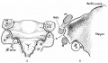
12 mm embryo
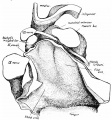
16 mm embryo
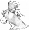
27mm embryo
35mm embryo
| Historic Disclaimer - information about historic embryology pages |
|---|
|
Reference
Frazer JE. The second visceral arch and groove in the tubo-tympanic region. (1914) J Anat Physiol. 48(4): 391-408. PMID 17233005
Cite this page: Hill, M.A. (2024, April 23) Embryology Paper - The second visceral arch and groove in the tubo-tympanic region. Retrieved from https://embryology.med.unsw.edu.au/embryology/index.php/Paper_-_The_second_visceral_arch_and_groove_in_the_tubo-tympanic_region
- © Dr Mark Hill 2024, UNSW Embryology ISBN: 978 0 7334 2609 4 - UNSW CRICOS Provider Code No. 00098G