Paper - The origin and development of the human extrinsic ocular muscles: Difference between revisions
mNo edit summary |
mNo edit summary |
||
| Line 9: | Line 9: | ||
==Figures== | ==Figures== | ||
===Plate 1=== | |||
[[File:Gilbert1957 plate01.jpg|800px]] | |||
===Plate 2=== | ===Plate 2=== | ||
Revision as of 00:58, 2 June 2016
| Embryology - 24 Apr 2024 |
|---|
| Google Translate - select your language from the list shown below (this will open a new external page) |
|
العربية | català | 中文 | 中國傳統的 | français | Deutsche | עִברִית | हिंदी | bahasa Indonesia | italiano | 日本語 | 한국어 | မြန်မာ | Pilipino | Polskie | português | ਪੰਜਾਬੀ ਦੇ | Română | русский | Español | Swahili | Svensk | ไทย | Türkçe | اردو | ייִדיש | Tiếng Việt These external translations are automated and may not be accurate. (More? About Translations) |
Gilbert PW. The origin and development of the human extrinsic ocular muscles. (1957) Carnegie Instn. Wash. Publ. 611, Contrib. Embryol., Carnegie Inst. Wash. 36: 59-78.
Extraocular Muscle Development
| Historic Disclaimer - information about historic embryology pages |
|---|
 |
The Origin and Development of the Human Extrinsic Ocular Muscles
Figures
Plate 1

Plate 2

Plate 3

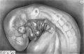
Fig. 11
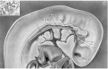
Fig. 12
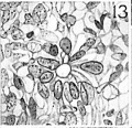
Fig. 13


Plate 4

Four drawings of a model of the eye, eye-muscle primordia, and associated nerves. Embryo No. 6258, horizon xvii.
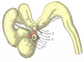
Fig. 14
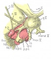
Fig. 15
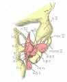
Fig. 16
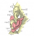
Fig. 17


Fig. 14. The eye-muscle primordia of embryo no. 6258 are superimposed on the brain of another embryo, no. 6520, of approximately the same age and size. The relation of the principal branches of cranial nerve V to the four peripheral condensations into which the four rectus muscles grow is illustrated. Lateral aspect. X 15.
Fig. 15. A portion of the brain, the eye, principal nerves, peripheral condensations, and the eye—muscle primordia. Dorso-lateral aspect. No. 6258, X 30.
Fig. 16. Dorsocranial aspect of the same model illustrated in figure 15. X30.
Fig. 17. Ventrocaudal aspect of the same model illustrated in figure 15. X30.
Attention is called to the four peripheral condensations, about the outer margin of the optic vesicle, into which the primordia of the four rectus muscles have grown. Cranial nerves III, IV, and VI have reached their respective eye-muscle primordia: the primordium of the inferior oblique has appeared as a conspicuous condensation at the distal end of the inferior rectus; a prominent bend (at the point where the trochlea will subsequently develop) has appeared near the distal end of the superior oblique primordium, and the proximal end of the superior oblique has begun to shift medially.
Cite this page: Hill, M.A. (2024, April 24) Embryology Paper - The origin and development of the human extrinsic ocular muscles. Retrieved from https://embryology.med.unsw.edu.au/embryology/index.php/Paper_-_The_origin_and_development_of_the_human_extrinsic_ocular_muscles
- © Dr Mark Hill 2024, UNSW Embryology ISBN: 978 0 7334 2609 4 - UNSW CRICOS Provider Code No. 00098G