Paper - The mammalian spermatozoon: Difference between revisions
mNo edit summary |
|||
| Line 7: | Line 7: | ||
As we approach the three hundredth anniversary of the discovery of the spermatozoon (1677) it seems timely to review what we know of the structure of this fascinating cell. Perhaps no better example could be found of the slow beginnings of biological science and of the rapid recent acceleration in the tempo of discovery resulting from advances in instrumentation. | As we approach the three hundredth anniversary of the discovery of the spermatozoon (1677) it seems timely to review what we know of the structure of this fascinating cell. Perhaps no better example could be found of the slow beginnings of biological science and of the rapid recent acceleration in the tempo of discovery resulting from advances in instrumentation. | ||
[[File:Fawcett1975 fig31.jpg|800px]] | |||
Fig. 31. A longitudinal thin section of the middle piece of a mammalian spermatozoon The circumferentially oriented mitochondria are cut transversely. Note how closely the celi membrane is apposed to the underlying mitochondria. | |||
Fig. 34. | |||
{| | |||
| [[File:Fawcett1975 fig34.jpg|600px]] | |||
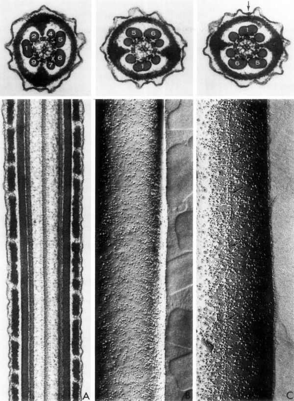
| Fig. 34. Spermatozoa Tail | |||
'''A''' - A longitudinal section through the principal piece of a sperm tail, approximally in the plane passing through fibers 2 and 7 or 9 and 4 of the cross-section shown above. The section includes two doublets and one of the central pair of axonemal microtubules, two outer fibers and groups of partially fused ribs of the fibrous sheath. | |||
'''B''' - A freeze cleaving preparation of the membrane on the side of the principal piece over the major compartment containing outer fibers 4 to 7. There is a high concentration of randomly distributed membrane intercalated particles. | |||
'''C''' - A freeze-cleave preparation of the membrane on the side of the minor compartment containing fibers 9, 1 and 2. A double row of large particles runs longitudinally within the membrane overlying fiber number one. A slight thickening of the membrane at this site is evident (at the arrow) in the cross-section shown above. | |||
|} | |||
{{Footer}} | {{Footer}} | ||
[[Category:Spermatozoa]][ | |||
[[Category:Draft]] | [[Category:Draft]] | ||
Revision as of 10:05, 20 August 2017
| Embryology - 16 Apr 2024 |
|---|
| Google Translate - select your language from the list shown below (this will open a new external page) |
|
العربية | català | 中文 | 中國傳統的 | français | Deutsche | עִברִית | हिंदी | bahasa Indonesia | italiano | 日本語 | 한국어 | မြန်မာ | Pilipino | Polskie | português | ਪੰਜਾਬੀ ਦੇ | Română | русский | Español | Swahili | Svensk | ไทย | Türkçe | اردو | ייִדיש | Tiếng Việt These external translations are automated and may not be accurate. (More? About Translations) |
Fawcett DW. The Mammalian Spermatozoon. (1975) Dev. Biol. 44, 394-436.
The Mammalian Spermatozoon
Don W. Fawcett
As we approach the three hundredth anniversary of the discovery of the spermatozoon (1677) it seems timely to review what we know of the structure of this fascinating cell. Perhaps no better example could be found of the slow beginnings of biological science and of the rapid recent acceleration in the tempo of discovery resulting from advances in instrumentation.
Fig. 31. A longitudinal thin section of the middle piece of a mammalian spermatozoon The circumferentially oriented mitochondria are cut transversely. Note how closely the celi membrane is apposed to the underlying mitochondria.
Cite this page: Hill, M.A. (2024, April 16) Embryology Paper - The mammalian spermatozoon. Retrieved from https://embryology.med.unsw.edu.au/embryology/index.php/Paper_-_The_mammalian_spermatozoon
- © Dr Mark Hill 2024, UNSW Embryology ISBN: 978 0 7334 2609 4 - UNSW CRICOS Provider Code No. 00098G[

