Paper - The form of the stomach in human embryos with notes upon the nomenclature of the stomach: Difference between revisions
mNo edit summary |
mNo edit summary |
||
| (18 intermediate revisions by the same user not shown) | |||
| Line 4: | Line 4: | ||
! Online Editor | ! Online Editor | ||
|- | |- | ||
| [[File:Mark_Hill.jpg|90px|left]] This historic 1912 paper by Lewis describes human embryonic stomach development using the [[Harvard Collection]] embryos. See the links below for current notes of development of the stomach. | | [[File:Mark_Hill.jpg|90px|left]] This historic 1912 paper by [[Embryology History - Frederic Lewis|Frederic Thomas Lewis (1875—1951)]] describes human embryonic stomach development using the [[Harvard Collection]] embryos. See the links below for current notes of development of the stomach. | ||
This paper uses models based on the [[Harvard Collection|Harvard Embryological Collection]]. | |||
Also by this author: | Also by this author: | ||
<br> | <br> | ||
'''Modern Notes:''' | {{Ref-LewisFT1902}} | ||
{{Ref-Lewis1912b}} | |||
<br><br> | |||
'''Modern Notes:''' [[Gastrointestinal Tract - Stomach Development|Stomach Development]] | |||
<br> | <br> | ||
{{Gastrointestinal | {{Gastrointestinal Tract Links}} | ||
|} | |} | ||
{{Historic Disclaimer}} | {{Historic Disclaimer}} | ||
| Line 27: | Line 31: | ||
Twelve Figures | Twelve Figures | ||
==Introduction== | |||
X-ray examinations of the stomach, in adults and especially in children, have led clinicians to inquire Whether the stomach has a characteristic embryonic form Which may sometimes persist. Figures of the typical embryonic stomach have, indeed, been published;‘but it must be remembered that the stomach changes in shape as the embryo grows older and, as Broman has found, its individual variations in embryos of the same stage of development is very great. Nevertheless certain fundamental subdivisions are strikingly distinct. These primary subdivisions, in which the embryologist is most interested, were keenly discussed by the early anatomists. In their writings many suggestive questions are raised, at the same time that the fundamental features of the organ are successively recognized and defined. In the following historical notes, taken from such Works as are at hand, provisional definitions are offered for certain terms adopted at Basle but at present loosely employed, and attention is called to the features of the adult stomach which will be examined in the embryos. | |||
==Stomach Nomenclature== | |||
The human stomach was first considered to be a simple sac with an orifice of entrance above and to the left, and an orifice of exit belowand to the right. Vesalius (1543) in his figures designates the orifices as the ‘superius ventriculi orificium’ and 'iI1ferius ventriculi orificium,’ respectively. In his text, however, both are said to be placed superiorly, so that food shall not escape by its own Weight, but when completely changed to chyme, shall be propelled by force of the discharging stomach into the intestine. Fabricius ab Aquapendente (1618) likewise states that the orificium inferius is n.ot inferior at all, and Spigelius (1627) places it in the highest part of the stomach; so that the term ‘orificium dextrum’ was preferred, and finally the less objectionable Greek name ‘pylorus’ (Latin, janitor), which had been. introduced by Galen, became the accepted designation. Winslow’, however, in 1732, insisted that the position of the orifices is such that “We ought with the’ ancient anatomists to call one of them superior, the other inferior.” | |||
The significance of ‘cardia’ (Latin, cor), as applied to the oesophageal orifice, was discussed by Fabricius, who cites Galen as stating that the upper orifice of the stomach is called the heart because the symptoms to which it gives rise are similar tothose which sometimes affect the heart, sometimes even the brain; but for Fabricius, cardia, as applied to this orifice, merely indicates a chief part of the body. Spigelius describes the cardia as consisting of circular fleshy fibers, so that the stomach, after having received food, may be closed perfectly, thus preventing fumes from rising, with consequent loss of heat. The cardia and pylorus are intimately associated with their respective sphincter muscles, but they do not include the adjacent portions of the stomach. | |||
For the stomach as a whole these anatomists use the Latin ‘ventriculus,’ rather than the Greek ‘''gaster''’ and the Latin term has been adopted at Basle. Since however, the adjective gastricus has been chosen instead of ventricularis, it seems desirable that gaster should be used in place of ventriculus, especially since cardia and pylorus are of Greek origin). | |||
* I am indebted to Prof. Albert A. Howard for the following note regarding these terms: ''Gaster'' is a Greek word meaning belly (the whole abdominal cavity) but was often used by the Greeks in the more restricted sense of stomach. It is not found in Latin with this meaning until very late (only after the literary period). Vcntriculus is used quite consistently for stomach by Celsus and at times by Pliny the Elder. Cicero in one passage speaks of ''ventriculus cordis'', but does not use ''ventriculus'' for stomach. If ''gaster'' is adopted I think the genitive ''gastri'' is preferable to ''gasteris'', though as a matter of fact the genitive does not happen to occur in any Latin that is preserved to our time. Petronius has used the ablative plural ''gastris'' which would be the reason for deciding as I have. | |||
The stomach, as described by Vesalius, is rounder and more spacious on the left side, and more slender on the right; to which Fabricius adds that it is not unlike a gourd with larger belly and narrower neck. On its dorsal side Vesalius found two swellings, separated by a vertical impression which was fitted against the trunks of the aorta and vena cava and the projecting bodies of the vertebrae. When the stomach was inflated, the impression and swellings were lost in an even rotundity. It was not until Willis (1674) described the pyloric antrum in the following passage, that a permanent subdivision of the stomach was established. | |||
The | The other orifice, commonly called the pylorus, on the right side of the stomach, having a capacious and long, gradually narrowed antrum, ends in a small foramcn and thence bent back is continued into the duodenum. Here the coats are much thicker than in any other part of the stomach. | ||
Indeed the long and capacious antrum seems to be a sort of recess and | Indeed the long and capacious antrum seems to be a sort of recess and diverticulum in the stomach, into which the more elaborated and perfected portion of the chylous mass may Withdraw and there remain, while the other cruder and more recently ingested portion may be further digested in the fundus of the stomach (ed. of 1680, p. 13-14).? | ||
diverticulum in the stomach, into which the more elaborated and perfected portion of the chylous mass may Withdraw and there remain, | |||
while the other cruder and more recently ingested portion may be further | |||
digested in the fundus of the stomach (ed. of 1680, p. 13-14).? | |||
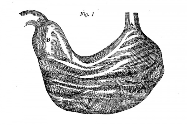
of the others, in which it has been stretched out so as to form | Accompanying this description Willis _published four lateral views of the stomach, with its coats successively removed. All of them show the antrum, but in a fifth figure, representing the everted stomach, its limits are most satisfactorily indicated (fig. 1). In this figure the antrum is shorter and broader than in one of the others, in which it has been stretched out so as to form a tube. | ||
In all of the figures it is clear that the antrum extends to the pylorus, which is referred to as its orifice. | In all of the figures it is clear that the antrum extends to the pylorus, which is referred to as its orifice. | ||
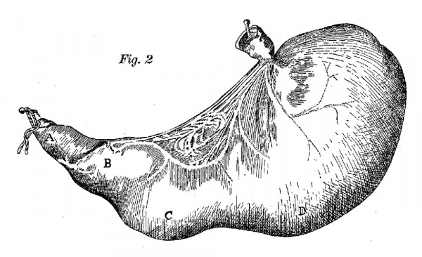
Bidloo (1685) published a Inore accurate figure of the stomach, here reproduced as figure 2, but he failed to describe it adequately. He states that the base is provided with two swellings, C and D. In another figure, showing the same stomach partly laid open, the portion of the duodenum near the stomach (A) is labelled pylorus, but Bidloo does not refer in any way to the subdivision which in figure 2 has been labelled B. Cowper (1698), who republished Bidloo’s plates, states that A is the part of the ‘duodenum arising from the pylorus and adds that B is the antrum pylori. | |||
* For verifying and revising the Latin translations, the author is under obligation to Mr. S. R. Meaker. | |||
In 1732 Winslow described the large arch running along the greatest convexity of the stomach, and the small one directly opposite, and named them the great and small curvatures. . Bichat (1802) states that “the great curvature ends simply at the pyloric orifice, without presenting anything of note unless it be the elbow (le coude) formed by this pyloric orifice, and named the small cul—de—sac ; but there is no particular swelling at this place and the bend is precisely in the direction of the pylorus.” Cloquet (1831) repeated this description and Cruveilhier (1834) made it more explicit. He states that at about 2 or 3 cm. from the pylorus “the stomach, bending sharply upon itself, forms a very pronounced elbow (coude de l’estomac) on the side of the greater curvature, and presents an ampulla, corresponding to an interior excavation, named by Willis the pyloric antrum, by others the small cul—de—sac.” As pointed out by Muller (1897), Cruveilhier was unjustified in identifying a pouch about an inch from the pylorus with the pyloric antrum of Willis; but he was correct in stating that “it is not rare to see a second ampulla beside the first, and a third but smaller one, on the side of the lesser curvature” (compare with figs. 2 and 3). These had not been recognized by Willis, but Cowper, in describing Bidloo’s plate, was confronted with the question whether one or more of these parts was to be regarded as the antruzn. In applying the term to the part adjacent to the pylorus but not extending to the bend of the stomach, Cowper was justified by Willis’s figure here reproduced as figure 1. According to Cunningham (1906) “no part of the organ is more definite and distinct” than the region which Cowper designated ‘antrum pylori’ and which, rediscovered by J onnesco (1895), was named the pyloric canal. It may be defined as the part of the stomach adj acent to-the pylorus, averaging 3 cm. in length, cylindrical when empty, bulbous when distended, separated from the remainder of the stomach by a groove on the greater curvature—the ‘sulcus intermedius’ of His (1903). For the small cul-de-sac of Cruveil— hier the term ‘pyloric vestibule’ (J onnesco, 1895) may be adopted. | |||
[[File:Lewis1912-fig01.jpg|600px]] | |||
'''Fig, 1''' Willis’s figure of the inverted stomach re-drawn and reduced one-half. "A, Orificium sinistrum, sive os ventrieuli. B, Pylori Antrum, in que, Tunicae crassiores exist-unt. C, Orificium ejus, cuo Duodenum annectitur.” | |||
[[File:Lewis1912-fig02.jpg|600px]] | |||
Bidloo’s | '''Fig. 2''' Bidloo’s figure of the unopened stomach, re-drawn and reduced twothirds, with lettering added from Bidloo’s drawing of the same stomach opened, and from Cowper’s edition of Bidloo’s plates. A, pylorus (Bidloo);portion of the intestinum duodenum (Cowper). B, antrum pylori (Cowper). C’, D, two bunchings out in the lower part or fundus of the stomach (Cowper) ; in funclo Gibbis ornatur duobus (Bidloo). | ||
from the | |||
[[File:Lewis1912-fig03.jpg|600px]] | |||
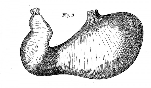
Fig | '''Fig. 3''' Home’s figure of “the human stomach inverted, to show the contraction which divides the cavity into two portions.” Re-drawn and reduced two-thirds. cm, the cardiac portion. 1), the contraction dividing the cardiac from the pyloric portion. c, the pyloric portion. 01, the pylorus. | ||
Unfortunately Cowper’s use of pyloric antrum has been overlooked by later anatomists, and the term has been so variously employed, as tabulated by Muller, that Muller, His and Cunningham have proposed to abandon it altogether. His has suggested an entirely new nomenclature for the pyloric region, as follows: for pyloric vestibule, camera princeps; for the swelling on the lesser curvature opposite the sulcus intermedius (fig. 3), camera minor; and for pyloric antrum, camera tertia. But these terms, as stated by Cunningham, are not in every respect satisfactory, and it may be well to retain the appropriate name ‘pyloric antrum’ in the sense of Cowper, following Meyer (1861) a11d Hasse and Strecker (1905). | |||
The normal division of the entire stomach into two parts, cardiac and pyloric (of which the latter presents the subdivisions just described), was first recognized by Home (1814). He wrote as follows: I found also, in the necessary examinations, that the dog’s stomach, while digestion is going on, is divided by a muscular contraction into two portions; that next the cardia is the largest, and usually containing a quantity of liquid, in which there was some solid food; but the other, which extended to the pylorus, being filled entirely with half—digested food of an ordinary consistence. I shall, therefore, in my future description call that part which constitutes the first cavity the cardiac portion, and that which constitutes the second the pyloric portion (p. 140). | |||
The cardiac portion is in length two-thirds of the whole, but in capacity much greater (p. 139). | |||
Home distinguished these two portions not only in the dog but, with varying distinctness and permanence, in many animals, including the horse, pig, rat, rabbit and man. He did not describe or label the subdivisions of the pars pylorica, which however are clearly shown in his figure of the everted human stomach (fig. 3). | |||
portions | |||
In connection with Home’s work, the following more recent physiological observations are of interest. Schiitz (1885) found that in the dog’s stomach, contraction waves travel from the cardiac end to a place about 1 cm. from the beginning of the pyloric antrum (pars pylorica?), which in the isolated resting stomach may be recognized by a shallow annular constriction about 2 cm. behind the pylorus, and there end with a deep ‘prae—antral constriction.’ The second phase, which follows the first immediately, concerns the antrum only. The motions of the two parts may take place independently of one another. Moritz (1895) determined the pressure within the two parts of the stomach, and stated that the difference between them was greater than would be inferred from merely observing their motions. Cannon (1898) found that the stomach of the cat, as shown by X-ray examinations, is composed of two physiologically distinct parts—a ‘busy antrum’ and a cardiac reservoir. In 1911 he states that during normal digestion “slight constrictions appear near the middle of the body of the stomach, and pressing deeper into the greater curvature, course towards the pyloric end. When a wave sweeps round the bend i11to the vestibule, the indentation made by it increases.” He adds that when vomiting occurs, a strong contraction at the angular incisure completely divides the gastric cavity into two parts. Thus the observations of Home have been amply confirmed and extended. Other X—ray observers, however, have considered that the antrum, or pars pylorica, of anatoinists is merely the part of the stomach marked off by a passing peri staltic wave (Hertz; Kaestle,Rieder and Rosenthal; Barclay). In this they follow Sappey (1874), who was of the opinion that Home’s subdivision of the stomach was based on fortuitous muscular contractions. This will be disproved by showing that the two divisions of the stomach are well marked in embryos in which the muscle-layers are still scarcely differentiated. | |||
When a peristaltic wave remains fixed after death, the stomach may appear as “two joined together” (Riolan 1618), in which case the subdivisions need not correspond with the anatomical parts already described. Usually the constriction is near the middle of the stomach, and falls within the cardiac portion. Morgagni (1761) observed five cases, all in women. One of the stomachs was from a patient who had been troubled with excessive vomiting since birth, but none of the stomachs showed any sign of disease. Since several cases had been reported in men, Morgagni concluded that the double stomach was not a deformity due to stays, but had existed from the first formation of the organ. Sandifort (1777-1781), as quoted by Bettman, described a typical case in a fetus, the age of which is not stated in the citation. Delamare and Dieulafé (1906) reported a case in a new-born syphilitie infant, in which they describe an hypertrophy of the circular muscle atthe place of constriction. The thickened muscle-layer may, however, be due to contraction, -as indicated by the folded and thickened overlying layers sho-wn in their figures. Cunningham (1906) holds that there is not an atom of evidence that the hour-glass stomach ever arises as a congenital deformity, but he is not prepared to state that the strictures which separate the two sacs of the bilocular stomach are always temporary and fleeting. The change in the direction of the lesser curvature is more dependable as a boundary between the pars pylorica and pars cardiaea, than the constriction which is present in certain cases but “not as a rule” (Huschkc). The lesser curvature, which is concave along the cardiac portion, becomes convex along the pars pylorica (Meekel 1820; Huschkc 1844). Retzius (1857) figured a deep stricture in the lesser curvature at the beginning of the bulbous pars‘ pylorica, where Luschka (1869) frequently found an acute angle directed toward the gastric cavity. This notch has been named by His (1903) the ‘incisura angularis,’ and it occurs between the two parts of the stomach. Along the greater curvature the boundary is less clearly marked. It is indicated by the change in direction already described as the elbow of the stomach, and referred to by Home as “an angle formed at the part where the temporary contraction takes place.” times the constriction is slightly to the cardiac side of the elbow, as shown in figure 3. | |||
cavity | |||
The angle which separates the two parts of the stomach is | The angle which separates the two parts of the stomach is obscure in the older drawings in which the organ is almost horizontally placed. According to Bichat (1802) “When the stomach is filled its obliquity increases considerably; often it appears almost perpendicular, so that the right extremity . . . . is strongly recurved upward, and forms a very acute angle with the body of the organ.” Luschka (1869) similarly found that the greater part of the stomach, as a rule, has a precisely vertical position, but that the pars pylorica is directed almost transversely. Both of these forms, with vertical body and transverse or ascending pars pylorica, will be seen in the embryos to be examined. | ||
obscure in the older drawings in which the organ is almost horizontally placed. According to Bichat (1802) “When the stomach | |||
is filled its obliquity increases considerably; often it appears almost | |||
perpendicular, so that the right extremity . . . . is strongly | |||
recurved upward, and forms a very acute angle with the body of | |||
the organ.” Luschka (1869) similarly found that the greater | |||
part of the stomach, as a rule, has a precisely vertical position, | |||
but that the pars pylorica is directed almost transversely. Both | |||
of these forms, with vertical body and transverse or ascending | |||
pars pylorica, will be seen in the embryos to be examined. | |||
The pars pylorica and its subdivisions having been described, the pars cardiaca may next be examined. It is divided into the ‘saccus caecus,’ now called the ‘fundus;’ the ‘corpus’ or body; and the gastric canal. The term fundus was appropriately applied by Vesalius to the lower part of the stomach, which in the transverse position of the organ, extends well toward the pyloric region. It was so used by Willis (1674); and by Cowper (1737), as seen in figure 2. Caldani (1804) makes fundus synonymous with greater curvature. The bulging left or upper extremity of the stomach received the special name ‘saccus caecus’ (Haller, 1764; Caldani, 1804). But Meckel (1820) considered fundus and saccus caecus as synonyms, and preferred fundus; Huschke (1844) likewise made them synonymous, but adopted saccus caecus, which Henle used in 1866. Nevertheless, fundus has become adopted for the highest part of the stomach and saccus caecus has been rejected. The fundus lies at the left of the cardia, being separated from the oesophagus by a notch, the ‘incisura cardiaca’ of His (1903). Below, as described by Cloquct, the fundus terminates almost imperceptibly in the greater curvature. It is therefore bounded arbitrarily by a horizontal plane at the level of the inferior border of the cardia (J onn'esco), or by a line prolonging the axis of the abdominal part of the oesophagus {Keith and Jones, 1902). | |||
According to Keith and Jones the fundus arises in human embryos as a localized outgrowth or diverticulum ofrthe stomach, and in its manner of origin has much in common with the caecum and Vermiform process. From numerous observations they conclude that “it is not uncommon to find in the stomach of the anthropoids, and to a lesser degree in that of the apes (especially in Mycetes) clear indications of three chambers, namely, a fundus, a body, and a pyloric part; and that therefore the stomach of the Primates (excluding the Lemuroidea) is probably tripartite in nature.” It should be noted that the fundus as defined by Keith and Jones is a larger part of the stomach than that set off by J onnesco, and that their boundary is justified by comparison with the stomach of Semnopithecus which they have figured. If the fundus corresponds in any way to a first stomach or rumen, it may be regarded as the globular upper end of the organ which is often marked off by the contraction of the corpus. | |||
and | |||
the | |||
and | |||
The body of the stomach (corpus gastri), as defined by Rudinger (1873), is its middle subdivision, situated between the fundus and the pars pylorica. Froriep (1907) proposed to rename it the pars intermedia; but since it is a portion of the pars carcliaca, and is not intermediate between the pars cardiaca and pars pylorica, the proposed term would lead to confusion. Jonnesco (1895) defined the body as including the pyloric vestibule, but in the same paragraph he described the boundary between the vestibule and “le corps proprement dit.” Muller (1897) included the fundus with the body, making corpus and pars cardiaca synonymous. It is only by accepting Rudinger’s earlier definition that corpus becomes a useful term. The corpus may be contracted at any point, as in the hour—glass stomach, in which case part of it appears to belong with the fundus and the remainder with the pars pylorica. Sometimes it is contracted as a whole, but more often it is relaxed, and its boundaries are then ill—defined. | |||
the | |||
in | |||
The gastric canal is a channel which follows the lesser curvature, appearing as a groove when seen from the inside of the stom— ach. It suggests a continuation of the oesophagus, split open toward the gastric cavity, and has been named the sulcus oesophageus, sulcus gastricus, sulcus salivalis and canalis salivalis. It is -confusing, however, to refer to this channel as a sulcus, since the external grooves of the stomach are so designated (sulcus inter—medius, sulcus pyloricus), and it is un.desirable to name a part of the stomach oesophageal or salival. Therefore the term gastric canal, ‘canalis gastricus,’ is here proposed, and canalis is used as in Latin for an open canal, which in this case may become a tube during its physiological activity, by the approximation of its lips. | |||
the | |||
of | |||
The | The gastric canal has long been known in ruminants, but in its less highly developed condition in the human stomach, it has attracted little attention. In man it is generally supposed to be due to the arrangement of the oblique muscle fibers, which were first described by Willis (1674), in connection with a figure of the stomach in the position shown in figure 1. The ‘top’ of the stomach is accordingly toward the lesser curvature, and the ‘fundus’ is toward the greater curvature. Willis wrote as follows: | ||
:These muscle fibers, which are seen to arise behind the cardia and to pass around its left margin, are carried forward to the right portion of the stomach. A notable bundle of them, proceeding in straight lines along the top of the stomach on either side, encounters the antrurn, and spreading over the length of its cavity in a scattered manner, terminates in the pylorus. Moreover the remaining fibers of this layer extend obliquely over the walls of the stomach on both sides, and then directly toward the fundus where they come together. The function of the former (the straight bundles) seems to be to bring one orifice toward the other in emptying, by making them lower and higher respectively (ed. of 1680, pp. 11-12). | |||
Gyllenskoeld (1862) | Retzius called attention to this description by Willis and, as reported by Gyllenskoeld (1862), he supplemented it as follows: | ||
to the | :The upper portion of the oblique fibers of the human stomach serves to form a sort of trough along the lesser curvature which, under the control of the motor nerves, becomes more or less closed ;_ along this path possibly fluids and soft things, saliva, etc., may proceed directly from the oesophagus to the pars pylorica, passing by the cardiac portion, which corresponds to the first two stomachs of ruminants and the non-glandular part of the stomach in rats. | ||
from the pars pylorica, | |||
The correctness of this conjecture concerning the passage of fluids was established by Cohnheim (1908), Who was surprised to find that water or salt—solution passed rapidly through the full stomach of a dog, without mixing with the gastric contents. | |||
Gyllenskoeld (1862) states that the oblique fibers extend only to the pars pylorica, and not to the pylorus as described by Willis. This has been confirmed by Kaufmann (1907). He found that there is no sphincter of circular fibers separating the pars cardiaca from the pars pylorica, but that the furrow between them has a special structure, since it is the place where the oblique fibers terminate and interlock with the circular fibers. | |||
Hasse and Strecker (1905) have named the folds which bound the gastric canal the ‘plica hepatica’ and ‘plica aortica’ respectively, and state that they are connected with one another by the ‘plica cardiaca’ which passes around the cardia, projecting into the stomach beneath the incisura cardiaca. According to Hasse and Strecker the plica cardiaca does not form a valve for the cardia, as Braune (1875) thought possible from the result of experiments on a cadaver. The hepatic, cardiac and aortic plieae together form a U-shaped structure, across the open end of which is the ‘plica angularis/o This is beneath the incisura angularis, at the beginning of the pars pylorica. | |||
Waldeyer, who describes the channel from cardia to pars pylorica as the ‘Magenstrasse’ (1908), considers that its formation depends upon the oblique muscles, rather than upon folds which arise in relation with adjacent organs. In the following pages evidence will be offered to showthat the gastric canal is a distinct epithelial structure, arising independently both of the muscle and the surrounding organs. | |||
There remain to be considered two structures which are beyond the limits of the stomach—the ‘antrum duodenale’ and the ‘antrum cardiacum’ . | |||
Retzius (1857) states that the beginning of the duodenum is often specially rounded, not only in man, but in a large proportion of mammals; in dolphins it has been considered a part of the stomach. Owen (1868) remarks that in all Artiodactyles the duodenum is dilated at its commencement; it there forms a distinct pouch in the camel. For this pouch Retzius proposed the names “antrum or atrium duodeni” but used the former in his figures. Luschka (1863) refers to a flask—shaped expansion at the beginning of the duodenum, which inhis figure is called the ‘antrum duodenale.’ This structure Will be seen to be far more distinct in human embryos than it appears to be in adults. | |||
The cardiac antrum was first described by Luschka (1863) as follows: | |||
:At the junction of fundus and lesser curvature the oesophagus enters the stomach, forming a funnel-shaped expansion—the cardia. Although ordinarily the cardia is continued into the rest of the stomach without definite boundary, in rare cases the funnel—like expansion is sharply marked off by an external depression and corresponding internal elevation, thus forming a sort of cardiac antrum (p. 179). | |||
In | In 1869 Luschka adds that if this funnel is to be regarded as part of the stomach, the beginning of which is not rather to be considered at the base of the funnel where the stratified epithelium ends in a zig-zag line (fig. 3), “then the funnel—shaped expansion must be specially designated as the pars cardiaca.” | ||
of the stomach, | |||
Thus Luschka proposed two names for a single structure; first, cardiac antrum; and later, in case the antrum is to be regarded as part of the stomach, pars cardiaca. The latter may be rejected, since it is generally agreed that the cardia is at the base of the cone, and that therefore ‘cardiac antrum’ is “merely another name for the intra—abdoIninal part of the oesophagus” (Cunningham). Moreover the earlier use of pars cardiaca, or cardiacfion tion, for the fundus and corpus taken together, was overlooked by Luschka, and by certain later anatomists Who have proposed to substitute Hauptmagen (His), saccus ventriculi (Hasse and Strecker) and pars digestoria (Froriep). | |||
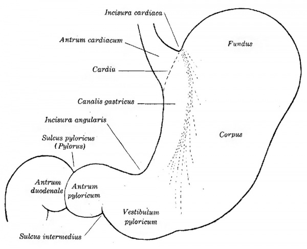
The fundamental subdivisions of the stomach and adjacent parts of the digestive tube, as they have been defined in the preceding pages, are presented in figure 4 and in the following table, With authority for certain of the definitions adopted: | |||
Canalis gastricus | * Antrum cardiacum (Luschka, 1863) | ||
* Gaster | |||
** Cardia | |||
** Pars cardiaca. gastri (Home 1814) | |||
*** Fundus (Meckel 1820) | |||
*** Corpus (Rudinger 1873) | |||
*** Canalis gastricus | |||
** Pars pylorica gastri (Home 1814) | |||
*** Vestibulum pyloricum (Jonnesco 1895) | |||
*** Antrum pyloricum (Willis 1674 CE); Cowper 1698) | |||
** Pylorus | |||
* Antrum duodenale (Retzius, 1857) | |||
As boundaries between these parts, the following may be recognized: Between the cardiac antrum and fundus, the ‘incisura cardiaca;’ between cardiac and pyloric parts, the ‘incisura angularis;’ between pyloric antrum and pyloric vestibule, the ‘sulcus intermedius’ (all of His 1903); at the pylorus,,the ‘sulcus pyloricus’ (Luschka 1863). | |||
==The Stomach in Human Embryos== | |||
The embryonic stomachs to be examined are five in number, from embryos between 10 mm and 45 mm in length. Thus they are all smaller than the specimens studied by Miiller, but similar stages have been described by Broman in his extensive work on the omental bursa. Broman modelled not only the gastric epithelium, but also entire stomachs, including the mesodermal portion. In the models to be described, only the epithelium has been included, since this is the portion having characteristic shape, to which the other layers subsequently conform. | |||
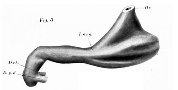
In the youngest embryo (10 mm, fig. 5) the stomach is no longer a simple sac with superior and inferior orifices, but is already divided into an expanded pars cardiaca and a tubular pars pylorica. Between the two, and almost exactly in the middle of the stomach, is the incisura angularis. Since" the incisure in the adult is perhaps twice as far from the cardia as from the pylorus, it is evident that the pars pylorica is relatively long in early stages. This is strikingly shown-in other models of the series (figs. 6-9). THE FORM or It is true also in the cat, if one may judge by comparing Thyng’s model of the stomach of a 10.7-mm. embryo (this Journal, vol. 7, p. 496) With Cannon’s tracings from the adult. In ruminants a constriction early separates the rumen and reticulum from the psalterium and abomasum; according to Ellcnberger and Baum the abomasum is larger than the rumen in embryos and very young animals, but later this relation is reversed. The relatively large size of the pars ipylorica in early stages is therefore not limited to human embryos. | |||
[[File:Lewis1912-fig04.jpg|600px]] | |||
'''Fig. 4''' Diagram showing the subdivisions of the human stomach. | |||
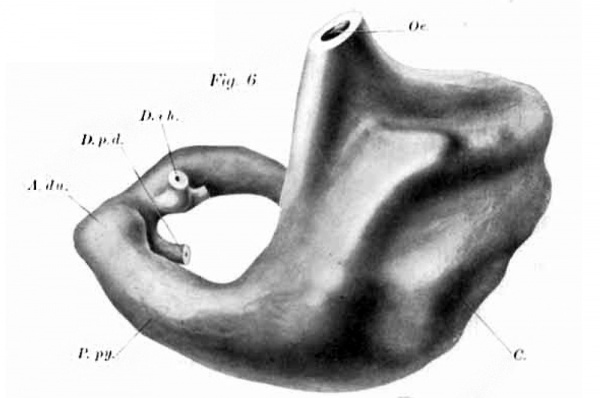
passes along this curvature from the oesophagus to the pars | The cardia cannot be definitely located in the 10-mm. embryo (fig. 5) since the oesophagus, in joining the stomach, expands into a flattened cone, one margin of which extends to the angular incisure. A similar extension of the oesophageal cone to the incisure is clearly seen in Broman’s model of the stomach of the seventh embryo in his series (11.7 mm.). At 16 mm. (fig. 6) the body of the stomach may be recognized along the lesser curvature, separating the oesophageal cone from the angular incisure; but a canal, distinctly marked out above and indicated below, passes along this curvature from the oesophagus to the pars pylorica. A more distinct canal in this position is seen in two of Broman’s models, from embryos of 10 mm. and 16.2 mm. respecgively. Apparently this canal has not been previously described in embryos, although Toldt (1879), referring to the general direction of the oesophagus in a 23—mm. specimen, states that it descends into the stomach “in such a Way that the lesser curvature forms, as it were, a continuation of the ventral border of the oesophagus.” . | ||
pylorica. A more distinct canal in this position is seen in two of | |||
Broman’s models, from embryos of 10 mm. and 16.2 mm. respecgively. Apparently this canal has not been previously described | |||
in embryos, although Toldt (1879), referring to the general | |||
direction of the oesophagus in a 23—mm. specimen, states that it | |||
descends into the stomach “in such a Way that the lesser curvature forms, as it were, a continuation of the ventral border of the | |||
oesophagus.” . | |||
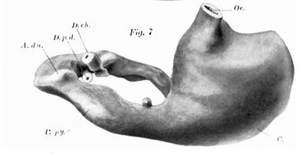
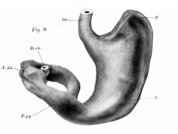
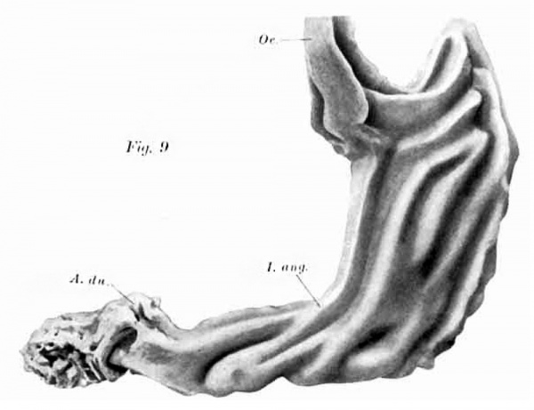
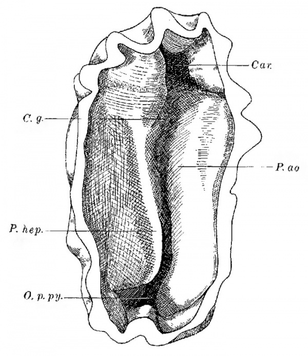
In the | In the embryos of 19.3 mm. and 19.0 mm. shown in figures 7 and 8 respectively, the canal is not seen. The first of these stomachs is abnormal, but the second specimen is unobjectionable. Moreover in Broman’s figure of the stomach from an embryo of 21 mm., there is no trace of the channel. Its obliteration, if normal, appears to be temporary however, for in the 44.3—mm. specimen shown in figure 9, it is more distinct than in preceding stages. It passes from the conical cardiac antrum to the angular incisure. This embryo, owing to its large size, was not perfectly preserved, and the epithelium has separated from the mese11— chyma; but whether 0110, or the other, or both of these tissues has shrunken is uncertain. The model may, however, be accepted as giving an essentially correct idea of the shape of the stomach, since the separated mesenchyma presents corresponding ridges and furrows. The distinetness of the gastric canal is strikingly shown when the model is viewed from the inside (fig. 10). It takes a slightly S—shaped course from the stellate cardia to the orifice of the pars pylorica, and is bounded by a rounded plica aortica, and a more prominent and angular plica hepatica. These folds are not formed, as described in theadult by Hasse and Strecker, through compression of the borderiof the stomach between the aorta behind and the caudate lobe of the liver in front; for the outer layers of ‘the stomach are not_ indented. Moreover at this stage there are no bands of oblique fibers to account for the canal. If the channel proves to be a constant feature of embryos of this stage, and it is present in an embryo of 37 mm. which was not modelled, it may be that the arrangement of the oblique fibers is a consequence rather than the cause of the gastric canal. | ||
the cardiac antrum | |||
This, | |||
the | |||
In the same way that the gastric canal accords with the ‘oesophageal sulcus’ of ruminants, which is described by comparative anatomists as a continuation of the oesophagus open on one side, the cardiac antrum may correspond to the ‘atrium ventriculi.’ This, according to Ellenberger and Baum is “a dome—shaped swelling on the dorsal side of the reticulum and thoracic end of the rumen, which is only indistinctly marked off from them by a shallow groove; ventrally its cavity passes directly into that of the reticulum, and caudo—ventrally into the vestibule of the rumen ; toward the thorax it rests against the diaphragm near the hiatus oesophagcus.” From the general ‘atrium’ seen in figure 5, the lower part is set off as the gastric canal, and the upper part remains as the cardiac antrum (figs. 8 and 9). From studies of the adult stomach it may be assumed that the cardia is at the base of this antrum, whi.ch therefore belongs with the oesophagus. | |||
is this | |||
The development of the fundus of the stomach has been described by Broman (1911) as follows: | |||
:By the beginning of the second month the cranial part of the greater curvature begins to bulge out. But not until the third month, or later, is this outpocketing generally directed so strongly craniad that its blind end comes to lie above the orifice of the oesophagus. Only from this time, therefore, can we speak of a distinct gastric fundus (pp. 326-328). | |||
of | |||
Similarly Keith and Jones state that the outgrowth is best marked in embryos of the third and fourth month. But as shown in figures 8 and 9, and by the fact that Toldt, in an embryo of 48 mm., found a well marked fundus projecting toward the concavity of the diaphragm, it is clear that the fundus may be well developed in the second month. In the model shown in figure 9, the fundus when seen from above, presents a curious appearance, since seven prominent ridges converge toward its apex. Two of them come from the cardiac antrum, sweeping in a semicircular curve beneath the cardiac incisure, thus resembling the ridges seen. in figure 8. There is normally no boundary between the fundus and corpus, but in an abnormal embryo of 18.5 mm., described by Broman, the fundus is cut off by a rather deep constriction. Broman states that this specimen suggests an l1our—glass stomach, from which, however, it is essentially different, since the oesophagus enters the part toward the pylorus. The fundus is best marked when the pars cardiaca is in an approximately vertical position, and this is the ease in figures 6 to 9. Broman, however, has found a greater variety of positions. In an embryo of 21 min. he figures the stomach as horizontal, so that both orifices are superior, as described in the adult by Vesalius; but this position must be regarded as exceptional. | |||
[[File:Lewis1912-fig05.jpg|600px]] | |||
[[File:Lewis1912-fig06.jpg|600px]] | |||
[[File:Lewis1912-fig07.jpg|600px]] | |||
[[File:Lewis1912-fig08.jpg|600px]] | |||
[[File:Lewis1912-fig09.jpg|600px]] | |||
'''Figs. 5 to 9''' Models of the gastric epithelium in human embryos, as follows: figure 5, 10 mm. [[Harvard Collection|Harvard Embryological Collection]], Series 1000, X 50 dia.m.; figure 6, 16.0 mm., H. C. 13:2, X 35 diam; figure 7, 19.3 mm., H. C. 1597, X 30 diam.;figurc 8, 19.0 mm., H. E. C. 819, X 26 diam.; figure 9, 44.3 mm., H. E. C. 1611, X 18 diam. | |||
H | |||
A.du., antrum duodenale. C., Corpus gastri. D.ch., ductus choledochus. D.p.d., ductus pancreatis dorsalis. F., fundus gastri. I.ang., incisum angularis. 0e., oesophagus. P.py., pars pylorica gastr. | |||
The | The body of the stomach requires no comment other than that its ridges appear to be rather definitely placed. The shelf-like prominence at the base of the oesophageal cone in figure 5, is evidently represented by the chief fold which extends horizontally across the base of the fundus, and bends down parallel with the lesser curvature in figure 6. Such an angular fold (With a subdividing furrow) is seen in figure 9, and it is clearly shown in embryos of 10 and 16.2 min. figured by Broinan. Why the ridges are absent from other specimens, as in figures 7 and 8, and in several of Broman’s embryos, is not apparent. | ||
[[File:Lewis1912-fig10.jpg|600px]] | |||
P. | '''Fig. 10''' Model of the interior of the stomach, from an embryo of 44.3 mm., [[Harvard Collection|Harvard Embryological Collection]], 1611, X 25 diam. Cara, carclia. (,‘.q., eanalis gastricus. O.p.py., orifieium partis pyloricae. P.a0., plica aortica. 1’./Lep., pliea hepatica. | ||
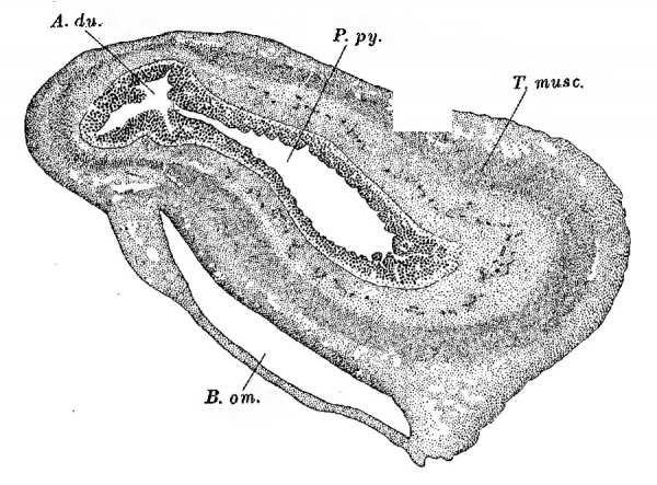
The position of the pylorus could not be determined with certainty in the 10-mm. embryo (fig. 5); and Tandler (1900) states that in an embryo of 11 mm. the pylorus is not marked. At 14.5 mm., “where the stomach passes into the duodenum, therefore at the place of the future pylorus” he saw “a considerable thickening of the epithelium.” The epithelial proliferation, which Tandler describes, is seen throughout the upper part of the duodenum. It is n.ot evident that he recognized the local swelling, chiefly on the upper side of the digestive tube, which is shown in figures 6 to 8. This swelling, which distinctly marks the position of the pylorus when the muscle-layers are still undifferentiated, and scarcely to be recognized, is apparently the duodcnal antrum of Retzius. In a frontal section through the pars pylorica of a 19—mm. embryo, it appears as shown in figure 11. At this stage the musculature of the pars pylorica is considerably thicker than that of the duodenum, but in this it conforms to the shape of the epithelial tube. In figure 9 the duodenal antrum is seen to be smoother than the more distal part of the duodenum, recalling the statement of Retzius that here, in the adult, the valvulae are absent and the villi are short. In this embryo the gastric epithelium is seen to be slightly invaginated into the duodenal tube, as observed by Cunningham at birth. Toldt found that the sulcus pyloricus could be seen externally in an embryo of 48 mm, and presumably it could have been found in this specimen by dissection. | |||
[[File:Lewis1912-fig11.jpg|600px]] | |||
'''Fig. 11.''' Frontal section through the pylorus of a 19 mm embryo, [[Harvard Collection|Harvard Embryological Collection]], 828, section 330, X 40 diam. A.du., antrum duodenale. B.o-m., bursa omentalis. P.py., pars pylorica. T.musc., tuniea muscularis. | |||
The pars pylorica, even in the 44.3 mm embryo, fails to show distinct subdivision into antrum an.d vestibule. Muller, who studied dissections of the embryonic stomach, states that in the ‘first fetal period’ the pyloric antrum is a direct continuation of the pyloric vestibule, but that later, when. the pars pylorica is bent convexly upward, the general direction of the vestibule is upward, and of the antrum, downward. In the earlier period the antrum is characterized by “its cylindrical form and the great development of its muscle-layer.” In the still earlier stages under discussion, neither distinction is applicable, for the entire pars pylorica is cylindrical, and as shown in figure 11, its musculature is thick. It is possible that the short and relatively smooth terminal portion of the pars pylorica, which in figure 9 is seen to be directed upward, represents the antrum; but this cannot be affirmed without further investigation. | |||
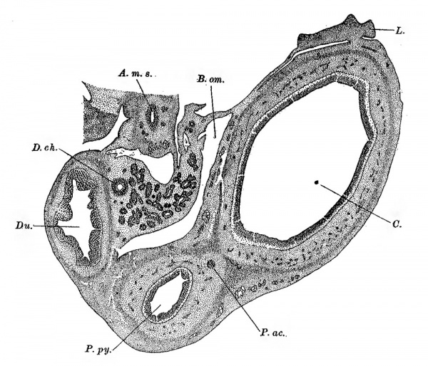
In conclusion, the abnormal stomach shown in figure 7 may be considered. It is of special interest since Gardiner (1907) has described the stomach of a child of three months, which presents a very similar condition. In the embryo there is a round nodule of epithelial cells near the angular incisure. In sections (figure 12) this nodule appears as a compact ring ‘of radiating cells arranged about a lumen.. Toward the gastric epithelium there is one section in which this structure fails to appear, so that it is apparently detached, but a short stem projects towards it from the adjacent epithelium, Both the nodule and its stalk are inside of the muscular coat. A comparable but larger structure was found by Lewis an.d Thyng in the duodenal region of a 20-mm. pig (figured in this Journal, vol. 7, p. 509). In that case, however, theldetached portion, which had become cystic, lay outside of the tunica muscularis. That the nodule in the human embryo is an accessory pancreas, is made certain by Gardiner’s specimen, in which a well developed gland with typical islands occurs in a corresponding position. Similar epithelial nodules were frequently found by Lewis and Thyng in young pig embryos, but they hesitated to interpret them as pancreases because of their abundance, and because they were never seen to branch like true pancreases. They may, however, as Elze has shown, be distinguished from the epithelial pockets of the gall-bladder and small intestine which these authors described, and which seem to be transient irregularities of the expanding tubes. Accepting the small, round, compact nodules as accessory pancreases, we may conclude that they arise at about the time when the normal pancreases become established, and usually at no great distance from them, either up or down the intestine. Subsequent elongation of the tube may carry them farther away. They may be assumed to develop slowly, since in the early stages they fail to produce branches like the adjacent normal pancreases; and as they are frequently seento be detached, probably many of them degenerate without becoming functional glands. | |||
[[File:Lewis1912-fig12.jpg|600px]] | |||
'''Fig. 12.''' Section through an abnormal stomach of an embryo of 19.3 mm.,[[Harvard Collection|Harvard Embryological Collection]], 1597, section 730, X 35 diam. A-.m.s., arteria mesenterica superior. B.om., bursa omentalis. C'., corpus gastri. D.ch., ductus choledochus. Du., duodenum. L., lien. P.ac., pancreas accessorium. P.py., pars pylorica gastri. | |||
Taken as a whole the stomach which Gardiner described is shaped like a retort. It has a globular cardiac end, 7 to 8 cm. in diameter; ‘a constriction about its middle,’ and a tubular pyloric portion, 3 to 4 cm. in diameter. If the cardiac half of the stomach shown in figure 7 should be pressed down, so that the lesser curvature became horizontal and the pars pylorica seemed to leave the upper portion of the corpus, then the form shown in Gardiner’s case would be duplicated. Although Gardiner describes his case as an hour—glass stomach, it should not be classed with those which are due to muscular contraction. It is an arrest of development, in which the pars pylorica remains clearly set off from the pars cardiaca, and as in the 19.3-mm. embryo, the line of separation is in the midd'le of the stomach. | |||
==Conclusions== | |||
In the | In addition to suggestions in regard to the nomenclature of the stomach, presented in tabular form on p. 490, the following conclusions may be drawn. | ||
latter is relatively long, constituting one-half the length of the | In the stomachs of embryos from 10 to 45 mm. in length, the division into pars cardiaca and pars pylorica is well marked; the latter is relatively long, constituting one-half the length of the stomach. The oesophagus in joining the stomach in 10-mm. embryos forms a cone extending to the angular incisure. Later this cone gives rise to the cardiac antrum above, and to a downward prolongation of the antrum below. This prolongation, which extends along the lesser curvature, constitutes the gastric canal (canalis gastricus). It was found to be well developed in an embryo of 44.3 mm. | ||
The fundus develops during the second month as a conical pouch; its boundary toward the corpus is arbitrary. | The fundus develops during the second month as a conical pouch; its boundary toward the corpus is arbitrary. | ||
The position of the pylorus is first indicated by the antrum | The position of the pylorus is first indicated by the antrum duodenale. The pylorus, like the gastric canal, is primarily an epithelial differentiation, to which the musculature conforms. | ||
duodenale. The pylorus, like the gastric canal, is primarily an | |||
epithelial differentiation, to which the musculature conforms. | |||
The occurrence of an accessory pancreas near the angular | The occurrence of an accessory pancreas near the angular incisure is shown in an embryo of 19.3 mm., in connection with a stomach which would probably have presented a permanent stricture between the pars cardiaca and the pars pylorica, thus giving rise to one form of the so-called hour-glass stomach. | ||
incisure is shown in an embryo of 19.3 mm., in connection with | |||
a stomach which would probably have presented a permanent | |||
stricture between the pars cardiaca and the pars pylorica, thus | |||
giving rise to one form of the so-called hour-glass stomach. | |||
==Bibliography== | ==Bibliography== | ||
(When translations or two editions are included in the following list, the earlier | (When translations or two editions are included in the following list, the earlier edition was not consulted). | ||
edition was not consulted). | |||
BARCLAY, A. E. 1910 The normal and pathological stomach as seen by the Xrays. Brit. Med. Journ., vol. 11, p. 537-541. | BARCLAY, A. E. 1910 The normal and pathological stomach as seen by the Xrays. Brit. Med. Journ., vol. 11, p. 537-541. | ||
BETTMANN, H. W. 1899 The shape and position of the stomach. Philadelphia | BETTMANN, H. W. 1899 The shape and position of the stomach. Philadelphia l\/Ionthly Med. Journ., vol. 1, p. 121—153. | ||
l\/Ionthly Med. Journ., vol. 1, p. 121—153. | |||
BICHAT, X. 1802 Traité d’anatornie descriptive. Tome 3. Paris. | BICHAT, X. 1802 Traité d’anatornie descriptive. Tome 3. Paris. BIDL00, G. 1685 Anatomia humani corporis. Amstelodami. BRAUNE, W. 1875 Topographiseh-anatomiseher Atlas. Leipzig. | ||
BIDL00, G. 1685 Anatomia humani corporis. Amstelodami. | |||
BRAUNE, W. 1875 Topographiseh-anatomiseher Atlas. Leipzig. | |||
BROMAN, I. 1904 Die Entwiekelungsgeschichtc der Bursa omentalis. Wiesbaden. | BROMAN, I. 1904 Die Entwiekelungsgeschichtc der Bursa omentalis. Wiesbaden. | ||
1911 Normale und abnorme Entwicklung des Menschen. Wiesbaden. | 1911 Normale und abnorme Entwicklung des Menschen. Wiesbaden. CALDAN1, L. M. A. AND CALDANI, F. 1804 Icones anatomicae, vol. 2, Venetiis. | ||
CALDAN1, L. M. A. AND CALDANI, F. 1804 Icones anatomicae, vol. 2, Venetiis. | |||
CANNON, W. B. 1898 The movements of the stomach studied by means of the | CANNON, W. B. 1898 The movements of the stomach studied by means of the Rontgen rays. Amer. Journ. _Physiol., Vol. 1, p. 359-382. | ||
Rontgen rays. Amer. Journ. _Physiol., Vol. 1, p. 359-382. | |||
1911 The mechanical factors of digestion. London. | 1911 The mechanical factors of digestion. London. CLOQUET, J. 1831 Anatomic de1’homme. Tome 5. Paris. | ||
CLOQUET, J. 1831 Anatomic de1’homme. Tome 5. Paris. | |||
COHNHEIM, O. 1907 Beobachtungen iiber Magenverdauung. Miinchen. med. | COHNHEIM, O. 1907 Beobachtungen iiber Magenverdauung. Miinchen. med. Wochenschr., Jahrg. 54, pp. 2581-2583. | ||
Wochenschr., Jahrg. 54, pp. 2581-2583. | |||
COWPER, W. 1698 The anatomy of human bodies. Oxford. (2d ed., Lcyden, | COWPER, W. 1698 The anatomy of human bodies. Oxford. (2d ed., Lcyden, 1737.) | ||
1737.) | |||
CRUvE1LH1ER,J. 1834 Anatomic descriptive. Tome 2. Paris. | CRUvE1LH1ER,J. 1834 Anatomic descriptive. Tome 2. Paris. | ||
CUNNINGHAM. D. J. 1906 The varying form of the stomach in man and the | CUNNINGHAM. D. J. 1906 The varying form of the stomach in man and the anthropoid ape. Trans. Roy. Soc. Edinburgh, vol. 45, Part I, pp. 1-47. | ||
anthropoid ape. Trans. Roy. Soc. Edinburgh, vol. 45, Part I, pp. 1-47. | |||
DELAMARE, G. AND DIEULAFE, 1906 Estomac de nouveau-né a te11dancebiloculaire. Journ. de 1’Anat. et de la Physiol., Vol. 42, pp. 624~629. | DELAMARE, G. AND DIEULAFE, 1906 Estomac de nouveau-né a te11dancebiloculaire. Journ. de 1’Anat. et de la Physiol., Vol. 42, pp. 624~629. 502 FREDERIC T. LEWIS | ||
502 FREDERIC T. LEWIS | |||
ELLENBERGER, W. AND BAUM, H. 1908 Handbuch der vergleichenden Anatoznie | ELLENBERGER, W. AND BAUM, H. 1908 Handbuch der vergleichenden Anatoznie der Haustiere. Zwolfte Auflagc. Berlin. | ||
der Haustiere. Zwolfte Auflagc. Berlin. | |||
ELZE, C. 1909 Beitfag zur Histologie des embryonalen Séiugetierdarmes. Inaug. | ELZE, C. 1909 Beitfag zur Histologie des embryonalen Séiugetierdarmes. Inaug. Diss. Freiburg. | ||
Diss. Freiburg. | |||
FABRICIUS AB AQUAPENDENTE, H. 1618 De gula, ventrieulo, intestinis tractatus. | FABRICIUS AB AQUAPENDENTE, H. 1618 De gula, ventrieulo, intestinis tractatus. Patetvii. (Opera omnia. Lugduni Batavorum, 1737.) | ||
Patetvii. (Opera omnia. Lugduni Batavorum, 1737.) | |||
FRORIEP, A. 1907 Ueber Form und Lage des menschlichen Magens. Verh. d. | FRORIEP, A. 1907 Ueber Form und Lage des menschlichen Magens. Verh. d. Gesellsch. deutsch. Natiirf. u. Aerzte., Vers. 78, Teil 2, 2 Hfilfte, pp. 312—314. | ||
Gesellsch. deutsch. Natiirf. u. Aerzte., Vers. 78, Teil 2, 2 Hfilfte, pp. | |||
312—314. | |||
GARDINER, J. P. 1907 A case of congenital hou1'—glass stomach with accessory | GARDINER, J. P. 1907 A case of congenital hou1'—glass stomach with accessory pancreas. Journ. Amer. Med. Assoc., vol. 49, pp. 15984600. | ||
pancreas. Journ. Amer. Med. Assoc., vol. 49, pp. 15984600. | |||
GYLLENSKOELD, O. 1862 Ueber die Fibrae obliqnae in dem Magen. Arch. f. | GYLLENSKOELD, O. 1862 Ueber die Fibrae obliqnae in dem Magen. Arch. f. Anat. Physio]. u. wiss. M’cd., Jahrg. 1862, pp. 132439. | ||
Anat. Physio]. u. wiss. M’cd., Jahrg. 1862, pp. 132439. | |||
HASSE, C. AND STRECKER, F. 1905 Der menschliche Magen. Arch. f. Anat. u. | HASSE, C. AND STRECKER, F. 1905 Der menschliche Magen. Arch. f. Anat. u. Entw., Jahrg. 1895, pp. 33434. | ||
Entw., Jahrg. 1895, pp. 33434. | |||
HERTZ, A. F. 1910 The motor functions of the stomach. Quart. Journ. Mod., | HERTZ, A. F. 1910 The motor functions of the stomach. Quart. Journ. Mod., Vol. 3, pp. 373-394. | ||
Vol. 3, pp. 373-394. | |||
Hrs, W. 1903 Studien an gehéirteten Leichen fiber Form und Lagerung des | Hrs, W. 1903 Studien an gehéirteten Leichen fiber Form und Lagerung des menschlichen Magens. Arch. f. Anat. u. Entw., Jahrg. 1903, pp. 345367 . | ||
menschlichen Magens. Arch. f. Anat. u. Entw., Jahrg. 1903, pp. 345367 . | |||
HOME, E. 1814 Lectures on comparative anatomy. Vol. 1. London. | HOME, E. 1814 Lectures on comparative anatomy. Vol. 1. London. | ||
H'USCIIKE, E. 1844 Lehre Von den Eingeweiden. Bd. 5 of Von Sommering’s | H'USCIIKE, E. 1844 Lehre Von den Eingeweiden. Bd. 5 of Von Sommering’s “Voni Bane des mensch.1i'chen Korpers.” Leipzig. | ||
“Voni Bane des mensch.1i'chen Korpers.” Leipzig. | |||
JoNNEsco, T. 1895 Apparcil digestif. Traité d’anatomie humaine, ed. by P. | JoNNEsco, T. 1895 Apparcil digestif. Traité d’anatomie humaine, ed. by P. Poirier. Tome 4, Paris. | ||
Poirier. Tome 4, Paris. | |||
KAESTLE, (1., RIEDER, II. AND ROSENTHAL, J. 1910 The bioroentgenography of | KAESTLE, (1., RIEDER, II. AND ROSENTHAL, J. 1910 The bioroentgenography of the internal organs. Arch. of the Roentgen Ray, vol. 15, pp. 3-18. | ||
the internal organs. Arch. of the Roentgen Ray, vol. 15, pp. 3-18. | |||
KAUFMANN, R. 1907 Ueber ,Kontraktionsph5.nomene am Magen. Weimklin. | KAUFMANN, R. 1907 Ueber ,Kontraktionsph5.nomene am Magen. Weimklin. Wochenschim, Jahrg. 20, pp. 1063-1069. | ||
Wochenschim, Jahrg. 20, pp. 1063-1069. | |||
KEITH, A. AND JONES, F. W. 1902 A note on the development of the fundus | KEITH, A. AND JONES, F. W. 1902 A note on the development of the fundus of the human stomach. Proc. Anat. Soc. Gr. Brit. and I1-e., pp. xxx1v— xxxvnr, in Journ. Anat. and Physiol., vol. 36. | ||
of the human stomach. Proc. Anat. Soc. Gr. Brit. and I1-e., pp. xxx1v— | |||
xxxvnr, in Journ. Anat. and Physiol., vol. 36. | |||
LEWIS, F. T. AND THYNG, F. W. 1908 _ The regular occurrence of intestinal diverticula in embryos of the pig, rabbit and man. Am. Jour. Anat., vol. | LEWIS, F. T. AND THYNG, F. W. 1908 _ The regular occurrence of intestinal diverticula in embryos of the pig, rabbit and man. Am. Jour. Anat., vol. 7, pp. 505-519. T | ||
7, pp. 505-519. T | |||
LUscH_K.A., H. 1863 Die Anatomie des Menschcn. Bd. 2, AM. 1. Tiibingen. | LUscH_K.A., H. 1863 Die Anatomie des Menschcn. Bd. 2, AM. 1. Tiibingen. | ||
1869 Die Luge des rnenschlichen Magens. Vierteljahrsch. f. d‘. prakt. | 1869 Die Luge des rnenschlichen Magens. Vierteljahrsch. f. d‘. prakt. Heilkunde, Prag., Bd. 101‘, pp. 114--126. | ||
Heilkunde, Prag., Bd. 101‘, pp. 114--126. | |||
MECKEL, J. F. 1820 Handbuch der menschlichen Anatomic. Bd. 4. Halle und Berlin. | MECKEL, J. F. 1820 Handbuch der menschlichen Anatomic. Bd. 4. Halle und Berlin. | ||
MEYER, G. H. 1861 Lehrbuch der Anatomic des Menschen. Zweite Aufl. | MEYER, G. H. 1861 Lehrbuch der Anatomic des Menschen. Zweite Aufl. Leipzig. ' | ||
Leipzig. ' | |||
MORGAGNI, J. B. 1761 De sedibus et causis mo1‘boru'm. Venetiis. (Tr.by B. | MORGAGNI, J. B. 1761 De sedibus et causis mo1‘boru'm. Venetiis. (Tr.by B. Alexander, London, 1769.) | ||
Alexander, London, 1769.) | |||
MORITZ, 1895 Sftudieniibe1' die motorische Théitigkeit des Magens. Zeitschr. | MORITZ, 1895 Sftudieniibe1' die motorische Théitigkeit des Magens. Zeitschr. f. Bio1., Bd. 32, pp. 313-360. | ||
f. Bio1., Bd. 32, pp. 313-360. | |||
MULLER, E. 1897 Beitriige .zur Anatomie des menschlichen Foetus. Kongl. | MULLER, E. 1897 Beitriige .zur Anatomie des menschlichen Foetus. Kongl. Evenska. Vetenskaps—A kad. Ha.ndi., Bd. 29, no. 2. Stockholm. | ||
Evenska. Vetenskaps—A kad. Ha.ndi., Bd. 29, no. 2. Stockholm. | |||
OWEN, R. 1868 On the anatomy of vertebrates. V01. 3. London. | OWEN, R. 1868 On the anatomy of vertebrates. V01. 3. London. | ||
RETZIUS, A. 1857 Bemerkungen fiber das Antrum Pylori beim Menschen und | RETZIUS, A. 1857 Bemerkungen fiber das Antrum Pylori beim Menschen und einigen Thieren. Arch. f. Anat. Physiol. u. W1SS. Med., Jahrg. 1857, pp. 74-87. | ||
einigen Thieren. Arch. f. Anat. Physiol. u. W1SS. Med., Jahrg. 1857, | |||
pp. 74-87. | |||
RIOLAN, J. 1618 Anthropographia. Parisiis. (De1’anth1'opographie. Tr. by | RIOLAN, J. 1618 Anthropographia. Parisiis. (De1’anth1'opographie. Tr. by P. Constant. Paris, 1629.) | ||
P. Constant. Paris, 1629.) | |||
RUDINGER, N. 1873 Topographisch-chirurgische Anatomie des Menschen. | RUDINGER, N. 1873 Topographisch-chirurgische Anatomie des Menschen. Stuttgart. | ||
Stuttgart. | |||
SANDIFORT, E. 1777-1781 Observationes an-atomico—pathologicae. T.3. Lugd. | SANDIFORT, E. 1777-1781 Observationes an-atomico—pathologicae. T.3. Lugd. Bat. (Cited by Bettman.) | ||
Bat. (Cited by Bettman.) | |||
SAPPEY, P. C. 1874 Traité d’an3.t0mie descriptive. 21110 édit., tome 4. Paris. | SAPPEY, P. C. 1874 Traité d’an3.t0mie descriptive. 21110 édit., tome 4. Paris. | ||
SCHULTZ, E. 1885 Zur Kenn‘:-ni-ss der motorischen Function des Ma.gons. Zeitschr. f. Heilkunde, Bd. 6, pp. 467-478. | |||
SPIGELIUS, A. 1627 De humani corporis f2LbI‘1C2L. Venetiis. | SPIGELIUS, A. 1627 De humani corporis f2LbI‘1C2L. Venetiis. | ||
TANDLER, J. 1900 Zur Entwicklungsgéschichte des menschlichcn Duodonum. | TANDLER, J. 1900 Zur Entwicklungsgéschichte des menschlichcn Duodonum. Morph. Jahrb., Bd. 29, pp. 187-216. | ||
Morph. Jahrb., Bd. 29, pp. 187-216. | |||
THYNG, F. W. 1908 Models of the pa.nc1'ca.s in embryos of the pig, rabbit, cat | THYNG, F. W. 1908 Models of the pa.nc1'ca.s in embryos of the pig, rabbit, cat and man. Am. Jour. Ana.t., vol. 7, pp. 489-503. | ||
and man. Am. Jour. Ana.t., vol. 7, pp. 489-503. | |||
TOLDT, C. 1879 Bau und Wachsthumsverétnderungen der Gekriise dos mesch1ichen_Da.rmkana1es. Denkschr. kais. Akad. Wiss., Wien, mathnaturw. Classe, B. 41, Abt. 2, pp. 1-56. | TOLDT, C. 1879 Bau und Wachsthumsverétnderungen der Gekriise dos mesch1ichen_Da.rmkana1es. Denkschr. kais. Akad. Wiss., Wien, mathnaturw. Classe, B. 41, Abt. 2, pp. 1-56. | ||
| Line 951: | Line 348: | ||
VESALIUS, A. 1543 De humani corporis fa.brica.. Liber 5, Caput 3. Basileae. | VESALIUS, A. 1543 De humani corporis fa.brica.. Liber 5, Caput 3. Basileae. | ||
WALDEYER, W. 1908 Die Magenstrasse. Sitz.-ber. d. kon. preuss. Akad. d. | WALDEYER, W. 1908 Die Magenstrasse. Sitz.-ber. d. kon. preuss. Akad. d. Wiss., Bd. 29, pp. 595-606. | ||
Wiss., Bd. 29, pp. 595-606. | |||
WILLIS, T. 1674 Pharmac-eutice mtionaiis. Hagae-Comitis. (Genevae, 1680). | WILLIS, T. 1674 Pharmac-eutice mtionaiis. Hagae-Comitis. (Genevae, 1680). | ||
WINSLOW, J. B. 1732 Exposition anatomique dc la structure du corps humain. | WINSLOW, J. B. 1732 Exposition anatomique dc la structure du corps humain. Paris. (Tr. by G. Douglas, London, 1734.) | ||
Paris. (Tr. by G. Douglas, London, 1734.) | |||
{{Footer}} | {{Footer}} | ||
[[Category:Stomach]][[Category:Historic Embryology]][[Category:1910's]] | [[Category:Stomach]][[Category:Historic Embryology]][[Category:1910's]][[Category:Harvard Collection]] | ||
[[Category:Draft]] | [[Category:Draft]] | ||
Latest revision as of 12:57, 21 May 2017
| Embryology - 19 Apr 2024 |
|---|
| Google Translate - select your language from the list shown below (this will open a new external page) |
|
العربية | català | 中文 | 中國傳統的 | français | Deutsche | עִברִית | हिंदी | bahasa Indonesia | italiano | 日本語 | 한국어 | မြန်မာ | Pilipino | Polskie | português | ਪੰਜਾਬੀ ਦੇ | Română | русский | Español | Swahili | Svensk | ไทย | Türkçe | اردو | ייִדיש | Tiếng Việt These external translations are automated and may not be accurate. (More? About Translations) |
Lewis FT. The form of the stomach in human embryos with notes upon the nomenclature of the stomach. (1912) Amer. J Anat. 13(4): 477-503.
| Historic Disclaimer - information about historic embryology pages |
|---|
| Pages where the terms "Historic" (textbooks, papers, people, recommendations) appear on this site, and sections within pages where this disclaimer appears, indicate that the content and scientific understanding are specific to the time of publication. This means that while some scientific descriptions are still accurate, the terminology and interpretation of the developmental mechanisms reflect the understanding at the time of original publication and those of the preceding periods, these terms, interpretations and recommendations may not reflect our current scientific understanding. (More? Embryology History | Historic Embryology Papers) |
The Form of the Stomach In Human Embryos with Notes upon the Nomenclature of the Stomach
By
Harvard Medical School, Boston, Massachusetts
Twelve Figures
Introduction
X-ray examinations of the stomach, in adults and especially in children, have led clinicians to inquire Whether the stomach has a characteristic embryonic form Which may sometimes persist. Figures of the typical embryonic stomach have, indeed, been published;‘but it must be remembered that the stomach changes in shape as the embryo grows older and, as Broman has found, its individual variations in embryos of the same stage of development is very great. Nevertheless certain fundamental subdivisions are strikingly distinct. These primary subdivisions, in which the embryologist is most interested, were keenly discussed by the early anatomists. In their writings many suggestive questions are raised, at the same time that the fundamental features of the organ are successively recognized and defined. In the following historical notes, taken from such Works as are at hand, provisional definitions are offered for certain terms adopted at Basle but at present loosely employed, and attention is called to the features of the adult stomach which will be examined in the embryos.
Stomach Nomenclature
The human stomach was first considered to be a simple sac with an orifice of entrance above and to the left, and an orifice of exit belowand to the right. Vesalius (1543) in his figures designates the orifices as the ‘superius ventriculi orificium’ and 'iI1ferius ventriculi orificium,’ respectively. In his text, however, both are said to be placed superiorly, so that food shall not escape by its own Weight, but when completely changed to chyme, shall be propelled by force of the discharging stomach into the intestine. Fabricius ab Aquapendente (1618) likewise states that the orificium inferius is n.ot inferior at all, and Spigelius (1627) places it in the highest part of the stomach; so that the term ‘orificium dextrum’ was preferred, and finally the less objectionable Greek name ‘pylorus’ (Latin, janitor), which had been. introduced by Galen, became the accepted designation. Winslow’, however, in 1732, insisted that the position of the orifices is such that “We ought with the’ ancient anatomists to call one of them superior, the other inferior.”
The significance of ‘cardia’ (Latin, cor), as applied to the oesophageal orifice, was discussed by Fabricius, who cites Galen as stating that the upper orifice of the stomach is called the heart because the symptoms to which it gives rise are similar tothose which sometimes affect the heart, sometimes even the brain; but for Fabricius, cardia, as applied to this orifice, merely indicates a chief part of the body. Spigelius describes the cardia as consisting of circular fleshy fibers, so that the stomach, after having received food, may be closed perfectly, thus preventing fumes from rising, with consequent loss of heat. The cardia and pylorus are intimately associated with their respective sphincter muscles, but they do not include the adjacent portions of the stomach.
For the stomach as a whole these anatomists use the Latin ‘ventriculus,’ rather than the Greek ‘gaster’ and the Latin term has been adopted at Basle. Since however, the adjective gastricus has been chosen instead of ventricularis, it seems desirable that gaster should be used in place of ventriculus, especially since cardia and pylorus are of Greek origin).
- I am indebted to Prof. Albert A. Howard for the following note regarding these terms: Gaster is a Greek word meaning belly (the whole abdominal cavity) but was often used by the Greeks in the more restricted sense of stomach. It is not found in Latin with this meaning until very late (only after the literary period). Vcntriculus is used quite consistently for stomach by Celsus and at times by Pliny the Elder. Cicero in one passage speaks of ventriculus cordis, but does not use ventriculus for stomach. If gaster is adopted I think the genitive gastri is preferable to gasteris, though as a matter of fact the genitive does not happen to occur in any Latin that is preserved to our time. Petronius has used the ablative plural gastris which would be the reason for deciding as I have.
The stomach, as described by Vesalius, is rounder and more spacious on the left side, and more slender on the right; to which Fabricius adds that it is not unlike a gourd with larger belly and narrower neck. On its dorsal side Vesalius found two swellings, separated by a vertical impression which was fitted against the trunks of the aorta and vena cava and the projecting bodies of the vertebrae. When the stomach was inflated, the impression and swellings were lost in an even rotundity. It was not until Willis (1674) described the pyloric antrum in the following passage, that a permanent subdivision of the stomach was established.
The other orifice, commonly called the pylorus, on the right side of the stomach, having a capacious and long, gradually narrowed antrum, ends in a small foramcn and thence bent back is continued into the duodenum. Here the coats are much thicker than in any other part of the stomach.
Indeed the long and capacious antrum seems to be a sort of recess and diverticulum in the stomach, into which the more elaborated and perfected portion of the chylous mass may Withdraw and there remain, while the other cruder and more recently ingested portion may be further digested in the fundus of the stomach (ed. of 1680, p. 13-14).?
Accompanying this description Willis _published four lateral views of the stomach, with its coats successively removed. All of them show the antrum, but in a fifth figure, representing the everted stomach, its limits are most satisfactorily indicated (fig. 1). In this figure the antrum is shorter and broader than in one of the others, in which it has been stretched out so as to form a tube.
In all of the figures it is clear that the antrum extends to the pylorus, which is referred to as its orifice.
Bidloo (1685) published a Inore accurate figure of the stomach, here reproduced as figure 2, but he failed to describe it adequately. He states that the base is provided with two swellings, C and D. In another figure, showing the same stomach partly laid open, the portion of the duodenum near the stomach (A) is labelled pylorus, but Bidloo does not refer in any way to the subdivision which in figure 2 has been labelled B. Cowper (1698), who republished Bidloo’s plates, states that A is the part of the ‘duodenum arising from the pylorus and adds that B is the antrum pylori.
- For verifying and revising the Latin translations, the author is under obligation to Mr. S. R. Meaker.
In 1732 Winslow described the large arch running along the greatest convexity of the stomach, and the small one directly opposite, and named them the great and small curvatures. . Bichat (1802) states that “the great curvature ends simply at the pyloric orifice, without presenting anything of note unless it be the elbow (le coude) formed by this pyloric orifice, and named the small cul—de—sac ; but there is no particular swelling at this place and the bend is precisely in the direction of the pylorus.” Cloquet (1831) repeated this description and Cruveilhier (1834) made it more explicit. He states that at about 2 or 3 cm. from the pylorus “the stomach, bending sharply upon itself, forms a very pronounced elbow (coude de l’estomac) on the side of the greater curvature, and presents an ampulla, corresponding to an interior excavation, named by Willis the pyloric antrum, by others the small cul—de—sac.” As pointed out by Muller (1897), Cruveilhier was unjustified in identifying a pouch about an inch from the pylorus with the pyloric antrum of Willis; but he was correct in stating that “it is not rare to see a second ampulla beside the first, and a third but smaller one, on the side of the lesser curvature” (compare with figs. 2 and 3). These had not been recognized by Willis, but Cowper, in describing Bidloo’s plate, was confronted with the question whether one or more of these parts was to be regarded as the antruzn. In applying the term to the part adjacent to the pylorus but not extending to the bend of the stomach, Cowper was justified by Willis’s figure here reproduced as figure 1. According to Cunningham (1906) “no part of the organ is more definite and distinct” than the region which Cowper designated ‘antrum pylori’ and which, rediscovered by J onnesco (1895), was named the pyloric canal. It may be defined as the part of the stomach adj acent to-the pylorus, averaging 3 cm. in length, cylindrical when empty, bulbous when distended, separated from the remainder of the stomach by a groove on the greater curvature—the ‘sulcus intermedius’ of His (1903). For the small cul-de-sac of Cruveil— hier the term ‘pyloric vestibule’ (J onnesco, 1895) may be adopted.
Fig, 1 Willis’s figure of the inverted stomach re-drawn and reduced one-half. "A, Orificium sinistrum, sive os ventrieuli. B, Pylori Antrum, in que, Tunicae crassiores exist-unt. C, Orificium ejus, cuo Duodenum annectitur.”
Fig. 2 Bidloo’s figure of the unopened stomach, re-drawn and reduced twothirds, with lettering added from Bidloo’s drawing of the same stomach opened, and from Cowper’s edition of Bidloo’s plates. A, pylorus (Bidloo);portion of the intestinum duodenum (Cowper). B, antrum pylori (Cowper). C’, D, two bunchings out in the lower part or fundus of the stomach (Cowper) ; in funclo Gibbis ornatur duobus (Bidloo).
Fig. 3 Home’s figure of “the human stomach inverted, to show the contraction which divides the cavity into two portions.” Re-drawn and reduced two-thirds. cm, the cardiac portion. 1), the contraction dividing the cardiac from the pyloric portion. c, the pyloric portion. 01, the pylorus.
Unfortunately Cowper’s use of pyloric antrum has been overlooked by later anatomists, and the term has been so variously employed, as tabulated by Muller, that Muller, His and Cunningham have proposed to abandon it altogether. His has suggested an entirely new nomenclature for the pyloric region, as follows: for pyloric vestibule, camera princeps; for the swelling on the lesser curvature opposite the sulcus intermedius (fig. 3), camera minor; and for pyloric antrum, camera tertia. But these terms, as stated by Cunningham, are not in every respect satisfactory, and it may be well to retain the appropriate name ‘pyloric antrum’ in the sense of Cowper, following Meyer (1861) a11d Hasse and Strecker (1905).
The normal division of the entire stomach into two parts, cardiac and pyloric (of which the latter presents the subdivisions just described), was first recognized by Home (1814). He wrote as follows: I found also, in the necessary examinations, that the dog’s stomach, while digestion is going on, is divided by a muscular contraction into two portions; that next the cardia is the largest, and usually containing a quantity of liquid, in which there was some solid food; but the other, which extended to the pylorus, being filled entirely with half—digested food of an ordinary consistence. I shall, therefore, in my future description call that part which constitutes the first cavity the cardiac portion, and that which constitutes the second the pyloric portion (p. 140).
The cardiac portion is in length two-thirds of the whole, but in capacity much greater (p. 139).
Home distinguished these two portions not only in the dog but, with varying distinctness and permanence, in many animals, including the horse, pig, rat, rabbit and man. He did not describe or label the subdivisions of the pars pylorica, which however are clearly shown in his figure of the everted human stomach (fig. 3).
In connection with Home’s work, the following more recent physiological observations are of interest. Schiitz (1885) found that in the dog’s stomach, contraction waves travel from the cardiac end to a place about 1 cm. from the beginning of the pyloric antrum (pars pylorica?), which in the isolated resting stomach may be recognized by a shallow annular constriction about 2 cm. behind the pylorus, and there end with a deep ‘prae—antral constriction.’ The second phase, which follows the first immediately, concerns the antrum only. The motions of the two parts may take place independently of one another. Moritz (1895) determined the pressure within the two parts of the stomach, and stated that the difference between them was greater than would be inferred from merely observing their motions. Cannon (1898) found that the stomach of the cat, as shown by X-ray examinations, is composed of two physiologically distinct parts—a ‘busy antrum’ and a cardiac reservoir. In 1911 he states that during normal digestion “slight constrictions appear near the middle of the body of the stomach, and pressing deeper into the greater curvature, course towards the pyloric end. When a wave sweeps round the bend i11to the vestibule, the indentation made by it increases.” He adds that when vomiting occurs, a strong contraction at the angular incisure completely divides the gastric cavity into two parts. Thus the observations of Home have been amply confirmed and extended. Other X—ray observers, however, have considered that the antrum, or pars pylorica, of anatoinists is merely the part of the stomach marked off by a passing peri staltic wave (Hertz; Kaestle,Rieder and Rosenthal; Barclay). In this they follow Sappey (1874), who was of the opinion that Home’s subdivision of the stomach was based on fortuitous muscular contractions. This will be disproved by showing that the two divisions of the stomach are well marked in embryos in which the muscle-layers are still scarcely differentiated.
When a peristaltic wave remains fixed after death, the stomach may appear as “two joined together” (Riolan 1618), in which case the subdivisions need not correspond with the anatomical parts already described. Usually the constriction is near the middle of the stomach, and falls within the cardiac portion. Morgagni (1761) observed five cases, all in women. One of the stomachs was from a patient who had been troubled with excessive vomiting since birth, but none of the stomachs showed any sign of disease. Since several cases had been reported in men, Morgagni concluded that the double stomach was not a deformity due to stays, but had existed from the first formation of the organ. Sandifort (1777-1781), as quoted by Bettman, described a typical case in a fetus, the age of which is not stated in the citation. Delamare and Dieulafé (1906) reported a case in a new-born syphilitie infant, in which they describe an hypertrophy of the circular muscle atthe place of constriction. The thickened muscle-layer may, however, be due to contraction, -as indicated by the folded and thickened overlying layers sho-wn in their figures. Cunningham (1906) holds that there is not an atom of evidence that the hour-glass stomach ever arises as a congenital deformity, but he is not prepared to state that the strictures which separate the two sacs of the bilocular stomach are always temporary and fleeting. The change in the direction of the lesser curvature is more dependable as a boundary between the pars pylorica and pars cardiaea, than the constriction which is present in certain cases but “not as a rule” (Huschkc). The lesser curvature, which is concave along the cardiac portion, becomes convex along the pars pylorica (Meekel 1820; Huschkc 1844). Retzius (1857) figured a deep stricture in the lesser curvature at the beginning of the bulbous pars‘ pylorica, where Luschka (1869) frequently found an acute angle directed toward the gastric cavity. This notch has been named by His (1903) the ‘incisura angularis,’ and it occurs between the two parts of the stomach. Along the greater curvature the boundary is less clearly marked. It is indicated by the change in direction already described as the elbow of the stomach, and referred to by Home as “an angle formed at the part where the temporary contraction takes place.” times the constriction is slightly to the cardiac side of the elbow, as shown in figure 3.
The angle which separates the two parts of the stomach is obscure in the older drawings in which the organ is almost horizontally placed. According to Bichat (1802) “When the stomach is filled its obliquity increases considerably; often it appears almost perpendicular, so that the right extremity . . . . is strongly recurved upward, and forms a very acute angle with the body of the organ.” Luschka (1869) similarly found that the greater part of the stomach, as a rule, has a precisely vertical position, but that the pars pylorica is directed almost transversely. Both of these forms, with vertical body and transverse or ascending pars pylorica, will be seen in the embryos to be examined.
The pars pylorica and its subdivisions having been described, the pars cardiaca may next be examined. It is divided into the ‘saccus caecus,’ now called the ‘fundus;’ the ‘corpus’ or body; and the gastric canal. The term fundus was appropriately applied by Vesalius to the lower part of the stomach, which in the transverse position of the organ, extends well toward the pyloric region. It was so used by Willis (1674); and by Cowper (1737), as seen in figure 2. Caldani (1804) makes fundus synonymous with greater curvature. The bulging left or upper extremity of the stomach received the special name ‘saccus caecus’ (Haller, 1764; Caldani, 1804). But Meckel (1820) considered fundus and saccus caecus as synonyms, and preferred fundus; Huschke (1844) likewise made them synonymous, but adopted saccus caecus, which Henle used in 1866. Nevertheless, fundus has become adopted for the highest part of the stomach and saccus caecus has been rejected. The fundus lies at the left of the cardia, being separated from the oesophagus by a notch, the ‘incisura cardiaca’ of His (1903). Below, as described by Cloquct, the fundus terminates almost imperceptibly in the greater curvature. It is therefore bounded arbitrarily by a horizontal plane at the level of the inferior border of the cardia (J onn'esco), or by a line prolonging the axis of the abdominal part of the oesophagus {Keith and Jones, 1902).
According to Keith and Jones the fundus arises in human embryos as a localized outgrowth or diverticulum ofrthe stomach, and in its manner of origin has much in common with the caecum and Vermiform process. From numerous observations they conclude that “it is not uncommon to find in the stomach of the anthropoids, and to a lesser degree in that of the apes (especially in Mycetes) clear indications of three chambers, namely, a fundus, a body, and a pyloric part; and that therefore the stomach of the Primates (excluding the Lemuroidea) is probably tripartite in nature.” It should be noted that the fundus as defined by Keith and Jones is a larger part of the stomach than that set off by J onnesco, and that their boundary is justified by comparison with the stomach of Semnopithecus which they have figured. If the fundus corresponds in any way to a first stomach or rumen, it may be regarded as the globular upper end of the organ which is often marked off by the contraction of the corpus.
The body of the stomach (corpus gastri), as defined by Rudinger (1873), is its middle subdivision, situated between the fundus and the pars pylorica. Froriep (1907) proposed to rename it the pars intermedia; but since it is a portion of the pars carcliaca, and is not intermediate between the pars cardiaca and pars pylorica, the proposed term would lead to confusion. Jonnesco (1895) defined the body as including the pyloric vestibule, but in the same paragraph he described the boundary between the vestibule and “le corps proprement dit.” Muller (1897) included the fundus with the body, making corpus and pars cardiaca synonymous. It is only by accepting Rudinger’s earlier definition that corpus becomes a useful term. The corpus may be contracted at any point, as in the hour—glass stomach, in which case part of it appears to belong with the fundus and the remainder with the pars pylorica. Sometimes it is contracted as a whole, but more often it is relaxed, and its boundaries are then ill—defined.
The gastric canal is a channel which follows the lesser curvature, appearing as a groove when seen from the inside of the stom— ach. It suggests a continuation of the oesophagus, split open toward the gastric cavity, and has been named the sulcus oesophageus, sulcus gastricus, sulcus salivalis and canalis salivalis. It is -confusing, however, to refer to this channel as a sulcus, since the external grooves of the stomach are so designated (sulcus inter—medius, sulcus pyloricus), and it is un.desirable to name a part of the stomach oesophageal or salival. Therefore the term gastric canal, ‘canalis gastricus,’ is here proposed, and canalis is used as in Latin for an open canal, which in this case may become a tube during its physiological activity, by the approximation of its lips.
The gastric canal has long been known in ruminants, but in its less highly developed condition in the human stomach, it has attracted little attention. In man it is generally supposed to be due to the arrangement of the oblique muscle fibers, which were first described by Willis (1674), in connection with a figure of the stomach in the position shown in figure 1. The ‘top’ of the stomach is accordingly toward the lesser curvature, and the ‘fundus’ is toward the greater curvature. Willis wrote as follows:
- These muscle fibers, which are seen to arise behind the cardia and to pass around its left margin, are carried forward to the right portion of the stomach. A notable bundle of them, proceeding in straight lines along the top of the stomach on either side, encounters the antrurn, and spreading over the length of its cavity in a scattered manner, terminates in the pylorus. Moreover the remaining fibers of this layer extend obliquely over the walls of the stomach on both sides, and then directly toward the fundus where they come together. The function of the former (the straight bundles) seems to be to bring one orifice toward the other in emptying, by making them lower and higher respectively (ed. of 1680, pp. 11-12).
Retzius called attention to this description by Willis and, as reported by Gyllenskoeld (1862), he supplemented it as follows:
- The upper portion of the oblique fibers of the human stomach serves to form a sort of trough along the lesser curvature which, under the control of the motor nerves, becomes more or less closed ;_ along this path possibly fluids and soft things, saliva, etc., may proceed directly from the oesophagus to the pars pylorica, passing by the cardiac portion, which corresponds to the first two stomachs of ruminants and the non-glandular part of the stomach in rats.
The correctness of this conjecture concerning the passage of fluids was established by Cohnheim (1908), Who was surprised to find that water or salt—solution passed rapidly through the full stomach of a dog, without mixing with the gastric contents.
Gyllenskoeld (1862) states that the oblique fibers extend only to the pars pylorica, and not to the pylorus as described by Willis. This has been confirmed by Kaufmann (1907). He found that there is no sphincter of circular fibers separating the pars cardiaca from the pars pylorica, but that the furrow between them has a special structure, since it is the place where the oblique fibers terminate and interlock with the circular fibers.
Hasse and Strecker (1905) have named the folds which bound the gastric canal the ‘plica hepatica’ and ‘plica aortica’ respectively, and state that they are connected with one another by the ‘plica cardiaca’ which passes around the cardia, projecting into the stomach beneath the incisura cardiaca. According to Hasse and Strecker the plica cardiaca does not form a valve for the cardia, as Braune (1875) thought possible from the result of experiments on a cadaver. The hepatic, cardiac and aortic plieae together form a U-shaped structure, across the open end of which is the ‘plica angularis/o This is beneath the incisura angularis, at the beginning of the pars pylorica.
Waldeyer, who describes the channel from cardia to pars pylorica as the ‘Magenstrasse’ (1908), considers that its formation depends upon the oblique muscles, rather than upon folds which arise in relation with adjacent organs. In the following pages evidence will be offered to showthat the gastric canal is a distinct epithelial structure, arising independently both of the muscle and the surrounding organs.
There remain to be considered two structures which are beyond the limits of the stomach—the ‘antrum duodenale’ and the ‘antrum cardiacum’ .
Retzius (1857) states that the beginning of the duodenum is often specially rounded, not only in man, but in a large proportion of mammals; in dolphins it has been considered a part of the stomach. Owen (1868) remarks that in all Artiodactyles the duodenum is dilated at its commencement; it there forms a distinct pouch in the camel. For this pouch Retzius proposed the names “antrum or atrium duodeni” but used the former in his figures. Luschka (1863) refers to a flask—shaped expansion at the beginning of the duodenum, which inhis figure is called the ‘antrum duodenale.’ This structure Will be seen to be far more distinct in human embryos than it appears to be in adults.
The cardiac antrum was first described by Luschka (1863) as follows:
- At the junction of fundus and lesser curvature the oesophagus enters the stomach, forming a funnel-shaped expansion—the cardia. Although ordinarily the cardia is continued into the rest of the stomach without definite boundary, in rare cases the funnel—like expansion is sharply marked off by an external depression and corresponding internal elevation, thus forming a sort of cardiac antrum (p. 179).
In 1869 Luschka adds that if this funnel is to be regarded as part of the stomach, the beginning of which is not rather to be considered at the base of the funnel where the stratified epithelium ends in a zig-zag line (fig. 3), “then the funnel—shaped expansion must be specially designated as the pars cardiaca.”
Thus Luschka proposed two names for a single structure; first, cardiac antrum; and later, in case the antrum is to be regarded as part of the stomach, pars cardiaca. The latter may be rejected, since it is generally agreed that the cardia is at the base of the cone, and that therefore ‘cardiac antrum’ is “merely another name for the intra—abdoIninal part of the oesophagus” (Cunningham). Moreover the earlier use of pars cardiaca, or cardiacfion tion, for the fundus and corpus taken together, was overlooked by Luschka, and by certain later anatomists Who have proposed to substitute Hauptmagen (His), saccus ventriculi (Hasse and Strecker) and pars digestoria (Froriep).
The fundamental subdivisions of the stomach and adjacent parts of the digestive tube, as they have been defined in the preceding pages, are presented in figure 4 and in the following table, With authority for certain of the definitions adopted:
- Antrum cardiacum (Luschka, 1863)
- Gaster
- Cardia
- Pars cardiaca. gastri (Home 1814)
- Fundus (Meckel 1820)
- Corpus (Rudinger 1873)
- Canalis gastricus
- Pars pylorica gastri (Home 1814)
- Vestibulum pyloricum (Jonnesco 1895)
- Antrum pyloricum (Willis 1674 CE); Cowper 1698)
- Pylorus
- Antrum duodenale (Retzius, 1857)
As boundaries between these parts, the following may be recognized: Between the cardiac antrum and fundus, the ‘incisura cardiaca;’ between cardiac and pyloric parts, the ‘incisura angularis;’ between pyloric antrum and pyloric vestibule, the ‘sulcus intermedius’ (all of His 1903); at the pylorus,,the ‘sulcus pyloricus’ (Luschka 1863).
The Stomach in Human Embryos
The embryonic stomachs to be examined are five in number, from embryos between 10 mm and 45 mm in length. Thus they are all smaller than the specimens studied by Miiller, but similar stages have been described by Broman in his extensive work on the omental bursa. Broman modelled not only the gastric epithelium, but also entire stomachs, including the mesodermal portion. In the models to be described, only the epithelium has been included, since this is the portion having characteristic shape, to which the other layers subsequently conform.
In the youngest embryo (10 mm, fig. 5) the stomach is no longer a simple sac with superior and inferior orifices, but is already divided into an expanded pars cardiaca and a tubular pars pylorica. Between the two, and almost exactly in the middle of the stomach, is the incisura angularis. Since" the incisure in the adult is perhaps twice as far from the cardia as from the pylorus, it is evident that the pars pylorica is relatively long in early stages. This is strikingly shown-in other models of the series (figs. 6-9). THE FORM or It is true also in the cat, if one may judge by comparing Thyng’s model of the stomach of a 10.7-mm. embryo (this Journal, vol. 7, p. 496) With Cannon’s tracings from the adult. In ruminants a constriction early separates the rumen and reticulum from the psalterium and abomasum; according to Ellcnberger and Baum the abomasum is larger than the rumen in embryos and very young animals, but later this relation is reversed. The relatively large size of the pars ipylorica in early stages is therefore not limited to human embryos.
Fig. 4 Diagram showing the subdivisions of the human stomach.
The cardia cannot be definitely located in the 10-mm. embryo (fig. 5) since the oesophagus, in joining the stomach, expands into a flattened cone, one margin of which extends to the angular incisure. A similar extension of the oesophageal cone to the incisure is clearly seen in Broman’s model of the stomach of the seventh embryo in his series (11.7 mm.). At 16 mm. (fig. 6) the body of the stomach may be recognized along the lesser curvature, separating the oesophageal cone from the angular incisure; but a canal, distinctly marked out above and indicated below, passes along this curvature from the oesophagus to the pars pylorica. A more distinct canal in this position is seen in two of Broman’s models, from embryos of 10 mm. and 16.2 mm. respecgively. Apparently this canal has not been previously described in embryos, although Toldt (1879), referring to the general direction of the oesophagus in a 23—mm. specimen, states that it descends into the stomach “in such a Way that the lesser curvature forms, as it were, a continuation of the ventral border of the oesophagus.” .
In the embryos of 19.3 mm. and 19.0 mm. shown in figures 7 and 8 respectively, the canal is not seen. The first of these stomachs is abnormal, but the second specimen is unobjectionable. Moreover in Broman’s figure of the stomach from an embryo of 21 mm., there is no trace of the channel. Its obliteration, if normal, appears to be temporary however, for in the 44.3—mm. specimen shown in figure 9, it is more distinct than in preceding stages. It passes from the conical cardiac antrum to the angular incisure. This embryo, owing to its large size, was not perfectly preserved, and the epithelium has separated from the mese11— chyma; but whether 0110, or the other, or both of these tissues has shrunken is uncertain. The model may, however, be accepted as giving an essentially correct idea of the shape of the stomach, since the separated mesenchyma presents corresponding ridges and furrows. The distinetness of the gastric canal is strikingly shown when the model is viewed from the inside (fig. 10). It takes a slightly S—shaped course from the stellate cardia to the orifice of the pars pylorica, and is bounded by a rounded plica aortica, and a more prominent and angular plica hepatica. These folds are not formed, as described in theadult by Hasse and Strecker, through compression of the borderiof the stomach between the aorta behind and the caudate lobe of the liver in front; for the outer layers of ‘the stomach are not_ indented. Moreover at this stage there are no bands of oblique fibers to account for the canal. If the channel proves to be a constant feature of embryos of this stage, and it is present in an embryo of 37 mm. which was not modelled, it may be that the arrangement of the oblique fibers is a consequence rather than the cause of the gastric canal.
In the same way that the gastric canal accords with the ‘oesophageal sulcus’ of ruminants, which is described by comparative anatomists as a continuation of the oesophagus open on one side, the cardiac antrum may correspond to the ‘atrium ventriculi.’ This, according to Ellenberger and Baum is “a dome—shaped swelling on the dorsal side of the reticulum and thoracic end of the rumen, which is only indistinctly marked off from them by a shallow groove; ventrally its cavity passes directly into that of the reticulum, and caudo—ventrally into the vestibule of the rumen ; toward the thorax it rests against the diaphragm near the hiatus oesophagcus.” From the general ‘atrium’ seen in figure 5, the lower part is set off as the gastric canal, and the upper part remains as the cardiac antrum (figs. 8 and 9). From studies of the adult stomach it may be assumed that the cardia is at the base of this antrum, whi.ch therefore belongs with the oesophagus.
The development of the fundus of the stomach has been described by Broman (1911) as follows:
- By the beginning of the second month the cranial part of the greater curvature begins to bulge out. But not until the third month, or later, is this outpocketing generally directed so strongly craniad that its blind end comes to lie above the orifice of the oesophagus. Only from this time, therefore, can we speak of a distinct gastric fundus (pp. 326-328).
Similarly Keith and Jones state that the outgrowth is best marked in embryos of the third and fourth month. But as shown in figures 8 and 9, and by the fact that Toldt, in an embryo of 48 mm., found a well marked fundus projecting toward the concavity of the diaphragm, it is clear that the fundus may be well developed in the second month. In the model shown in figure 9, the fundus when seen from above, presents a curious appearance, since seven prominent ridges converge toward its apex. Two of them come from the cardiac antrum, sweeping in a semicircular curve beneath the cardiac incisure, thus resembling the ridges seen. in figure 8. There is normally no boundary between the fundus and corpus, but in an abnormal embryo of 18.5 mm., described by Broman, the fundus is cut off by a rather deep constriction. Broman states that this specimen suggests an l1our—glass stomach, from which, however, it is essentially different, since the oesophagus enters the part toward the pylorus. The fundus is best marked when the pars cardiaca is in an approximately vertical position, and this is the ease in figures 6 to 9. Broman, however, has found a greater variety of positions. In an embryo of 21 min. he figures the stomach as horizontal, so that both orifices are superior, as described in the adult by Vesalius; but this position must be regarded as exceptional.
Figs. 5 to 9 Models of the gastric epithelium in human embryos, as follows: figure 5, 10 mm. Harvard Embryological Collection, Series 1000, X 50 dia.m.; figure 6, 16.0 mm., H. C. 13:2, X 35 diam; figure 7, 19.3 mm., H. C. 1597, X 30 diam.;figurc 8, 19.0 mm., H. E. C. 819, X 26 diam.; figure 9, 44.3 mm., H. E. C. 1611, X 18 diam.
A.du., antrum duodenale. C., Corpus gastri. D.ch., ductus choledochus. D.p.d., ductus pancreatis dorsalis. F., fundus gastri. I.ang., incisum angularis. 0e., oesophagus. P.py., pars pylorica gastr.
The body of the stomach requires no comment other than that its ridges appear to be rather definitely placed. The shelf-like prominence at the base of the oesophageal cone in figure 5, is evidently represented by the chief fold which extends horizontally across the base of the fundus, and bends down parallel with the lesser curvature in figure 6. Such an angular fold (With a subdividing furrow) is seen in figure 9, and it is clearly shown in embryos of 10 and 16.2 min. figured by Broinan. Why the ridges are absent from other specimens, as in figures 7 and 8, and in several of Broman’s embryos, is not apparent.
Fig. 10 Model of the interior of the stomach, from an embryo of 44.3 mm., Harvard Embryological Collection, 1611, X 25 diam. Cara, carclia. (,‘.q., eanalis gastricus. O.p.py., orifieium partis pyloricae. P.a0., plica aortica. 1’./Lep., pliea hepatica.
The position of the pylorus could not be determined with certainty in the 10-mm. embryo (fig. 5); and Tandler (1900) states that in an embryo of 11 mm. the pylorus is not marked. At 14.5 mm., “where the stomach passes into the duodenum, therefore at the place of the future pylorus” he saw “a considerable thickening of the epithelium.” The epithelial proliferation, which Tandler describes, is seen throughout the upper part of the duodenum. It is n.ot evident that he recognized the local swelling, chiefly on the upper side of the digestive tube, which is shown in figures 6 to 8. This swelling, which distinctly marks the position of the pylorus when the muscle-layers are still undifferentiated, and scarcely to be recognized, is apparently the duodcnal antrum of Retzius. In a frontal section through the pars pylorica of a 19—mm. embryo, it appears as shown in figure 11. At this stage the musculature of the pars pylorica is considerably thicker than that of the duodenum, but in this it conforms to the shape of the epithelial tube. In figure 9 the duodenal antrum is seen to be smoother than the more distal part of the duodenum, recalling the statement of Retzius that here, in the adult, the valvulae are absent and the villi are short. In this embryo the gastric epithelium is seen to be slightly invaginated into the duodenal tube, as observed by Cunningham at birth. Toldt found that the sulcus pyloricus could be seen externally in an embryo of 48 mm, and presumably it could have been found in this specimen by dissection.
Fig. 11. Frontal section through the pylorus of a 19 mm embryo, Harvard Embryological Collection, 828, section 330, X 40 diam. A.du., antrum duodenale. B.o-m., bursa omentalis. P.py., pars pylorica. T.musc., tuniea muscularis.
The pars pylorica, even in the 44.3 mm embryo, fails to show distinct subdivision into antrum an.d vestibule. Muller, who studied dissections of the embryonic stomach, states that in the ‘first fetal period’ the pyloric antrum is a direct continuation of the pyloric vestibule, but that later, when. the pars pylorica is bent convexly upward, the general direction of the vestibule is upward, and of the antrum, downward. In the earlier period the antrum is characterized by “its cylindrical form and the great development of its muscle-layer.” In the still earlier stages under discussion, neither distinction is applicable, for the entire pars pylorica is cylindrical, and as shown in figure 11, its musculature is thick. It is possible that the short and relatively smooth terminal portion of the pars pylorica, which in figure 9 is seen to be directed upward, represents the antrum; but this cannot be affirmed without further investigation.
In conclusion, the abnormal stomach shown in figure 7 may be considered. It is of special interest since Gardiner (1907) has described the stomach of a child of three months, which presents a very similar condition. In the embryo there is a round nodule of epithelial cells near the angular incisure. In sections (figure 12) this nodule appears as a compact ring ‘of radiating cells arranged about a lumen.. Toward the gastric epithelium there is one section in which this structure fails to appear, so that it is apparently detached, but a short stem projects towards it from the adjacent epithelium, Both the nodule and its stalk are inside of the muscular coat. A comparable but larger structure was found by Lewis an.d Thyng in the duodenal region of a 20-mm. pig (figured in this Journal, vol. 7, p. 509). In that case, however, theldetached portion, which had become cystic, lay outside of the tunica muscularis. That the nodule in the human embryo is an accessory pancreas, is made certain by Gardiner’s specimen, in which a well developed gland with typical islands occurs in a corresponding position. Similar epithelial nodules were frequently found by Lewis and Thyng in young pig embryos, but they hesitated to interpret them as pancreases because of their abundance, and because they were never seen to branch like true pancreases. They may, however, as Elze has shown, be distinguished from the epithelial pockets of the gall-bladder and small intestine which these authors described, and which seem to be transient irregularities of the expanding tubes. Accepting the small, round, compact nodules as accessory pancreases, we may conclude that they arise at about the time when the normal pancreases become established, and usually at no great distance from them, either up or down the intestine. Subsequent elongation of the tube may carry them farther away. They may be assumed to develop slowly, since in the early stages they fail to produce branches like the adjacent normal pancreases; and as they are frequently seento be detached, probably many of them degenerate without becoming functional glands.
Fig. 12. Section through an abnormal stomach of an embryo of 19.3 mm.,Harvard Embryological Collection, 1597, section 730, X 35 diam. A-.m.s., arteria mesenterica superior. B.om., bursa omentalis. C'., corpus gastri. D.ch., ductus choledochus. Du., duodenum. L., lien. P.ac., pancreas accessorium. P.py., pars pylorica gastri.
Taken as a whole the stomach which Gardiner described is shaped like a retort. It has a globular cardiac end, 7 to 8 cm. in diameter; ‘a constriction about its middle,’ and a tubular pyloric portion, 3 to 4 cm. in diameter. If the cardiac half of the stomach shown in figure 7 should be pressed down, so that the lesser curvature became horizontal and the pars pylorica seemed to leave the upper portion of the corpus, then the form shown in Gardiner’s case would be duplicated. Although Gardiner describes his case as an hour—glass stomach, it should not be classed with those which are due to muscular contraction. It is an arrest of development, in which the pars pylorica remains clearly set off from the pars cardiaca, and as in the 19.3-mm. embryo, the line of separation is in the midd'le of the stomach.
Conclusions
In addition to suggestions in regard to the nomenclature of the stomach, presented in tabular form on p. 490, the following conclusions may be drawn.
In the stomachs of embryos from 10 to 45 mm. in length, the division into pars cardiaca and pars pylorica is well marked; the latter is relatively long, constituting one-half the length of the stomach. The oesophagus in joining the stomach in 10-mm. embryos forms a cone extending to the angular incisure. Later this cone gives rise to the cardiac antrum above, and to a downward prolongation of the antrum below. This prolongation, which extends along the lesser curvature, constitutes the gastric canal (canalis gastricus). It was found to be well developed in an embryo of 44.3 mm.
The fundus develops during the second month as a conical pouch; its boundary toward the corpus is arbitrary.
The position of the pylorus is first indicated by the antrum duodenale. The pylorus, like the gastric canal, is primarily an epithelial differentiation, to which the musculature conforms.
The occurrence of an accessory pancreas near the angular incisure is shown in an embryo of 19.3 mm., in connection with a stomach which would probably have presented a permanent stricture between the pars cardiaca and the pars pylorica, thus giving rise to one form of the so-called hour-glass stomach.
Bibliography
(When translations or two editions are included in the following list, the earlier edition was not consulted).
BARCLAY, A. E. 1910 The normal and pathological stomach as seen by the Xrays. Brit. Med. Journ., vol. 11, p. 537-541.
BETTMANN, H. W. 1899 The shape and position of the stomach. Philadelphia l\/Ionthly Med. Journ., vol. 1, p. 121—153.
BICHAT, X. 1802 Traité d’anatornie descriptive. Tome 3. Paris. BIDL00, G. 1685 Anatomia humani corporis. Amstelodami. BRAUNE, W. 1875 Topographiseh-anatomiseher Atlas. Leipzig.
BROMAN, I. 1904 Die Entwiekelungsgeschichtc der Bursa omentalis. Wiesbaden.
1911 Normale und abnorme Entwicklung des Menschen. Wiesbaden. CALDAN1, L. M. A. AND CALDANI, F. 1804 Icones anatomicae, vol. 2, Venetiis.
CANNON, W. B. 1898 The movements of the stomach studied by means of the Rontgen rays. Amer. Journ. _Physiol., Vol. 1, p. 359-382.
1911 The mechanical factors of digestion. London. CLOQUET, J. 1831 Anatomic de1’homme. Tome 5. Paris.
COHNHEIM, O. 1907 Beobachtungen iiber Magenverdauung. Miinchen. med. Wochenschr., Jahrg. 54, pp. 2581-2583.
COWPER, W. 1698 The anatomy of human bodies. Oxford. (2d ed., Lcyden, 1737.)
CRUvE1LH1ER,J. 1834 Anatomic descriptive. Tome 2. Paris.
CUNNINGHAM. D. J. 1906 The varying form of the stomach in man and the anthropoid ape. Trans. Roy. Soc. Edinburgh, vol. 45, Part I, pp. 1-47.
DELAMARE, G. AND DIEULAFE, 1906 Estomac de nouveau-né a te11dancebiloculaire. Journ. de 1’Anat. et de la Physiol., Vol. 42, pp. 624~629. 502 FREDERIC T. LEWIS
ELLENBERGER, W. AND BAUM, H. 1908 Handbuch der vergleichenden Anatoznie der Haustiere. Zwolfte Auflagc. Berlin.
ELZE, C. 1909 Beitfag zur Histologie des embryonalen Séiugetierdarmes. Inaug. Diss. Freiburg.
FABRICIUS AB AQUAPENDENTE, H. 1618 De gula, ventrieulo, intestinis tractatus. Patetvii. (Opera omnia. Lugduni Batavorum, 1737.)
FRORIEP, A. 1907 Ueber Form und Lage des menschlichen Magens. Verh. d. Gesellsch. deutsch. Natiirf. u. Aerzte., Vers. 78, Teil 2, 2 Hfilfte, pp. 312—314.
GARDINER, J. P. 1907 A case of congenital hou1'—glass stomach with accessory pancreas. Journ. Amer. Med. Assoc., vol. 49, pp. 15984600.
GYLLENSKOELD, O. 1862 Ueber die Fibrae obliqnae in dem Magen. Arch. f. Anat. Physio]. u. wiss. M’cd., Jahrg. 1862, pp. 132439.
HASSE, C. AND STRECKER, F. 1905 Der menschliche Magen. Arch. f. Anat. u. Entw., Jahrg. 1895, pp. 33434.
HERTZ, A. F. 1910 The motor functions of the stomach. Quart. Journ. Mod., Vol. 3, pp. 373-394.
Hrs, W. 1903 Studien an gehéirteten Leichen fiber Form und Lagerung des menschlichen Magens. Arch. f. Anat. u. Entw., Jahrg. 1903, pp. 345367 .
HOME, E. 1814 Lectures on comparative anatomy. Vol. 1. London.
H'USCIIKE, E. 1844 Lehre Von den Eingeweiden. Bd. 5 of Von Sommering’s “Voni Bane des mensch.1i'chen Korpers.” Leipzig.
JoNNEsco, T. 1895 Apparcil digestif. Traité d’anatomie humaine, ed. by P. Poirier. Tome 4, Paris.
KAESTLE, (1., RIEDER, II. AND ROSENTHAL, J. 1910 The bioroentgenography of the internal organs. Arch. of the Roentgen Ray, vol. 15, pp. 3-18.
KAUFMANN, R. 1907 Ueber ,Kontraktionsph5.nomene am Magen. Weimklin. Wochenschim, Jahrg. 20, pp. 1063-1069.
KEITH, A. AND JONES, F. W. 1902 A note on the development of the fundus of the human stomach. Proc. Anat. Soc. Gr. Brit. and I1-e., pp. xxx1v— xxxvnr, in Journ. Anat. and Physiol., vol. 36.
LEWIS, F. T. AND THYNG, F. W. 1908 _ The regular occurrence of intestinal diverticula in embryos of the pig, rabbit and man. Am. Jour. Anat., vol. 7, pp. 505-519. T
LUscH_K.A., H. 1863 Die Anatomie des Menschcn. Bd. 2, AM. 1. Tiibingen.
1869 Die Luge des rnenschlichen Magens. Vierteljahrsch. f. d‘. prakt. Heilkunde, Prag., Bd. 101‘, pp. 114--126.
MECKEL, J. F. 1820 Handbuch der menschlichen Anatomic. Bd. 4. Halle und Berlin.
MEYER, G. H. 1861 Lehrbuch der Anatomic des Menschen. Zweite Aufl. Leipzig. '
MORGAGNI, J. B. 1761 De sedibus et causis mo1‘boru'm. Venetiis. (Tr.by B. Alexander, London, 1769.)
MORITZ, 1895 Sftudieniibe1' die motorische Théitigkeit des Magens. Zeitschr. f. Bio1., Bd. 32, pp. 313-360.
MULLER, E. 1897 Beitriige .zur Anatomie des menschlichen Foetus. Kongl. Evenska. Vetenskaps—A kad. Ha.ndi., Bd. 29, no. 2. Stockholm.
OWEN, R. 1868 On the anatomy of vertebrates. V01. 3. London.
RETZIUS, A. 1857 Bemerkungen fiber das Antrum Pylori beim Menschen und einigen Thieren. Arch. f. Anat. Physiol. u. W1SS. Med., Jahrg. 1857, pp. 74-87.
RIOLAN, J. 1618 Anthropographia. Parisiis. (De1’anth1'opographie. Tr. by P. Constant. Paris, 1629.)
RUDINGER, N. 1873 Topographisch-chirurgische Anatomie des Menschen. Stuttgart.
SANDIFORT, E. 1777-1781 Observationes an-atomico—pathologicae. T.3. Lugd. Bat. (Cited by Bettman.)
SAPPEY, P. C. 1874 Traité d’an3.t0mie descriptive. 21110 édit., tome 4. Paris.
SCHULTZ, E. 1885 Zur Kenn‘:-ni-ss der motorischen Function des Ma.gons. Zeitschr. f. Heilkunde, Bd. 6, pp. 467-478.
SPIGELIUS, A. 1627 De humani corporis f2LbI‘1C2L. Venetiis.
TANDLER, J. 1900 Zur Entwicklungsgéschichte des menschlichcn Duodonum. Morph. Jahrb., Bd. 29, pp. 187-216.
THYNG, F. W. 1908 Models of the pa.nc1'ca.s in embryos of the pig, rabbit, cat and man. Am. Jour. Ana.t., vol. 7, pp. 489-503.
TOLDT, C. 1879 Bau und Wachsthumsverétnderungen der Gekriise dos mesch1ichen_Da.rmkana1es. Denkschr. kais. Akad. Wiss., Wien, mathnaturw. Classe, B. 41, Abt. 2, pp. 1-56.
VESALIUS, A. 1543 De humani corporis fa.brica.. Liber 5, Caput 3. Basileae.
WALDEYER, W. 1908 Die Magenstrasse. Sitz.-ber. d. kon. preuss. Akad. d. Wiss., Bd. 29, pp. 595-606.
WILLIS, T. 1674 Pharmac-eutice mtionaiis. Hagae-Comitis. (Genevae, 1680).
WINSLOW, J. B. 1732 Exposition anatomique dc la structure du corps humain. Paris. (Tr. by G. Douglas, London, 1734.)
Cite this page: Hill, M.A. (2024, April 19) Embryology Paper - The form of the stomach in human embryos with notes upon the nomenclature of the stomach. Retrieved from https://embryology.med.unsw.edu.au/embryology/index.php/Paper_-_The_form_of_the_stomach_in_human_embryos_with_notes_upon_the_nomenclature_of_the_stomach
- © Dr Mark Hill 2024, UNSW Embryology ISBN: 978 0 7334 2609 4 - UNSW CRICOS Provider Code No. 00098G


