Paper - The Embryological Basis of Pathology: Difference between revisions
m (→Introduction) |
|||
| (5 intermediate revisions by the same user not shown) | |||
| Line 7: | Line 7: | ||
|- | |- | ||
|[[File:Mark_Hill.jpg|50px|left]] This paper was published in 1901. | |[[File:Mark_Hill.jpg|50px|left]] This paper was published in 1901. | ||
<br><br> | |||
'''Links:''' [[Embryology History - Charles Minot|Charles Minot]] | '''Links:''' [[Embryology History - Charles Minot|Charles Minot]] | ||
|} | |} | ||
| Line 15: | Line 15: | ||
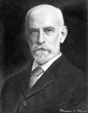
[[File:Charles minot.jpg|thumb|300px|link=Embryology History - Charles Minot|Charles Sedgwick Minot (1852–1914)]] | [[File:Charles minot.jpg|thumb|300px|link=Embryology History - Charles Minot|Charles Sedgwick Minot (1852–1914)]] | ||
The Middleton Goldsmith lecture delivered before the New York Pathological Society, March 26, 1901. | |||
==Introduction== | |||
Embryology is the basis upon which pathological science must be erected. Pathology is even more a superstructure upon embryology than is anatomy. Anatomy, in its descriptive form, may stand by itself and have usefulness. Pathology cannot be built up as a merely descriptive science. It fails of its true purpose unless it discovers the causes of diseases. Now since function is dependent on structure, the aim of the pathologist must be first to discover the causes of morbid structure. In brief, pathology at the present time deals chiefly with problems of the development of anatomical forms. Pathology and embryology might almost be united in a single comprehensive study — morphogeny. Let us then try, for this evening at least, to free ourselves from the conception of an essential difference between normal and abnormal structure, a conception which, I believe, domineers too largely over our daily thoughts. This belief of mine I hope to justify to-night. | |||
Simple description is indispensable, it furnishes the virgin facts ; but facts do not develop by parthenogenesis into science ; they must be saturated with the stimulus of study, with the stimulus of knowledge of their history, their antecedents, their causation, then we may see them evolving themselves into new orders, which we call natural laws. As little as a description of the people of the United States with no information as to their history could satisfy a serious thinker, so little can descriptions of fully developed structures satisfy an earnest pathologist. An innate, intense mental impulse is continually driving us forward in the search for causes, and obedience to this impulse is one of the main factors in scientific progress. All this is familiar, trite even, but may serve to fix our starting thought, namely, that we are to study causes. | |||
themselves into new orders, which we call natural laws. As little as a description of the people of the United States with no information as to their history could satisfy a serious thinker, so little can descriptions of fully developed structures satisfy an earnest pathologist. An innate, intense mental impulse is continually driving us forward in the search for causes, and obedience to this impulse is one of the main factors in scientific progress. All this is familiar, trite even, but may serve to fix our starting thought, namely, that we are to study causes. | |||
| Line 30: | Line 27: | ||
This plan will exclude from our discussion many of the aspects of embryology which appeal most strongly to pathologists. We must omit from our study at least three groups of interesting phenomena, to wit: First, the arrests of development; second, the teratological formations, monstrosities and mis-developments, which Will, however, have to be included ultimately even in the precise field we are about to study; third, the so-called teratoma, or to use a more recent term, embryoma. I may say in passing that I find it very difficult to accept the hypothesis that these remarkable structures arise by a parthenogenetic development of ova, retained in the parent body. | This plan will exclude from our discussion many of the aspects of embryology which appeal most strongly to pathologists. We must omit from our study at least three groups of interesting phenomena, to wit: First, the arrests of development; second, the teratological formations, monstrosities and mis-developments, which Will, however, have to be included ultimately even in the precise field we are about to study; third, the so-called teratoma, or to use a more recent term, embryoma. I may say in passing that I find it very difficult to accept the hypothesis that these remarkable structures arise by a parthenogenetic development of ova, retained in the parent body. Professor Bonnet’s hypothesis is more legitimate, but towards that also my attitude is one of sceptical agnosis. Bonnet suggests that one of the early segmentation cells (blastomeres) may become isolated and retarded in its development, remaining as an inclusion in the foetal tissues, and afterwards develop and produce a variety of tissues, as isolated blastomeres have been shown by experiments on the lower animals to be capable of doing. | ||
Teratological formations fall, it seems to me, naturally into three fairly definite divisions: | |||
Teratological formations fall, it seems to me, naturally into three fairly definite divisions: | # those due to necrosis of the tissues, which apparently rarely if ever takes place uniformly throughout the embryo; | ||
# those due to gross mechanical disturbances of the development, consequent upon failure of the proper correlation of the growth of parts ; monstrosities of this division are probably the most common; | |||
# errors in the differentiation of the tissue or pathological histogenesis. It is only phenomena of the first and second divisions of teratology which we can safely drop from view, while those of the third division—errors in differentiation—we must bear uninterruptedly in mind. | |||
| Line 44: | Line 40: | ||
Histogenesis is the common territory in which the pathologist and embryologist have —-to borrow a legal phrase—an undivided interest. It is unfortunate that our tendency has so long been to attempt an unnatural and impossible partition of the territory, which has resulted only in a division of our forces into two camps, between which has reigned little interest and less sympathy. I venture to regard your invitation to address you to-night as a wish, which I fully share, to secure fuller cooperation between the two camps of workers, who are both striving to lay bare the laws which govern the differentiation of cells. | Histogenesis is the common territory in which the pathologist and embryologist have —-to borrow a legal phrase—an undivided interest. It is unfortunate that our tendency has so long been to attempt an unnatural and impossible partition of the territory, which has resulted only in a division of our forces into two camps, between which has reigned little interest and less sympathy. I venture to regard your invitation to address you to-night as a wish, which I fully share, to secure fuller cooperation between the two camps of workers, who are both striving to lay bare the laws which govern the differentiation of cells. | ||
After these preliminary explanations it is possible to define the evening’s task with precision. It is twofold. First, to present some of the more important conceptions derived from embryological study in regard to the processes of cell differentiation. Second, to suggest some of the bearings of these conceptions on the problems of pathology. | After these preliminary explanations it is possible to define the evening’s task with precision. It is twofold. First, to present some of the more important conceptions derived from embryological study in regard to the processes of cell differentiation. Second, to suggest some of the bearings of these conceptions on the problems of pathology. | ||
==Part I. Normal Differentiation== | ==Part I. Normal Differentiation== | ||
Under this head I propose to discuss three fundamental ideas : | Under this head I propose to discuss three fundamental ideas : | ||
First, of the undifferentiated cell. | * First, of the undifferentiated cell. | ||
* Second, of the progress of differentiation. | |||
Second, of the progress of differentiation. | * Third, of the -changes which may succeed differentiation. | ||
Third, of the -changes which may succeed differentiation. | |||
The fertilized ovum is an undifferentiated being, although it has a very complex organization, and contains besides the protoplasm a store of nutritive material, the so-called yolk or deutoplasm. As there is only one nucleus, there can be no variety of nuclei ; the term undifferentiated, therefore, applies to the protoplasm, which seems to have a uniform essential structure throughout, although the masses and strands of protoplasm may exhibit characteristic peculiarities, especially in relation to the distribution of the yolk. In the adult, on the contrary, the protoplasm of the cells of different tissues offers many varieties of essential structure, which can often be readily distinguished under the microscope. It is a legitimate conclusion that the absence of visible peculiarities of the intimate structure of egg protoplasm, by which one part may be distinguished from another, corresponds to uniformity of structure throughout the egg, excepting, of course, certain special characteristic arrangements, as, for example, the centering about the centrosome, which may occur in any cell. | The fertilized ovum is an undifferentiated being, although it has a very complex organization, and contains besides the protoplasm a store of nutritive material, the so-called yolk or deutoplasm. As there is only one nucleus, there can be no variety of nuclei ; the term undifferentiated, therefore, applies to the protoplasm, which seems to have a uniform essential structure throughout, although the masses and strands of protoplasm may exhibit characteristic peculiarities, especially in relation to the distribution of the yolk. In the adult, on the contrary, the protoplasm of the cells of different tissues offers many varieties of essential structure, which can often be readily distinguished under the microscope. It is a legitimate conclusion that the absence of visible peculiarities of the intimate structure of egg protoplasm, by which one part may be distinguished from another, corresponds to uniformity of structure throughout the egg, excepting, of course, certain special characteristic arrangements, as, for example, the centering about the centrosome, which may occur in any cell. | ||
We have also direct experimental proof that the egg is uniform throughout, or to use a better phrase, that the egg is isotropic. Pfluger, in 1884, proved that the side of the frog’s egg, which normally develops into the ventral surface of the embryo, can be made to develop into a perfectly typical dorsal surface. The frog’s egg has a small white area, which normally lies underneath, the larger darkly pigmented area of the egg alone showing from above. Out of the dark area the back with the nervous system and other parts takes its origin. If the eggs, freshly fertilized, are fastened with the white side up, then the white side produces an absolutely normal back and nervous system, normal as to form and function, though lacking the typical pigmentation. These observations were confirmed by Born, who further discovered’ that the segmentation nucleus always rises towards the upper side of the egg, and that the position of the nucleus determines which part of the ovum shall become the ‘dorsal side of the embryo. Another set of experiments by Oskar Schultze demonstrated that both the unpigmented and the pigmented sides of the same egg could be made to produce dorsal structures. Another class of experiments, which were first made by Hans Driesch, have demon strated that the earliest cells (segmentation spheres, blastomeres or cleavage cells, as they are variously called) produced by the ovum preserve the undifferentiated qualities of the parent egg, and may develop in one way or another according to circumstances. The egg of a sea urchin divides into two cells, each of which multiplies and normally gives rise to half | We have also direct experimental proof that the egg is uniform throughout, or to use a better phrase, that the egg is isotropic. Pfluger, in 1884, proved that the side of the frog’s egg, which normally develops into the ventral surface of the embryo, can be made to develop into a perfectly typical dorsal surface. The frog’s egg has a small white area, which normally lies underneath, the larger darkly pigmented area of the egg alone showing from above. Out of the dark area the back with the nervous system and other parts takes its origin. If the eggs, freshly fertilized, are fastened with the white side up, then the white side produces an absolutely normal back and nervous system, normal as to form and function, though lacking the typical pigmentation. These observations were confirmed by Born, who further discovered’ that the segmentation nucleus always rises towards the upper side of the egg, and that the position of the nucleus determines which part of the ovum shall become the ‘dorsal side of the embryo. Another set of experiments by Oskar Schultze demonstrated that both the unpigmented and the pigmented sides of the same egg could be made to produce dorsal structures. Another class of experiments, which were first made by Hans Driesch, have demon strated that the earliest cells (segmentation spheres, blastomeres or cleavage cells, as they are variously called) produced by the ovum preserve the undifferentiated qualities of the parent egg, and may develop in one way or another according to circumstances. The egg of a sea urchin divides into two cells, each of which multiplies and normally gives rise to half<ref>It would be safer to say supposedly about half.</ref> of the body of the animal. By somewhat violent shaking the two cells may be artificially separated ; each cell may then develop into a complete larval sea-urchin, but of half the normal size only. Similar experiments have since been made by several investigators, who have obtained like results with other animals, vertebrate as well as invertebrate. Even more remarkable larvae have been raised from blastomeres of the four cell and eight cell stages of segmentation, producing larvae of one-fourth and one-eighth the normal size. Zoja. claims to have repeated the experiment successfully on the eggs of Olytia and to have obtained one-sixteenth larvae. | ||
| Line 77: | Line 65: | ||
The eggs of all animals | The eggs of all animals<ref>The protozoa are obviously excluded from the present discussion.</ref> pass through two well-marked phases of development. | ||
During the earlier and much shorter phase, the nuclei are multiplying rapidly, while the cytoplasm is growing but little, if at all. This period includes the time of segmentation, as commonly described, and somewhat longer. During this period the total bulk of the nuclei in proportion to the protoplasm is fundamentally changed. The ovum arises from a cell, the ovocyte, which, as its last act, grows rapidly ; this enlarged cell, by the process of maturation gives rise to the female sexual element, which has a single nucleus. After the fertilization we have an ovum with much protoplasm and deutoplasm, but again with only the single segmentation nucleus, The development of each individual begins, therefore, with a cell in which the extreme disproportion between the size of the nucleus and of the whole cell-body occurs. The first effort of development is to correct this disproportion by the enormously rapid increase of the nuclei, which continues until cells of the embryonic type are produced, that is to say, cells each with a minimal amount of protoplasm around the nucleus. With the production of cells of the embryonic type, the first phase of development is completed. The limits of this phase are very indefinite for we observe often that the prodution of cells of the type defined maybe far advanced in one part of the germ, while it is still in early progress in another. In fact the phase has no exact boundary in time. | During the earlier and much shorter phase, the nuclei are multiplying rapidly, while the cytoplasm is growing but little, if at all. This period includes the time of segmentation, as commonly described, and somewhat longer. During this period the total bulk of the nuclei in proportion to the protoplasm is fundamentally changed. The ovum arises from a cell, the ovocyte, which, as its last act, grows rapidly ; this enlarged cell, by the process of maturation gives rise to the female sexual element, which has a single nucleus. After the fertilization we have an ovum with much protoplasm and deutoplasm, but again with only the single segmentation nucleus, The development of each individual begins, therefore, with a cell in which the extreme disproportion between the size of the nucleus and of the whole cell-body occurs. The first effort of development is to correct this disproportion by the enormously rapid increase of the nuclei, which continues until cells of the embryonic type are produced, that is to say, cells each with a minimal amount of protoplasm around the nucleus. With the production of cells of the embryonic type, the first phase of development is completed. The limits of this phase are very indefinite for we observe often that the prodution of cells of the type defined maybe far advanced in one part of the germ, while it is still in early progress in another. In fact the phase has no exact boundary in time. | ||
| Line 92: | Line 77: | ||
After segmentation there follows the formation of the germ layers, a gradual arrangement of the cells in three distinct primary strata—at least in all vertebrates there are always three strata, never more | After segmentation there follows the formation of the germ layers, a gradual arrangement of the cells in three distinct primary strata—at least in all vertebrates there are always three strata, never more<ref>Hertwig and some other German embryologists divide the mesoderm into two layers ; the division is contrary to the actual development, and is made, in my opinion, quite arbitrarily to satisfy the needs of an erroneous theory.</ref> nor less. The outer and inner layers, ectoderm and entoderm, very early "become distinctly epithelial. The middle layers become partly epithelial, partly of a special character, that is mesenchymal. At first one is inclined to think of the difference between epithelium and mesenchyma as a fundamental one, an early and unalterable separation of cells into classes. ‘This view finds support in the fact that the mesenchyma, and it only, produces in the course of further development the connective tissue and supporting tissues of the adult. More attentive study of the germ layers in early stages reveals, however, that the mesenchymal cells arise from the epithelium, single epithelial cells migrating from the parent territory, while on the other hand groups of mesenchymal cells rearrange themselves so as to form an epithelial covering of some surface, as for example, in synovial cavities, arachnoid spaces, the inner surface of the cornea, lymph vessels, etc. Such observations teach us that we must not assume that either the form or the arrangement of cells is necessarily and always a sign of true differentiation, but that instead we are to conceive of differentiation as a change in the intimate and essential structure of the individual cell, more specifically of its protoplasm, and perhaps of its nucleus. The role of nuclei in histogenesis "is a topic which unfortunately is still awaiting serious investigation. To resume: it seems to me probable that the cells of the germ layers are at first quite indifferent, so that if it were possible to graft a young mesodermal cell on to the ectoderm or entoderm, it would become a true ectodermal or entodermal cell, as the case might be. | ||
| Line 124: | Line 106: | ||
B. MESODERMAL. | B. MESODERMAL. | ||
a. Epithelium | a. Epithelium of 2. | ||
C. ENTODERMAL. | C. ENTODERMAL. | ||
| Line 174: | Line 156: | ||
The two types of | The two types of differentiation produce essentially unlike conditions. The pathologist may not overlook such unlikeness with impunity. The two types pass into one another with many intergrades. Hence when we consider the possibilities of pathological alteration we must in each case seek to determine how far the condition of the tissue involved permits cell multiplication, as well as differentiation. | ||
| Line 186: | Line 168: | ||
Finally as to the mesoderm, in which layer variety of differentiation attains its maximum. To follow the genesis of this variety is most instructive. | Finally as to the mesoderm, in which layer variety of differentiation attains its maximum. To follow the genesis of this variety is most instructive. The mesoderm is found very early to include in vertebrate embryos, four kinds of cells, of which the most numerous are undifferentiated cells: the other three kinds being (1) endothelial cells of blood vessels; (2) blood cells; (3) sexual cells; all these are precociously specialized; they are few in number, yet they are probably the parents of all the cells which are produced of their kind throughout life. Our present knowledge does not permit us to speak with entire certainty, but the evidence is strongly in favor of the following three conceptions: | ||
| Line 216: | Line 198: | ||
Thus far I have expressed myself somewhat as if there were two sharply defined conditions, the differentiated and the undifferentiated. To give such an impression would be to create error, since differentiation is a slowly progressive and wholly gradual change in the cell. We must look upon each step in the process of dif1“erentia.tion as establishing narrower limits for future changes. Thus, when in the spinal cord neuroblasts diverge from the glia cells, they are not specialized into different classes of neuroblasts; such specialization comes later. So in the mesenchyma after the embryonic cells have changed and large numbers of them have become connective tissue cells, these last still are capable of various further differentiations, and may, therefore, be said to have been arrested in | Thus far I have expressed myself somewhat as if there were two sharply defined conditions, the differentiated and the undifferentiated. To give such an impression would be to create error, since differentiation is a slowly progressive and wholly gradual change in the cell. We must look upon each step in the process of dif1“erentia.tion as establishing narrower limits for future changes. Thus, when in the spinal cord neuroblasts diverge from the glia cells, they are not specialized into different classes of neuroblasts; such specialization comes later. So in the mesenchyma after the embryonic cells have changed and large numbers of them have become connective tissue cells, these last still are capable of various further differentiations, and may, therefore, be said to have been arrested in their development at a stage of partial differentiation. This quality of the connective tissue cells is, from the pathological standpoint, one of the most important facts known to us concerning the structure of the body. | ||
| Line 233: | Line 212: | ||
The normal and pathological changes associated with the death of cells, and consequently also of the tissues which are formed by cells, are so nearly identical that they may be combined in a single discussion. | The normal and pathological changes associated with the death of cells, and consequently also of the tissues which are formed by cells, are so nearly identical that they may be combined in a single discussion. | ||
For the more convenient presentation of the subject the | For the more convenient presentation of the subject the following table has been prepared. Concerning the table little explanation is necessary. I | ||
A few special points need mention. The distinction made between necrobiosis and degeneration corresponds to recognizable differences, but our present knowledge is insufficient to provide clear definitions for the two closely related types of indirect cell death. I feel much doubt as to the propriety of including atrophy in the table at all, since it seems to me that we ought, perhaps, not to regard atrophy as a phenomenonof a distinct class, but merely as a result of necrobiotic or degenerative alterations in cells and organs. Under the heading ‘degeneration’ the division into ‘ cytoplasmic’ and ‘paraplasmic’ takes us beyond our present knowledge, while the division ‘ nuclear’ is added rather to satisfy a biological conscience than to represent a part of our knowledge. | A few special points need mention. The distinction made between necrobiosis and degeneration corresponds to recognizable differences, but our present knowledge is insufficient to provide clear definitions for the two closely related types of indirect cell death. I feel much doubt as to the propriety of including atrophy in the table at all, since it seems to me that we ought, perhaps, not to regard atrophy as a phenomenonof a distinct class, but merely as a result of necrobiotic or degenerative alterations in cells and organs. Under the heading ‘degeneration’ the division into ‘ cytoplasmic’ and ‘paraplasmic’ takes us beyond our present knowledge, while the division ‘ nuclear’ is added rather to satisfy a biological conscience than to represent a part of our knowledge. | ||
| Line 300: | Line 279: | ||
B. Hg/pertrophic degeneration. I | B. Hg/pertrophic degeneration. I | ||
1. Cytoplasmic. | 1. Cytoplasmic.<ref name=degen>I cannot venture to assert that these two divisions are valid, and not arbitrary.</ref> | ||
a. granular. | a. granular. | ||
b. cornifying. | b. cornifying. | ||
0. hyaline. | 0. hyaline. | ||
2. Paraplasmic. | 2. Paraplasmic.<ref name=degen>I cannot venture to assert that these two divisions are valid, and not arbitrary.</ref> | ||
a. fatty. | a. fatty. | ||
b. pigmentary. | b. pigmentary. | ||
| Line 315: | Line 294: | ||
We begin, therefore, with necrobiosis. We may appropriately mention first those organs of which the existence is limited in time, such as the thymus and the foetal kidney (mesonephros or Wolffian body). These organs attain first their full differentiation; their elements during the next phase die off, and finally are resorbed, most of the organ disappearing. In the same category of change belong the histories of the senile ovary and testis. Another familiar illustration is offered by the notochord, which in the mammals totally disappears during the foetal period. The notochord cells undergo peculiar characteristic modifications, hence it is difficult to say whether or not there is degeneration in the strict sense. Cell-death on a large scale is a common phenomenon of the tissues. It occurs in cartilage both when the cartilage is permanent and even more conspicuously when cartilage gives way to bone, the disintegration of the cartilage cells preceding the irruption of the bone forming tissues. It occurs among the bone cells after they are imbedded in their calcified matrix. It occurs in the ovary, where we designate its result as atresia of the follicles. It occurs in the sebaceous glands as an accompaniment of the process of their secretion. It occurs among the glands of the intestine as discovered by Stohr, and occurs normally, though not constantly, in the appendix, as recorded by Ribbert. It occurs in the epithelium of the human pregnant uterus and in all the tissues of the human decidua reflexa. Other examples could be enumerated, but we may content ourselves with citing the constant destruction of blood corpuscles, both red and white. | We begin, therefore, with necrobiosis. We may appropriately mention first those organs of which the existence is limited in time, such as the thymus and the foetal kidney (mesonephros or Wolffian body). These organs attain first their full differentiation; their elements during the next phase die off, and finally are resorbed, most of the organ disappearing. In the same category of change belong the histories of the senile ovary and testis. Another familiar illustration is offered by the notochord, which in the mammals totally disappears during the foetal period. The notochord cells undergo peculiar characteristic modifications, hence it is difficult to say whether or not there is degeneration in the strict sense. Cell-death on a large scale is a common phenomenon of the tissues. It occurs in cartilage both when the cartilage is permanent and even more conspicuously when cartilage gives way to bone, the disintegration of the cartilage cells preceding the irruption of the bone forming tissues. It occurs among the bone cells after they are imbedded in their calcified matrix. It occurs in the ovary, where we designate its result as atresia of the follicles. It occurs in the sebaceous glands as an accompaniment of the process of their secretion. It occurs among the glands of the intestine as discovered by Stohr, and occurs normally, though not constantly, in the appendix, as recorded by Ribbert. It occurs in the epithelium of the human pregnant uterus and in all the tissues of the human decidua reflexa. Other examples could be enumerated, but we may content ourselves with citing the constant destruction of blood corpuscles, both red and white. | ||
| Line 323: | Line 299: | ||
-ought also to be regarded a.s casesiof hyaline degeneration. That fatty degeneration takes place normally has long been taught. There seems no reason for regarding the development of ordinary or mesenchymal fat-cells otherwise than as instances of normal degeneration. In old age a more or less marked fatty degeneration may be wide spread and occur in many different kinds of cells. The same.is true of the deposit of pigment, as we see it in the liver cells and motor nerve cells of adults. Finally, mucoid and colloid degeneration are so obviously normal, that we commonly think of their pathological occurrence as merely an exaggeration of a normal state. | |||
| Line 374: | Line 315: | ||
The cycle of changes through which cells pass is obviously longer than the period of development and the differentiation, yet its phases all belong together as members of a single series. We lack a word to designate the entire series of changes, and for the lack of such a word often fail to appreciate the essential unity of progressive and regressive modification of cell-structure. Accordingly, I wish to propose the new term cytomorphosis to designate comprehensively all the structural alterations which cells, or successive generations of cells, may undergo from the earliest undifferentiated stage to their final destruction. | The cycle of changes through which cells pass is obviously longer than the period of development and the differentiation, yet its phases all belong together as members of a single series. We lack a word to designate the entire series of changes, and for the lack of such a word often fail to appreciate the essential unity of progressive and regressive modification of cell-structure. Accordingly, I wish to propose the new term cytomorphosis to designate comprehensively all the structural alterations which cells, or successive generations of cells, may undergo from the earliest undifferentiated stage to their final destruction. | ||
==Part II. Pathological Differentiation== | ==Part II. Pathological Differentiation== | ||
| Line 380: | Line 320: | ||
We have now completed our brief reviews of the four fundamental successive stages of cytomorphosis. These stages are: | We have now completed our brief reviews of the four fundamental successive stages of cytomorphosis. These stages are: | ||
First | * First - Undifferentiated. | ||
* Second - Progressive differentiation, which itself often comprises many successive stages. | |||
Second | * Third - Regression (necrobiosis or degeneration) . | ||
* Fourth - Removal of the dead material. | |||
Third | |||
Fourth | |||
Let us now apply some of the conceptions won to the interpretation of pathological differentiation, remembering all the time that the interpretation of disease is a distinct and different problem. "Although presumably pathological differentiation is the sole and exclusive cause of disease and no disease arises from any other immediate cause, yet the disease must be regarded as the result and, owing to the physiological correlation of the organs, this result may include many secondary effects, some of which are often of the greatest diagnostic value, and therefore likely to divert attention from the primary structural cause. | Let us now apply some of the conceptions won to the interpretation of pathological differentiation, remembering all the time that the interpretation of disease is a distinct and different problem. "Although presumably pathological differentiation is the sole and exclusive cause of disease and no disease arises from any other immediate cause, yet the disease must be regarded as the result and, owing to the physiological correlation of the organs, this result may include many secondary effects, some of which are often of the greatest diagnostic value, and therefore likely to divert attention from the primary structural cause. | ||
Our review of normal conditions furnishes us with three general conceptions, which are valuable for their pathological applications, | Our review of normal conditions furnishes us with three general conceptions, which are valuable for their pathological applications,—namely : | ||
First. That each germ layer has a specific and exclusive share in the production of tissues. | * First. That each germ layer has a specific and exclusive share in the production of tissues. | ||
* Second. That undifferentiated cells, characterized by having only a small amount of unspecialized protoplasm, exist not only in the embryo, but also throughout life in certain parts of all three germ—layers. | |||
* Third. That differentiated cells characterized by having a larger amount of specialized protoplasm, form most of the organs of the adult and are incapable of undergoing any new unlike differentiation, though they are still capable of completing their cytomorphosis, by necrobiosis or degeneration. | |||
We must apply these conceptions, according to my belief, as rigidly to pathological as to normal development. Thus, as to the germ-layers, it ought to be possible even with our present knowledge, to show their pathogenetic values, so that every elementary student as a matter of course can be taught to classify accurately most pathological differentiations, and to accept such such a classification as the basis of all his further study of the science. How much this reform is needed is indicated by the many writers who put glioma under the head of connective tissue tumors, although gliomata arise from the ectoderm, and connective tissue arises from the mesoderm. Such a classification is on a par with the ancient system which put the whales among the fish, for it is not going too far to say that it is impossible that connective tissue should produce a glioma, because the two things belong in different classes. Another noteworthy violation of embryological law is offered by the classification of all muscle tumors under one head, ‘myoma,’ although smooth and striated muscle fibers are genetically and structurally’ distinct, with no intermediate or connecting forms of tissue, and with only a slight physiological resemblance. As regards epitheliomata ; they should be studied in relation to their layership, and it is reasonable, in my judgment, to expect that they will be found to have very distinctive characteristics according to the germ-layer from which they take origin, for the layership of a tissue governs the normal differentiation and therefore probably also the abnormal. I believe that the first competent investigation in this field will mark a new epoch of pathological science. When the epoch comes our morphological sense will no longer be shocked, as for instance by the application of the name adenoma to an epithelioma of an organ like the kidney, which is in no sense a gland. | |||
explain the extraordinarily rapid disappearance of the degenerated material in the obplacenta the only available hypothesis seems to be that of a chemical change by which the material becomes soluble or is dissolved, for we see the disappearance of the substance taking place in the very heart of the layer, and not merely at the surface. Sloughing is impossible and there are no phagocytes, leaving the chemical explanation as the only one I have been able to conceive. The contemplation of the described phenomena of the rabbit’s obplacenta inevitably raises the question—do we not tend in our explanations of the removal of necrosed and degenerated tissues to attribute too much to phagocytes and too little to direct chemical action ? May it not be that the body produces histolytic toxins, which can destroy tissues somewhat as do snake-poisons ? | |||
The cycle of changes through which cells pass is obviously longer than the period of development and the differentiation, yet its phases all belong together as members of a single series. We lack a word to designate the entire series of changes, and for the lack of such a word often fail to appreciate the essential unity of progressive and regressive modification of cell-structure. Accordingly, I wish to propose the new term cytomorphosis to designate comprehensively all the structural alterations which cells, or successive generations of cells, may undergo from the earliest undifferentiated stage to their final destruction. | The cycle of changes through which cells pass is obviously longer than the period of development and the differentiation, yet its phases all belong together as members of a single series. We lack a word to designate the entire series of changes, and for the lack of such a word often fail to appreciate the essential unity of progressive and regressive modification of cell-structure. Accordingly, I wish to propose the new term cytomorphosis to designate comprehensively all the structural alterations which cells, or successive generations of cells, may undergo from the earliest undifferentiated stage to their final destruction. | ||
| Line 464: | Line 369: | ||
* Our very brief discussion of pathological differentiation seems to justify the following conclusions : first, the process in its essential features is identical with the process of normal differentiation; second, the character of a tumor depends primarily upon the layership of the cells producing it; third, normal differentiation impedes and limits the formation of tumors, precisely as it does of further normal structures; so that tumors arise most readily from undifferentiated tissues and may then be heteroplastic; and arise readily from differentiated tissues and are then always homoplastic; and arise unreadily or not at all from the most highly specialized tissues. | |||
Latest revision as of 15:56, 12 January 2017
| Embryology - 16 Apr 2024 |
|---|
| Google Translate - select your language from the list shown below (this will open a new external page) |
|
العربية | català | 中文 | 中國傳統的 | français | Deutsche | עִברִית | हिंदी | bahasa Indonesia | italiano | 日本語 | 한국어 | မြန်မာ | Pilipino | Polskie | português | ਪੰਜਾਬੀ ਦੇ | Română | русский | Español | Swahili | Svensk | ไทย | Türkçe | اردو | ייִדיש | Tiếng Việt These external translations are automated and may not be accurate. (More? About Translations) |
Minot CS. The embryological basis of pathology. (1901) Science 13(326);481-98. PMID 17791097
| Online Editor |
|---|

|
| Historic Disclaimer - information about historic embryology pages |
|---|
|
The Embryological Basis of Pathology
The Middleton Goldsmith lecture delivered before the New York Pathological Society, March 26, 1901.
Introduction
Embryology is the basis upon which pathological science must be erected. Pathology is even more a superstructure upon embryology than is anatomy. Anatomy, in its descriptive form, may stand by itself and have usefulness. Pathology cannot be built up as a merely descriptive science. It fails of its true purpose unless it discovers the causes of diseases. Now since function is dependent on structure, the aim of the pathologist must be first to discover the causes of morbid structure. In brief, pathology at the present time deals chiefly with problems of the development of anatomical forms. Pathology and embryology might almost be united in a single comprehensive study — morphogeny. Let us then try, for this evening at least, to free ourselves from the conception of an essential difference between normal and abnormal structure, a conception which, I believe, domineers too largely over our daily thoughts. This belief of mine I hope to justify to-night.
Simple description is indispensable, it furnishes the virgin facts ; but facts do not develop by parthenogenesis into science ; they must be saturated with the stimulus of study, with the stimulus of knowledge of their history, their antecedents, their causation, then we may see them evolving themselves into new orders, which we call natural laws. As little as a description of the people of the United States with no information as to their history could satisfy a serious thinker, so little can descriptions of fully developed structures satisfy an earnest pathologist. An innate, intense mental impulse is continually driving us forward in the search for causes, and obedience to this impulse is one of the main factors in scientific progress. All this is familiar, trite even, but may serve to fix our starting thought, namely, that we are to study causes.
Our attention is to be directed to the consideration of what embryology can teach us in regard to the causation of organization, and then to the application of those teachings to pathology.
This plan will exclude from our discussion many of the aspects of embryology which appeal most strongly to pathologists. We must omit from our study at least three groups of interesting phenomena, to wit: First, the arrests of development; second, the teratological formations, monstrosities and mis-developments, which Will, however, have to be included ultimately even in the precise field we are about to study; third, the so-called teratoma, or to use a more recent term, embryoma. I may say in passing that I find it very difficult to accept the hypothesis that these remarkable structures arise by a parthenogenetic development of ova, retained in the parent body. Professor Bonnet’s hypothesis is more legitimate, but towards that also my attitude is one of sceptical agnosis. Bonnet suggests that one of the early segmentation cells (blastomeres) may become isolated and retarded in its development, remaining as an inclusion in the foetal tissues, and afterwards develop and produce a variety of tissues, as isolated blastomeres have been shown by experiments on the lower animals to be capable of doing.
Teratological formations fall, it seems to me, naturally into three fairly definite divisions:
- those due to necrosis of the tissues, which apparently rarely if ever takes place uniformly throughout the embryo;
- those due to gross mechanical disturbances of the development, consequent upon failure of the proper correlation of the growth of parts ; monstrosities of this division are probably the most common;
- errors in the differentiation of the tissue or pathological histogenesis. It is only phenomena of the first and second divisions of teratology which we can safely drop from view, while those of the third division—errors in differentiation—we must bear uninterruptedly in mind.
One more preliminary explanation is necessary. The range of pathological changes is not so great as to reach equality with embryological; developments. In the normal embryo we deal with the evolution of complete organs together with all their accompanying varied and complex modifications of tissue; In pathology, on the contrary, we deal not with organs, but with modifications of tissues, with histogenesis. The statement Will not seem too absolute if it is recalled that we have excluded arrests of development and monstrosities from our discussion.
Histogenesis is the common territory in which the pathologist and embryologist have —-to borrow a legal phrase—an undivided interest. It is unfortunate that our tendency has so long been to attempt an unnatural and impossible partition of the territory, which has resulted only in a division of our forces into two camps, between which has reigned little interest and less sympathy. I venture to regard your invitation to address you to-night as a wish, which I fully share, to secure fuller cooperation between the two camps of workers, who are both striving to lay bare the laws which govern the differentiation of cells.
After these preliminary explanations it is possible to define the evening’s task with precision. It is twofold. First, to present some of the more important conceptions derived from embryological study in regard to the processes of cell differentiation. Second, to suggest some of the bearings of these conceptions on the problems of pathology.
Part I. Normal Differentiation
Under this head I propose to discuss three fundamental ideas :
- First, of the undifferentiated cell.
- Second, of the progress of differentiation.
- Third, of the -changes which may succeed differentiation.
The fertilized ovum is an undifferentiated being, although it has a very complex organization, and contains besides the protoplasm a store of nutritive material, the so-called yolk or deutoplasm. As there is only one nucleus, there can be no variety of nuclei ; the term undifferentiated, therefore, applies to the protoplasm, which seems to have a uniform essential structure throughout, although the masses and strands of protoplasm may exhibit characteristic peculiarities, especially in relation to the distribution of the yolk. In the adult, on the contrary, the protoplasm of the cells of different tissues offers many varieties of essential structure, which can often be readily distinguished under the microscope. It is a legitimate conclusion that the absence of visible peculiarities of the intimate structure of egg protoplasm, by which one part may be distinguished from another, corresponds to uniformity of structure throughout the egg, excepting, of course, certain special characteristic arrangements, as, for example, the centering about the centrosome, which may occur in any cell.
We have also direct experimental proof that the egg is uniform throughout, or to use a better phrase, that the egg is isotropic. Pfluger, in 1884, proved that the side of the frog’s egg, which normally develops into the ventral surface of the embryo, can be made to develop into a perfectly typical dorsal surface. The frog’s egg has a small white area, which normally lies underneath, the larger darkly pigmented area of the egg alone showing from above. Out of the dark area the back with the nervous system and other parts takes its origin. If the eggs, freshly fertilized, are fastened with the white side up, then the white side produces an absolutely normal back and nervous system, normal as to form and function, though lacking the typical pigmentation. These observations were confirmed by Born, who further discovered’ that the segmentation nucleus always rises towards the upper side of the egg, and that the position of the nucleus determines which part of the ovum shall become the ‘dorsal side of the embryo. Another set of experiments by Oskar Schultze demonstrated that both the unpigmented and the pigmented sides of the same egg could be made to produce dorsal structures. Another class of experiments, which were first made by Hans Driesch, have demon strated that the earliest cells (segmentation spheres, blastomeres or cleavage cells, as they are variously called) produced by the ovum preserve the undifferentiated qualities of the parent egg, and may develop in one way or another according to circumstances. The egg of a sea urchin divides into two cells, each of which multiplies and normally gives rise to half[1] of the body of the animal. By somewhat violent shaking the two cells may be artificially separated ; each cell may then develop into a complete larval sea-urchin, but of half the normal size only. Similar experiments have since been made by several investigators, who have obtained like results with other animals, vertebrate as well as invertebrate. Even more remarkable larvae have been raised from blastomeres of the four cell and eight cell stages of segmentation, producing larvae of one-fourth and one-eighth the normal size. Zoja. claims to have repeated the experiment successfully on the eggs of Olytia and to have obtained one-sixteenth larvae.
The facts offered suflice to illustrate the two aspects of our conception of the undifferentiated condition of living matter. The first aspect is morphological and presents to us the apparent uniformity of the visibly minute structure of protoplasm. While we readily admit that the uniformity may be only apparent in the sense that we fail to observe fine differences, yet we none the less maintain that the uniformity is real, because there is an absence of variations of structure comparable to the variations which we can observe in the cells of adult tissues. The second aspect is physiological and oifers to our view the wide range of possibilities in the future developmental history and growth of the protoplasm. The fate of the protoplasm of any given part of the ovum is not fixed, but if its conditions of development are changed its fate is changed. A few years ago the mosaic hypothesis was advanced by W. Roux and has been vigorously defended by him. According to the mosaic theory, the egg is a mosaic pattern, each member of which has its predestined history. It is fortunate for our comprehension of pathological process that‘ we are already able to say that Roux’s hypothesis is erroneous.
We must start then with the right conception of the ovum, every part of the protoplasm of which is to be regarded as potentially capable of producing any or all the tissues of the adult.
We turn next to the consideration of the progress of differentiation in order to establish a second fundamental idea, namely, that it acts as a progressive restriction of the further development. Each successive stage of differentiation puts a narrower limitation upon the possibilities of further advance. Applied to pathology, this law means that the range of possible pathological changes is determined not merely by the nature or kind, but also by the stage or degree, of the previous differentiation of the tissue.
The eggs of all animals[2] pass through two well-marked phases of development.
During the earlier and much shorter phase, the nuclei are multiplying rapidly, while the cytoplasm is growing but little, if at all. This period includes the time of segmentation, as commonly described, and somewhat longer. During this period the total bulk of the nuclei in proportion to the protoplasm is fundamentally changed. The ovum arises from a cell, the ovocyte, which, as its last act, grows rapidly ; this enlarged cell, by the process of maturation gives rise to the female sexual element, which has a single nucleus. After the fertilization we have an ovum with much protoplasm and deutoplasm, but again with only the single segmentation nucleus, The development of each individual begins, therefore, with a cell in which the extreme disproportion between the size of the nucleus and of the whole cell-body occurs. The first effort of development is to correct this disproportion by the enormously rapid increase of the nuclei, which continues until cells of the embryonic type are produced, that is to say, cells each with a minimal amount of protoplasm around the nucleus. With the production of cells of the embryonic type, the first phase of development is completed. The limits of this phase are very indefinite for we observe often that the prodution of cells of the type defined maybe far advanced in one part of the germ, while it is still in early progress in another. In fact the phase has no exact boundary in time.
During the second, later and much longer period or phase of development, the multiplication of nuclei lags behind the growth of the protoplasm, the increase is gradual and often shows itself through successive generations of cells, sometimes, however, in a single cell, which no longer multiplies. Of the first method of protoplasmic growth embryonic blood-cells offer a good illustration, of the second the neuroblasts or young nerve cells afford a striking example. Now cells of the embryonic type show little if any capacity for differentiation, and the increase of the protoplasm in the single cell is, so far as we can judge, a necessary preliminary step to cell differentiation. The embryonic cells have yet another characteristic of basal significance ; they-are capable of rapid multiplication. Hence we conclude that the growth of the cytoplasm impedes the multiplication of cells, and, therefore, ultimately retards the growth of the body, as a whole, while on the other hand it favors differentiation. Accordingly the growth of cells and their differentiation are essentially antagonistic processes, which are necessarily more or less mutually exclusive. This conclusion which I published in 1890, has since been amply confirmed by further observation. It is probably applicable alike to animals and plants, alike to normal and abnormal tissues. It is one of the many conclusions of embryology which are sure to throw a revealing light upon some of the dark problems of pathology.
During the first phase of development, as just defined, we encounter preparatory changes which we may characterize summarily as the manufacture of embryonic cells. During the second phase, though the production of embryonic cells is doubtless continued in certain regions, there supervenes the process of differentiation, the true histogenesis.
After segmentation there follows the formation of the germ layers, a gradual arrangement of the cells in three distinct primary strata—at least in all vertebrates there are always three strata, never more[3] nor less. The outer and inner layers, ectoderm and entoderm, very early "become distinctly epithelial. The middle layers become partly epithelial, partly of a special character, that is mesenchymal. At first one is inclined to think of the difference between epithelium and mesenchyma as a fundamental one, an early and unalterable separation of cells into classes. ‘This view finds support in the fact that the mesenchyma, and it only, produces in the course of further development the connective tissue and supporting tissues of the adult. More attentive study of the germ layers in early stages reveals, however, that the mesenchymal cells arise from the epithelium, single epithelial cells migrating from the parent territory, while on the other hand groups of mesenchymal cells rearrange themselves so as to form an epithelial covering of some surface, as for example, in synovial cavities, arachnoid spaces, the inner surface of the cornea, lymph vessels, etc. Such observations teach us that we must not assume that either the form or the arrangement of cells is necessarily and always a sign of true differentiation, but that instead we are to conceive of differentiation as a change in the intimate and essential structure of the individual cell, more specifically of its protoplasm, and perhaps of its nucleus. The role of nuclei in histogenesis "is a topic which unfortunately is still awaiting serious investigation. To resume: it seems to me probable that the cells of the germ layers are at first quite indifferent, so that if it were possible to graft a young mesodermal cell on to the ectoderm or entoderm, it would become a true ectodermal or entodermal cell, as the case might be.
But although we may, so far as we now see, regard the cells in the germ layers as originally wholly indifferent as individual cells, nevertheless, we must not forget that as members of a germ layer their potential fate is already restricted by the conditions of their very layership—if I may coin a word for the occasion. Each layer produces its own group of tissues and never any other. There are indeed apparent exceptions to the rule, as, for example, the stratified horny epithelium of the oesophagus, which is strikingly similar to the epidermis, although in one case the tissue is ectodermal, in the other entodermal. We have, however, to do only with a resemblance, and the development in the two cases is quite unlike—the oesophageal epithelium in the mammalian embryo being ciliated at one stage and exhibiting then little resemblance to any stage of the epidermis.
Each germ layer has its specific function, its exclusive share in the work of differentiation. The work of one layer is not done by another nor shared by another. It is true that attempts are made from time to time to upset the validity of this fundamental doctrine, but they have hitherto failed to find support or recognition from any leading embryologist, and I deem these attempts unworthy of serious consideration. We are able now to assign almost every cell of the normal adult human body to its proper germ layer. Our only great uncertainty is where two layers meet, as do the ectoderm and entoderm in the pharynx, or as do the mesoderm and entoderm, where the ureter opens into the bladder. With these and perhaps a very few other small exceptions everything in ‘adult anatomy can be correctly stated in terms of germ layers. The layership of every organ is known, save that in the cases of the thymus gland, the tonsils and the adrenals authorities are not yet agreed.
A remarkable attempt to upset the doctrine of the germ layers deserves a brief consideration. It was first maintained by Goronovitch that the cells forming at least a part of the skeleton arose from the ectoderm. The same opinion was expressed also on the basis of their own investigations by H. Klaatsch and by Miss Platt. Confirmation of these views has not followed, but on the contrary, C. Rabl,one of the most trustworthy of living observers, maintains that essential parts of Goronovitch’s and Klaatsch’s evidence are simply errors of observation. Klaatsch’s views were based partly on the study of the developing teleost fins. R. G. Harrison has shown that here the German worker is in error. Miss Platt’s observations were made in the head region of embryo Necturus. An examination of afinumber of series and stages has not enabled me to find the slightest evidence in favor of Miss Platt’s conclusions. H. K. Corning has pointed out that the processes alleged by Miss Platt to occur in Necturus do not take place in the frog, Roma temporaria. We may, therefore, I think, safely regard this attempt to overthrow the morphological value of the germ layers as unsuccessful. I know of no other attempt of suflicient importance to be even mentioned. '
The importance to pathologists of a thorough knowledge of the genesis of the tissues from their germ layers can hardly be emphasized too strongly, for it is more than probable that all pathological tissues are as strictly governed by the law of the specific value of germ-layers as are the normal tissues. Are there not many pathologists whose knowledge of embryology is wholly insufficient to meet the practical needs of their professional study even in this one direction?
The accompanying table presents the principal tissues classified according to their layership. There have been classi-‘ fications of organs on the layership basis published before, but inasmuch as organs usually contain cells from two layers, we get a more correct presentation of the actual genetic relationships by restricting our tabulation to the tissues. Leucocytes do not appear in the table for the reason that erto been clearly recognized or defined. For both types the starting point is the same, the undifferentiated embryonic cell. In one type we find that as the cells proliferate, a portion of them only undergoes dii‘1"erentiation, and another portion remains more -or less undifferentiated and retains more or less fully the power of continued proliferation. The epidermis is a good representative of this type. Its basal layer consists of embryonic cells, which multiply; some of these cells move into the upper layers, their first origin is uncertain. Blood cells arise very early, before the clear separation of mesoderm and entoderm has occurred it is possible that they are entodermal. With these two limitations, the table presents our present knowledge.
Classification Of The Tissues
A. ECTODERMAL.
1. Epiderntis.
a. Epidermal appendages.
1. Mesothelium.
B. MESODERMAL.
a. Epithelium of 2.
C. ENTODERMAL.
1. Notochord.
Epithelium of
b. Lens of eye. peritonaeum, a. Digestive tract,
2. Epithelium of pericardium, oesophagus,
a. cornea. pleura, stomach,
b. olfactory chamber. urogenital organs. liver,
o. auditory organ. b. Striated muscles. pancreas,
d. mouth
2. Mesenchyma. , small intestine, (oral glands),
a. Connective tissue, yolk-sack, (enamel organ), Smooth muscle, large intestine, (hypophysis). Pseudo-endothelium, caacum,
e. anus. Fat-cell, vermix,
f. chorion, Pigment cells. rectum‘,
Foetal placenta. b. Blood. Allantois (bladder).
g. amnion. 0. Blood vessels. b. Pharynx,
3. Nervous system. (1. Lymphatics. Eustachian tube,
a. Brain, e. Spleen. Tousils, optic nerve, 1. Supporting tissues, Thymus, retina. cartilage, Parathyroids,
b. Spinal cord. bone. Thyroid.
is. Ganglia. g. Marrow. 0. Respiratory tract,
(1. Neuraxons. Larynx, Traohea, Lungs.
We will now turn to the analysis of the differential process in each germ layer. We have to deal with changes in cells.
There are two distinct types of cell differentiation, which. I think have not with enlarge and differentiate themselves into horny cells; others remain in the basal layer and continue to multiply. The progeny of a given basal epidermal cell do not all have the same fate, but divide themselves into two kinds of cells, one kind retaining the ancestral character, the other becoming something new and unlike the parent cell. Differentiation according to the second type is characterized by its inclusion of all the cells. This type has its culminating and most perfect illustration in the central nervous system, where comparatively early in embryonic life all the cells become specialized, and with the acquisition of specialization they forfeit their power of multiplication, the neuroglia cells partly, the nerve cells wholly. The growth of the brain after early stages depends not on proliferation of cells, but chiefly upon the increase in size of the individual cell. The correctness of this statement is not affected, in my belief, by the fact that epithelial portions of the medullary tube in comparatively late stages may be added to the nervous portion, the cells multiplying rapidly, as we see at the growing edge of the young cerebellum. The brain here grows by the addition of cells in the indifferent stage, but as soon as these cells are differentiated they conform to the general law and divide no more (neurones) or slowly (glia cells).
The two types of differentiation produce essentially unlike conditions. The pathologist may not overlook such unlikeness with impunity. The two types pass into one another with many intergrades. Hence when we consider the possibilities of pathological alteration we must in each case seek to determine how far the condition of the tissue involved permits cell multiplication, as well as differentiation.
Just as the segmentating ovum divides itself into parts, which we name germ layers, each of which has its special and exclusive share in developing the adult tissues, so does each of the three germ layers divide into parts, each part having its special and exclusive roll, and these parts again subdivide until, after the final partition, the adult variety is produced. During all these changes there is no exchange of roles. It will be profitable to let the phenomena pass before us in rapid review.
first, then, the ectoderm. This layer early separates into two parts ; one to form the central nervous system, the second the epidermis; the nervous part thereafter never forms epidermal structures, the epidermal part never forms a nervous system. The central nervous system retains in part a simple epithelial character, but most of its walls become nervous tissue; its cells pass from the indifferent stage and become neuroglia. cells or young nerve cells (neuroblasts). Neuroglia cells never become anything else, and the nerve cells are always nerve cells to the end. The primitive epidermis forms a series of special sensory areas and the permanent epidermis. The sensory areas, which belong to the olfactory, auditory and gustatory organs, soon become well defined and never produce any cell arrangements like those of the epidermis. This last, on the contrary, remains as before stated, rich in undifferentiated cells, and gradually produces a great variety of structures. Most of these, namely, the hairs and glands, are small and very numerous, while a few like the nails, enamel organs and epithelium of the lips are larger. No one of these special structures, however, converts itself into another. The basal layer of the general epidermis may perhaps preserve a true embryonic quality and have wide differential possibilities.
Next, as to the entoderm, which undergoes less differentiation than either of the other two germ layers, since over a large part of its extent it remains throughout life a simple epithelium with many cells very slightly modified in structure. Wherever i_n it specialization takes place, as in the tonsil, thymus, thyroid, oesophagus, liver or pancreas, each territory of cells keeps its characteristics and never assumes those of another territory.
Finally as to the mesoderm, in which layer variety of differentiation attains its maximum. To follow the genesis of this variety is most instructive. The mesoderm is found very early to include in vertebrate embryos, four kinds of cells, of which the most numerous are undifferentiated cells: the other three kinds being (1) endothelial cells of blood vessels; (2) blood cells; (3) sexual cells; all these are precociously specialized; they are few in number, yet they are probably the parents of all the cells which are produced of their kind throughout life. Our present knowledge does not permit us to speak with entire certainty, but the evidence is strongly in favor of the following three conceptions:
First. That all the endothelium of the blood vessels of the adult is descended directly from the endothelium of the first blood vessels differentiated in the extraembryonic portion of the germinal area.
Second. ' That all the red blood corpuscles are descendants from the red-blood-cells of the blood-islands of the area vasculosa. According to this view the blood forming organs, as they are called, merely provide sites, where the red cells can multiply, as for instance in the mammalian embryonic liver or in the adult marrow.
Third. That the primitive sexual cells by their multiplication produce all the cells from which the genoblasts, or sexual elements proper, male and female, are evolved.
The future will decide the validity of these conceptions.- They are very significant, because they assume that there are cells which form exclusive classes, and are characterized by a special combination of qualities, so that while they retain so much of the embryonic character as to have still the power of rapid multiplication, they yet are so specialized that they can only produce their like. If the three conceptions are established, We shall regard these three sorts of cells as almost the first to be fully differentiated. We shall also have to regard the vascular endothelium as distinct not only from the epithelial lining of the body _cavity, but also from that of the lymphatic system. The immense importance of such a discovery as bearing upon pathological researches and interpretations is obvious.
The next important change in the mesoderm is the development of the main body-cavity, which the embryologist designates comprehensively as the coelom. The cells, which lie next the body. cavity and border it, assume an epithelial arrangement ; this epithelial layer around the coelom is properly named ‘mesothelium,’ and the loose cells about it constitute the ‘ mesenchyma.’ We do not have, however, at first a true differentiation of mesothelial and mesenchymal cells ; all are undifferentiated, and we can readily demonstrate that the cells are interchangeable, differing during early stages by their positions in relation to one another and to the body cavity, but not differing in their essential structures or qualities. Thus we find that the mesothelium constantly gives oil“ cells which join the mesenchyma, and we find later that mesenchymal cells may take on a.n epithelial arrangement around any of the cavities—and there are many such-—Which arise within the mesenchyma itself in the course of further development.
But, although difference of arrangement does not necessarily indicate differentiation of the cells, it does affect the character of the differentiation which ensues. As every text-book states, the mesothelium gives rise to the striated muscles and to the epithelial portions of the entire genito-urinary tract, and is permanently retained, with slighter modifications, as the epithelium of the pericardium, pleura and peritoneum. The mesenchyma produces an even greater variety since it is the parent of not only all the connective and supporting tissue, but also of the lymphatic system. .
I venture to turn aside for a moment to urge upon you the adoption of the term mesothelium as the correct designation for the epithelial lining for the cavities of the thorax and abdomen. It is literally the same epithelium in the four cavities, for they were originally one, with a single continuous epithelium. It is well also in our nomenclature to recognize the important fact that the epithelium is radically, because genetically, distinct from the endothelium of the blood vessels and lymphatics, and the application of the term ‘endothelium’ to the covering of, for instance, the peritoneum, leads and can lead only to confused bad thinking. If mesothelium be employed as suggested," clearness will be gained.
Coming back now to the subject of the mesoderm, let us note that when a striated muscle fiber is produced a striated muscle fiber it always remains, and it never becomes anything else; the ovary never changes. In short, with the mesoderm as with the ectoderm and entoderm, we see the fate of the cells once fixed to be thereafter unchangeable as to the kind of differentiation.
Our hasty review is worse than imperfect, yet is sufficient to impress upon us the great law that differentiation in any direction terminates the possibility of differentiation in any other direction. In accordance with this law we encounter no instances, either in normal or in pathological development,-.->f the transformation of a cell of one kind of tissue into a cell of another kind of tissue, and further we encounter no instance of a differentiated cell being transformed back into an undifferentiated cell of the embryonic type, with varied potentialities.
Thus far I have expressed myself somewhat as if there were two sharply defined conditions, the differentiated and the undifferentiated. To give such an impression would be to create error, since differentiation is a slowly progressive and wholly gradual change in the cell. We must look upon each step in the process of dif1“erentia.tion as establishing narrower limits for future changes. Thus, when in the spinal cord neuroblasts diverge from the glia cells, they are not specialized into different classes of neuroblasts; such specialization comes later. So in the mesenchyma after the embryonic cells have changed and large numbers of them have become connective tissue cells, these last still are capable of various further differentiations, and may, therefore, be said to have been arrested in their development at a stage of partial differentiation. This quality of the connective tissue cells is, from the pathological standpoint, one of the most important facts known to us concerning the structure of the body.
Having now elaborated, as far as time permits, our conception of the nature of differentiation, let us turn to our third fundamental idea, which concerns the changes which succeed differentiation. These changes are very unlike the constructive changes which precede them, for they are destructive. They fall into three main groups:
- Changes of direct cell death.
- Necrobiosis,[4] or indirect cell death preceded by changes in cell structure.
- Hypertrophic degeneration or indirect cell death preceded by growth and structural change of the cell.
Of direct cell death no discussion is here necessary, for the fundamental idea, which I wish to emphasize, is that necrobiosis and hypertrophic degeneration are normal processes, which invariably occur in the normal body and play in many cases important roles in the life history of the individual. Without necrobiosis and degeneration on a large scale, the normal round of human life would be impossible. It is singular that in treatises on normal anatomy and histology these two subjects are generally neglected or at most appear only as matters of incidental reference. The force of tradition makes us apply these terms as if they correspond exclusively to pathological conditions. This tradition might still prescribe our mental attitude, were it not that the -studies of the last dozen years have made us familiar with the enormous extent, variety and rapidity of the destructive degenerationswhich go on in the pregnant uterus of placental mammals, a degeneration which ’ takes place without affording a trace or a suggestion of any pathological modification whatsoever of the organ. prejudices the uterine phenomena alluded to are startling, but their evidence before the tribunal of biology has settled the case in favor of the plea that hypertrophic degeneration is a normal factor in typical healthy development.
The normal and pathological changes associated with the death of cells, and consequently also of the tissues which are formed by cells, are so nearly identical that they may be combined in a single discussion.
For the more convenient presentation of the subject the following table has been prepared. Concerning the table little explanation is necessary. I
A few special points need mention. The distinction made between necrobiosis and degeneration corresponds to recognizable differences, but our present knowledge is insufficient to provide clear definitions for the two closely related types of indirect cell death. I feel much doubt as to the propriety of including atrophy in the table at all, since it seems to me that we ought, perhaps, not to regard atrophy as a phenomenonof a distinct class, but merely as a result of necrobiotic or degenerative alterations in cells and organs. Under the heading ‘degeneration’ the division into ‘ cytoplasmic’ and ‘paraplasmic’ takes us beyond our present knowledge, while the division ‘ nuclear’ is added rather to satisfy a biological conscience than to represent a part of our knowledge.
Death of Cells
First—Causes of death.
a. External to the organism.
1. Physical (mechanical, chemical, thermal, etc.).
2. Parasites.
b. Changes in intercellular substances (probably primarily due to cells). 1. Hypertrophy. 2. Induration. 3. Calcification. 4. Amyloid degeneration.
c. Changes inherent in cells.
Second—Morphological changes of dying cells. a. Direct death of cells. 1. Atrophy. 4 2. Disintegration and resorption. b. Indirect death of cells.
1. Necrobiosis (structural change precedes final death). 2. Hypertrophic degeneration (growth and structural change often with nuclear proliferation precede final death).
Third—Removal of cells. a. By mechanical means (sloughing or shedding). b. By chemical means (solution). o. By phagocytes.
The preceding table represents the only attempt of the kind known to me, and like other first attempts is undoubtedly very imperfect. facts. But, because it is frequently a scientific gain to systematize our information, I hope the table may be useful, and it will certainly serve its immediate purpose, namely, to guide our discussion of the normal changes which follow after cellular differentiation.
As the time at our command is brief let us pass by the consideration of the causes of cell death. I will remark only thatI think amyloid degeneration may be found to occur in the placental decidua of the human pregnant uterus and perhaps in other normal structures. No positive information
It embodies obviously no new on this point is known to me. For the reasons stated a few minutes ago atrophy may also be omitted here. We pass by also the direct forms of cell death, to reach at once the consideration of the indirect forms.
The accompanying table offers an analysis of some of the principal varieties of structural change, which occur during indirect cell death.
Indirect Death of Cells
A. Necrobiosis.
1. Cytoplasmic changes. a. granulation. b. hyaline transformation. c. imbibition. d. desiccation. e. clasmatosis.
2. Nuclear changes. a. karyorhexis. b. karyolysis.
B. Hg/pertrophic degeneration. I
1. Cytoplasmic.[5] a. granular. b. cornifying. 0. hyaline.
2. Paraplasmic.[5] a. fatty. b. pigmentary. c. mucoid. d. colloid, etc.
3. Nuclear (? increase of chromatine).
We begin, therefore, with necrobiosis. We may appropriately mention first those organs of which the existence is limited in time, such as the thymus and the foetal kidney (mesonephros or Wolffian body). These organs attain first their full differentiation; their elements during the next phase die off, and finally are resorbed, most of the organ disappearing. In the same category of change belong the histories of the senile ovary and testis. Another familiar illustration is offered by the notochord, which in the mammals totally disappears during the foetal period. The notochord cells undergo peculiar characteristic modifications, hence it is difficult to say whether or not there is degeneration in the strict sense. Cell-death on a large scale is a common phenomenon of the tissues. It occurs in cartilage both when the cartilage is permanent and even more conspicuously when cartilage gives way to bone, the disintegration of the cartilage cells preceding the irruption of the bone forming tissues. It occurs among the bone cells after they are imbedded in their calcified matrix. It occurs in the ovary, where we designate its result as atresia of the follicles. It occurs in the sebaceous glands as an accompaniment of the process of their secretion. It occurs among the glands of the intestine as discovered by Stohr, and occurs normally, though not constantly, in the appendix, as recorded by Ribbert. It occurs in the epithelium of the human pregnant uterus and in all the tissues of the human decidua reflexa. Other examples could be enumerated, but we may content ourselves with citing the constant destruction of blood corpuscles, both red and white.
Degeneration, in the stricter sense of an ante-mortem hypertrophic change of cell structure, is also of widespread occurrence in the healthy body. No case of so-called granular degeneration under strictly normal conditions is known to me, though it seems quite credible that such cases should On the other hand, the cornifying degeneration is very important and does occur in all three germ-layers, for we observe it in the ectoderm of the skin, the entoderm of the oesophagus and the mesoderm of the vagina. Hyaline degeneration of so striking a character as to have been termed pseudo-pathologicial takes place regularly in the ectoderm (outer epithelium) of the placental chorion. In the rabbit it occurs in the uterine glands, during pregnancy, causing most rapid histolysis, and it seems to me probable that some of the changes, which can be observed in the decidua of the pregnant human uterus on this point is known to me. For the reasons stated a few minutes ago atrophy may also be omitted here. We pass by also the direct forms of cell death, to reach at once the consideration of the indirect forms.
-ought also to be regarded a.s casesiof hyaline degeneration. That fatty degeneration takes place normally has long been taught. There seems no reason for regarding the development of ordinary or mesenchymal fat-cells otherwise than as instances of normal degeneration. In old age a more or less marked fatty degeneration may be wide spread and occur in many different kinds of cells. The same.is true of the deposit of pigment, as we see it in the liver cells and motor nerve cells of adults. Finally, mucoid and colloid degeneration are so obviously normal, that we commonly think of their pathological occurrence as merely an exaggeration of a normal state.
The various kinds of changes in dying cells, with which the pathologist is most familiar, recur in healthy tissues. In the preceding table seven forms of change are enumerated under the heading ‘necroticsis.’ ,Every one of these seven occurs normally. Granulation of the bodies of the cartilage cells and of the notochord cells may be observed to precede their resorption. Hyaline transformation is conspicuous in the decidua reflexa. Imbibition or cellular oedema occurs in the epidermis of the lips, in the cells of the uterine’ glands during pregnancy after they have detached themselves from the gland walls, and in the «endothelium of placental blood vessels of the rabbit. Desiccation is the usual accompaniment of cornification. Clasmatosis has given its name to the clasmatocytes of Ranvier, and we may well apply the same term to the cells of the secreting milk gland, and also, as an unpublished research indicates, to the cells of the secreting glands of the cervix uteri. Karyolysis is, according to present probabilities, the method by which nucleated red blood cells are converted into non-nucleated blood-corpuscles. Karyorhexis, or the fragmentation of the nucleus, occurs in the cells of the disappearing follicles of the ovary.
Lastly as to the removal of cells. The sloughing off of cells is one of the most familiar phenomena, since it occurs incessantly over the epidermis and with the hairs; its part in menstruation and its colossal role in the afterbirth are known to all, and every practitioner is accustomed to look for shed cells in urinary sediment. Large numbers of cells are lost by the intestinal epithelium. Cells without access to the external world must be got rid of by resorption, which seems to take place either with or without the cooperation of leucocytes. In the latter case we must for lack of a better hypothesis attribute the resorption to chemical means. Of resorption with the aid of leucocytes the necrosed human decidua reflexa offers a perfect illustration. Of resorption Without leucocytes the masses of degenerated epithelium in the placenta, periplacenta and obplacenta of the rabbit afford by far the most impressive demonstration I have ever seen. At nine days after conception the epithelium is profoundly changed, being very much thickened, and where thickened transformed into a syncytium Without cell boundaries, but with an enormously increased number of nuclei. In the obplacenta (or portion of the uterine wall opposite the placenta proper) portions of the epithelium from the fundus of the glands remain, but the upper stratum has not only undergone syncytial degeneration, but has become vacuolated and partly resorbed without being directly attacked by either leucocytes or epithelium or any other kind of cells. At eleven days the resorption has progressed still farther, so that the degenerated part is almost gone, but meanwhile the isolated’ patches of epithelium have spread until they have united and so reformed a continuous epithelium. vAt thirteen days the epithelium has reconstituted new glands or follicles, very unlike, however, those of the resting uterus. To exought also to be regarded as cases of hyaline degeneration. That fatty degeneration takes place normally has long been taught. There seems no reason for regarding the development of ordinary or mesenchymal fat-cells otherwise than as instances of normal degeneration. In old age a more or less marked fatty degeneration may be wide spread and occur in many different kinds of cells. The same.is true of the deposit of pigment, as we see it in the liver cells and motor nerve cells of adults. Finally, mucoid and colloid degeneration are so obviously normal, that we commonly think of their pathological occurrence as merely an exaggeration of a normal state.
The various kinds of changes in dying cells, with which the pathologist is most familiar, recur in healthy tissues. In the preceding table seven forms of change are enumerated under the heading ‘necroticsis.’ ,Every one of these seven occurs normally. Granulation of the bodies of the cartilage cells and of the notochord cells may be observed to precede their resorption. Hyaline transformation is conspicuous in the decidua reflexa. Imbibition or cellular oedema occurs in the epidermis of the lips, in the cells of the uterine’ glands during pregnancy after they have detached themselves from the gland walls, and in the «endothelium of placental blood vessels of the rabbit. Desiccation is the usual accompaniment of cornification. Clasmatosis has given its name to the clasmatocytes of Ranvier, and we may well apply the same term to the cells of the secreting milk gland, and also, as an unpublished research indicates, to the cells of the secreting glands of the cervix uteri. Karyolysis is, according to present probabilities, the method by which nucleated red blood cells are converted into non-nucleated blood-corpuscles. Karyorhexis, or the fragmentation of the nucleus, occurs in the cells of the disappearing follicles of the ovary.
Lastly as to the removal of cells. The sloughing off of cells is one of the most familiar phenomena, since it occurs incessantly over the epidermis and with the hairs; its part in menstruation and its colossal role in the afterbirth are known to all, and every practitioner is accustomed to look for shed cells in urinary sediment. Large numbers of cells are lost by the intestinal epithelium. Cells without access to the external world must be got rid of by resorption, which seems to take place either with or without the cooperation of leucocytes. In the latter case we must for lack of a better hypothesis attribute the resorption to chemical means. Of resorption with the aid of leucocytes the necrosed human decidua reflexa offers a perfect illustration. Of resorption Without leucocytes the masses of degenerated epithelium in the placenta, periplacenta and obplacenta of the rabbit afford by far the most impressive demonstration I have ever seen. At nine days after conception the epithelium is profoundly changed, being very much thickened, and where thickened transformed into a syncytium Without cell boundaries, but with an enormously increased number of nuclei. In the obplacenta (or portion of the uterine wall opposite the placenta proper) portions of the epithelium from the fundus of the glands remain, but the upper stratum has not only undergone syncytial degeneration, but has become vacuolated and partly resorbed without being directly attacked by either leucocytes or epithelium or any other kind of cells. Ateleven days the resorption has progressed still farther, so that the degenerated part is almost gone, but meanwhile the isolated’ patches of epithelium have spread until they have united and so reformed a continuous epithelium. vAt thirteen days the epithelium has reconstituted new glands or follicles, very unlike, however, those of the resting uterus. To explain the extraordinarily rapid disappearance of the degenerated material in the obplacenta the only available hypothesis seems to be that of a chemical change by which the material becomes soluble or is dissolved, for we see the disappearance of the substance taking place in the very heart of the layer, and not merely at the surface. Sloughing is impossible and there are no phagocytes, leaving the chemical explanation as the only one I have been able to conceive. The contemplation of the described phenomena of the rabbit’s obplacenta inevitably raises the question—do we not tend in our explanations of the removal of necrosed and degenerated tissues to attribute too much to phagocytes and too little to direct chemical action ? May it not be that the body produces histolytic toxins, which can destroy tissues somewhat as do snake-poisons ?
The cycle of changes through which cells pass is obviously longer than the period of development and the differentiation, yet its phases all belong together as members of a single series. We lack a word to designate the entire series of changes, and for the lack of such a word often fail to appreciate the essential unity of progressive and regressive modification of cell-structure. Accordingly, I wish to propose the new term cytomorphosis to designate comprehensively all the structural alterations which cells, or successive generations of cells, may undergo from the earliest undifferentiated stage to their final destruction.
Part II. Pathological Differentiation
We have now completed our brief reviews of the four fundamental successive stages of cytomorphosis. These stages are:
- First - Undifferentiated.
- Second - Progressive differentiation, which itself often comprises many successive stages.
- Third - Regression (necrobiosis or degeneration) .
- Fourth - Removal of the dead material.
Let us now apply some of the conceptions won to the interpretation of pathological differentiation, remembering all the time that the interpretation of disease is a distinct and different problem. "Although presumably pathological differentiation is the sole and exclusive cause of disease and no disease arises from any other immediate cause, yet the disease must be regarded as the result and, owing to the physiological correlation of the organs, this result may include many secondary effects, some of which are often of the greatest diagnostic value, and therefore likely to divert attention from the primary structural cause.
Our review of normal conditions furnishes us with three general conceptions, which are valuable for their pathological applications,—namely :
- First. That each germ layer has a specific and exclusive share in the production of tissues.
- Second. That undifferentiated cells, characterized by having only a small amount of unspecialized protoplasm, exist not only in the embryo, but also throughout life in certain parts of all three germ—layers.
- Third. That differentiated cells characterized by having a larger amount of specialized protoplasm, form most of the organs of the adult and are incapable of undergoing any new unlike differentiation, though they are still capable of completing their cytomorphosis, by necrobiosis or degeneration.
We must apply these conceptions, according to my belief, as rigidly to pathological as to normal development. Thus, as to the germ-layers, it ought to be possible even with our present knowledge, to show their pathogenetic values, so that every elementary student as a matter of course can be taught to classify accurately most pathological differentiations, and to accept such such a classification as the basis of all his further study of the science. How much this reform is needed is indicated by the many writers who put glioma under the head of connective tissue tumors, although gliomata arise from the ectoderm, and connective tissue arises from the mesoderm. Such a classification is on a par with the ancient system which put the whales among the fish, for it is not going too far to say that it is impossible that connective tissue should produce a glioma, because the two things belong in different classes. Another noteworthy violation of embryological law is offered by the classification of all muscle tumors under one head, ‘myoma,’ although smooth and striated muscle fibers are genetically and structurally’ distinct, with no intermediate or connecting forms of tissue, and with only a slight physiological resemblance. As regards epitheliomata ; they should be studied in relation to their layership, and it is reasonable, in my judgment, to expect that they will be found to have very distinctive characteristics according to the germ-layer from which they take origin, for the layership of a tissue governs the normal differentiation and therefore probably also the abnormal. I believe that the first competent investigation in this field will mark a new epoch of pathological science. When the epoch comes our morphological sense will no longer be shocked, as for instance by the application of the name adenoma to an epithelioma of an organ like the kidney, which is in no sense a gland.
explain the extraordinarily rapid disappearance of the degenerated material in the obplacenta the only available hypothesis seems to be that of a chemical change by which the material becomes soluble or is dissolved, for we see the disappearance of the substance taking place in the very heart of the layer, and not merely at the surface. Sloughing is impossible and there are no phagocytes, leaving the chemical explanation as the only one I have been able to conceive. The contemplation of the described phenomena of the rabbit’s obplacenta inevitably raises the question—do we not tend in our explanations of the removal of necrosed and degenerated tissues to attribute too much to phagocytes and too little to direct chemical action ? May it not be that the body produces histolytic toxins, which can destroy tissues somewhat as do snake-poisons ?
The cycle of changes through which cells pass is obviously longer than the period of development and the differentiation, yet its phases all belong together as members of a single series. We lack a word to designate the entire series of changes, and for the lack of such a word often fail to appreciate the essential unity of progressive and regressive modification of cell-structure. Accordingly, I wish to propose the new term cytomorphosis to designate comprehensively all the structural alterations which cells, or successive generations of cells, may undergo from the earliest undifferentiated stage to their final destruction.
I should like to urge especially the study of the layership of the various cancers. Can we safely assume that there is only one kind of cancer? May it not well be that ectodermal, mesothelial and entodermal cancers are separate kinds ‘P ,
Next as to undifferentiated cells. The cells of this sort have the power of multiplication in a high degree, and they have the possibilities of increasing their size and of undergoing further differentiation, and their occurrence in the adult is of the utmost pathological significance. Such cells exist in four important parts, (1)' in the basal layer of the epidermis and in corresponding portions of the epidermal appendages ; (2) in the adult mesenchyma or connective tissue; (3)in many parts of the adult mesothelium, especially of the epithelia of the genito-urinary tracts ; (4) in the entodermal epithelium of the gastro-intestinal tract. It is significant that it is precisely from these parts that the development of many rapidly growing tumors takes place, and it is further significant that the least differentiated or specialized of all, namely, the mesenchymal cells, are the ones which produce the greatest variety of tumors——as the following list recalls : myxoma, myoma (but not rhabdomyoma), fibroma, lipoma, chondroma, osteoma and sarcoma. ably belongs in a different category. The mesenchyma still exhibits, by the formation of its characteristic tumors in the adult, its embryonic capacity to transform itself in varied ways.
Further insight into pathological development may be gained from the tissues or cells which have undergone differentiation,’ but do not attain a high grade of specialization. The endothelium of blood-vessels, the endothelium of lymph-vessels, the red blood cells, the leucocytes and the neuroglia are examples of this class. All the cells of the kind just enumerated have advanced in organization beyond the embryonic state, but have retained the power of cell multiplication. When they multiply they produce cells like themselves, so that we might describe them as so many histological species each capable of reproducing its own kind. In accordance with this conception, derived from the normal development, is the pathological fact that each of these species of cells produces tumors of its own kind. This is a familiar conclusion as regards the endothelium, both of the blood vessels and of the lymph-vessels, and also as regards the neuroglia. It seems to me that the excessive multiplication of leucocytes may properly be classed in the same category as the growths resulting in angioma and glioma. I do not know whether or not an excessive and abnormally rapid production of red blood cells may occur so as to occasion a special and distinct disease. Increased production of red blood cells (erythrocytes) is, of course, well known to occur, but I understand that a distinct disease of this origin is, as yet at least, not recognized. It is not improbable, however, that such a‘disease exists—we should, I suppose, name it erg/throcytosis or haematoma.
We can now distinguish two main groups of new formations ; first, those with marked cytomorphosis, or changes in cell structure, as, for example, myoma, lipoma, chondroma, etc.; second, those without marked cytomorphosis, the cells of the new growth resembling those of the parent tissue as, for example, angioma and glioma.
Members of the first group have been termed heteroplastic.
Members of the second group have been termed homoplastic.
Accepting these terms,we may say tumors are either heteroplastic or homoplastic. From the standpoint of the embryologist these terms are much more than convenient adjectives; on the contrary, they denote differences of a fundamental character, upon which we must base a large part of our notions about pathological differentiation.
Finally, as to the differentiated cells. We have just considered cells which have reached a low degree of differentiation, and therefore will now give our attention only to the most highly differentiated. Of these the nerve cells, or as they are now termed, the neurones first and highest and are characterized not only by the great specialization of their organization, but also by the complete loss of their ability to multiply by cell-division. The neurones are then extremely unlike the embryonic cells and they represent the extreme end of that scale of which the undifferentiated cell is the beginning. It is, therefore, very significant that neurones do not form tumors. Neuronoma, as such a tumor would be called, does not occur, so far as hitherto recorded—and if, as is possible, a neuronoma should be found, we should have to explain it not as a tumor-growth of neurones, but as the result of proliferation of indifferent cells, which subsequently became differentiated into neurones. The so-called neuromata of pathologists do not here come into consideration because they are merely accumulations of growing axiscylinders.
Liver cells and striated muscles also represent a very high differentiation. It is possible that with more exact knowledge we shall be able to state that these elements also cannot produce tumors, although there may be tumors of the liver and of striated muscle-fibers. Possible, because Cohnheim’s famous theory of tumor origin from persistent embryonic tissue may be, though not generally applicable, available in these two instances. The adoption of this view would furnish an explanation of several familiar facts; of the fact that we do not find tumors formed by differentiated liver cells; of the fact that cancer of the liver arises usually from the bile-ducts, which have a simple and little differentiated epithelium ; of the fact that myoma of the cardiac and of the developed skeletal muscles is exceedingly rare; of the fact that rhabdomyomas so occur that their origin may be attributed to inclusions of portions of embryonic muscle plates. As regards primary epithelioma of the liver, it is claimed that it arises usually from the bile ducts, but the liver-cells are also involved, but how it comes about that the liver-cells participate is, so far as I have been able to learn, by no means clear. From analogy with other tissues, we infer that it is improbable that the large and specialized livercells ever resume an embryonic character. In short, I deem our understanding of the pathological differentiation of hepatic cells and of striated muscle fibers too imperfect to support a judgment. We can only say that the rarity of such differentiation concords with the degree of normal specialization of the cells and fibers in question.
- Our very brief discussion of pathological differentiation seems to justify the following conclusions : first, the process in its essential features is identical with the process of normal differentiation; second, the character of a tumor depends primarily upon the layership of the cells producing it; third, normal differentiation impedes and limits the formation of tumors, precisely as it does of further normal structures; so that tumors arise most readily from undifferentiated tissues and may then be heteroplastic; and arise readily from differentiated tissues and are then always homoplastic; and arise unreadily or not at all from the most highly specialized tissues.
Each of these three conclusions might be advanced as a law of normal development, if we ‘substitute the term ‘differentiated tissue’ for ‘ tumor.’
We now pass on to the final stage of cytomorphosis, necrobiosis and hypertrophic degeneration in their pathological manifestations. The consideration of the direct or simple death of cells need not detain us, nor need we pause long over the indirect forms of cell death. In fact, the analysis made earlier this evening of normal necrobiosis and degeneration forced us to recognize that all, or nearly all, the modes of indirect cell death which the pathologist encounters in morbid tissue recur under healthy normal conditions. To put the conclusion in its correct form, we need only to reverse it, saying :
Most, and probably all, pathological necrobiosis and degenerations of cells are essentially identical with normal processes, and are pathological, owing to the abnormality of their occurrence in time and site.
Death of a cell may, of course, occur at any moment as a consequence of conditions external to itself. To a given cell, as such, it is of no _moment whether the term ‘physiological ’ or ‘ pathological’ be applied by us to the conditions which cause its death. The cell has its own inherent qualities, and its own cytomorphic possibilities. All that the environment of the cell can do, so far as we can at present understand, is to evoke, and perhaps to a minor degree modify, one of the possible structural changes of the cell. Hence we find actually that the processes of cellular necrobiosis appear to us identical in normal and pathological cases. This affirmation does not imply that a given cell has only one kind of possible necrobiosis before it. Quite otherwise, it being reasonable to believe that any one of several forms of necrobiosis, according to the circumstances, may ensue.
All that has just been said might be repeated in reference to hypertrophic degeneration. One of the investigations which is most needed at the present time, and which promises results of extreme interest and importance, is the investigation of neerobiotic and degenerative cytomorphosis, carried out as a research upon cell structure. At present We cannot discuss the subject except in terms the very vagueness of which is a mortifying confession of ignorance.
Time forbids the prolongation of the discussion. But, although a more detailed study is thus for the present excluded, we have, nevertheless, dealt with the subject with sufficient fullness, I hope, to convince you, if you were not already convinced, that the fundamental problems of pathology and embryology are alike, not only in being problems of cell life, but also in being similar and even identical problems of cell life. Widely as the two sciences differ, they rest on a common foundation.
To complete our subject it would be necessary to summarize our present knowledge as to the causes of cell differentiation. Physiological morphology is a new science; we have barely crossed its threshhold, and are not yet at home in it. To the physician this new science promises to far surpass in practical importance even the bacteriology of our time, since it is not presumptuous to hope that when we understand the physiological factors, thermal, chemical stimulant, mechanical and other, which bring about structure, which cause cytomorphosis, we can acquire control over cellular differentiation, and ultimately be able to prevent some of the most formidable diseases, over which we now have little or no power. The diseases which we may attack in the future in this way are diseases which may be designated as morphogenetic, because they are due to errors of morphological differentiation. At this vast topic it is impossible now to more than hint.
Here we may stop, not because all the great host of relations between embryology and pathology have been marshaled before us, but because enough of these relations have passed us in review to present a conclusive body of arguments. As we follow their march, we find ourselves led to the attack upon the problem of the causes of the specialization of cells, of histogenesis.
To conquer this problem our only hope lies in the junction of all our forces.
Before closing, a personal word first, of sincere thanks for the honor you have conferred upon me both by your invitation and by your attention, and then a word to express the great diffidence with which I have undertaken to deal with pathological phenomena. A man of science ranks according to the number of details which he has mastered, and his ability to drill them into coherent battalions. By no such system of ranking can I hope to be included among pathologists. I offer, therefore, only the thoughts of an outsider, derived from the long pursuit of a cognate science. Such external suggestions, being independent to some degree of pathological tradition, may contribute to vivify the conception of the unity of the biological phenomena and, therefore, of all forms of biological investigation. It will be a service rendered if my words recall the great truth that biology is not a congery of sciences, but a single science, which we artificially divide and subdivide until the parts are commensurate with our mental capacity. In the truest‘ sense we are fellow-workers. Let us, therefore, work together.
CHARLES SEDGWICK MINOT.
Cite this page: Hill, M.A. (2024, April 16) Embryology Paper - The Embryological Basis of Pathology. Retrieved from https://embryology.med.unsw.edu.au/embryology/index.php/Paper_-_The_Embryological_Basis_of_Pathology
- © Dr Mark Hill 2024, UNSW Embryology ISBN: 978 0 7334 2609 4 - UNSW CRICOS Provider Code No. 00098G
- ↑ It would be safer to say supposedly about half.
- ↑ The protozoa are obviously excluded from the present discussion.
- ↑ Hertwig and some other German embryologists divide the mesoderm into two layers ; the division is contrary to the actual development, and is made, in my opinion, quite arbitrarily to satisfy the needs of an erroneous theory.
- ↑ It is a matter for regret that so awkward a term as ‘necrobiosis’ should have become current.
- ↑ 5.0 5.1 I cannot venture to assert that these two divisions are valid, and not arbitrary.