Paper - The Comparative Behavior of Mammalian Eggs in Vivo and in Vitro
| Embryology - 16 Apr 2024 |
|---|
| Google Translate - select your language from the list shown below (this will open a new external page) |
|
العربية | català | 中文 | 中國傳統的 | français | Deutsche | עִברִית | हिंदी | bahasa Indonesia | italiano | 日本語 | 한국어 | မြန်မာ | Pilipino | Polskie | português | ਪੰਜਾਬੀ ਦੇ | Română | русский | Español | Swahili | Svensk | ไทย | Türkçe | اردو | ייִדיש | Tiếng Việt These external translations are automated and may not be accurate. (More? About Translations) |
Pincus G. and Enzmann EV. The comparative behavior of mammalian eggs in vivo and in vitro. (1935) J Exp Med. 62(5): 665-75. PMID 19870440
| Online Editor |
|---|

|
| Historic Disclaimer - information about historic embryology pages |
|---|
|
The Comparative Behavior of Mammalian Eggs In Vivo And In Vitro
I. The Activation of Ovarian Eggs
By Gregory Pincus, S.D., And E. V. Enzmann, Ph.D.
(From the Biological Laboratories, Harvard University, Cambridge)
- This investigation has been aided by a grant from the National Research Council Committee for Problems of Sex.
PLATES 29 AND 30
(Received for publication, July 17, 1935)
The eggs of most mammals are shed from the ovary with the first polar body formed. The mechanism controlling this stage of maturation has never been investigated in detail. Furthermore, under normal conditions only shed ova are fertilized. Does this indicate that the first maturation division is an essential prelude to fertilization? Or may ovarian eggs in fact be activated before the first meiotic division?
This investigation concerns itself with these problems, and falls into two parts dealing with: (1) the mechanism controlling the first meiotic division; (2) the capacity for fertilization of ovarian eggs. Superficially unrelated, these two studies are aspects of the broad problem of the fundamental nature of the activation process.
Experimental
The rabbit is especially favorable material for this study since it ovulates only after copulation. It has been established that copulation results in a stimulation of pituitary secretion, and that the amount of anterior pituitary secretion necessary to induce ovulation occurs during the 1st hour after copulation (Deansley, Fee, and Parkes, 1930). The injection of pituitary extracts or of prolan induces ovulation (Friedman, 1929); and furthermore, ovulation induced by stimulating hormones occurs at 10 hours after injection (Bellerby, 1929). Ova are normally shed with the first polar body at 10 hours after copulation. According to Heape (1905) both polar bodies are formed at 9 hours after copulation.
Heape’s statement is but partially correct. Only one polar body is formed. We have investigated this situation in detail, and our data are summarized in Table I. Before copulation occurs the ovum contains a single large vesicular nucleus about 30 microns in diameter (Fig. 1). At 2 hours after copulation some of the ripe ova show signs of the initiation of maturation. The diakinesis-like chromatin begins to condense into tetrads, but the nuclear membrane remains intact (Fig. 2). The separation of strands of follicle cells adjacent to the corona radiata begins to be manifest. By 4 hours after copulation the tetrads of the first polar spindle are formed and the nuclear membrane is ordinarily dissolved (Figs. 3, 4, and 5). The metaphase plate is found in all maturing ova by 6 hours after copulation, and the freeing of the egg and corona from the connecting follicular strands is almost complete. The first polar body is given off between 7 and 8 hours after copulation (Fig. 6). The second polar spindle is formed during the 9th hour post coitum and the ripe ovum (Fig. 7) is shed between 9.5 and 10.5 hours.
Table I
| Table 1 - Progressive Changes in Maturing Ovarian Eggs during the Time Interval between Capulaticm and Ovulation | |||
| Time elapsed since copulation (hrs.) | No. of cases observed | Condition of the egg | Condition of the follicles |
|---|---|---|---|
| 0 | 10 | Egg fully grown. Nucleus vesicular, in some cases vesicular tetrads present | Average diameter 970 um. The follicular epithelium forms a spider web. In some cases the egg is in a cumulus |
| 2 | 9 | The vesicular nucleus present in most cases. In all cases tetrads present | Spider web arrangement of granulosa. Liquor folliculi increasing in amount. Disintegration of follicle cells adjacent to
corona begins |
| 4 | 7 | Vesicular membranes have disappeared. Only traces present. Tetrads free in cytoplasm type | Average diameter 1045 um. Follicular epithelium changing from spider web to cumulus |
| 6 | 6 | Chromatic material decreases very much in size and forms the first spindle. No trace of nuclear membrane left | Most follicles in cumulus type. The liquor folliculi becomes increasingly viscous |
| 7 | 4 | First polar body extruded in many cases. The remaining chromatin moves sideways | As in preceding type |
| 8 | 10 | First polar body present in all cases | Average diameter 1125 um |
| 9 | 4 | First polar body present. Second spindle in place and ready to form the second polar body | Average diameter 1310 um. Egg almost free in follicle |
There is also a definite follicular enlargement during this period, our measurements of fixed material giving a maximum follicular diameter of 970 microns (average of 10 follicles) before copulation and an increase to 1125 microns (average of 10 follicles) by the 8th hour after copulation.
When pregnancy urine, antuitrin-S,‘ or saline pituitary extracts, are injected intravenously, exactly the same sequence of events ensues.
We attempted to determine whether the maturation process involved in the production of the first polar body was due to the direct action of pituitary hormones.
Ova were taken from the large follicles of unmated does and cultivated in vitro. The culture medium consisted of sterile rabbit blood plasma, to which was added in the control series, several drops of a phosphate-buffered Ringer’s solution. In the experimental series we substituted for the Ringer’s solution extracts of beef pituitary glands made in Ringer’s or a preparation of maturity hormone? The ova were rapidly dissected in a Ringer-serum solution, care being taken to remove the viscous liquor folliculi that often surrounds the cumulus oophorus. The cumulus cells were not dissected away to any extent, since it seemed desirable to reduce handling to a minimum. The ova were cultured for varying periods at 38°C. in hollow ground slides sealed with paraflin, fixed in Bouin’s solution, and prepared for microscopic examination.
The data of this experiment are summarized in Table II. It is evident that the control series difiers in no way from the experimental series. All the cultured ova had formed tetrads and in some cases the nuclear membrane had dissolved. In certain instances the nucleus resembled the fusion nucleus of normally fertilized eggs (Fig. 8), as though chromosome division had taken place without polar body formation but with subsequent refusion of the nuclear elements.
These data indicate that polar body formation as a result of direct stimulation by pituitary hormones is improbable. It is possible however, that the hormone concentration in vitro was too low to effect any stimulation of the ova. In order to test this we determined the efiect of varying concentrations of the two preparations injected intravenously. The data are summarized in Table III.
It will be seen that a dosage of 1/4 cc. of maturity hormones was sufiicient to cause polar body formation, and 2 cc. of Ringer’s extract
1 We are indebted to Dr. Oliver Kamm of Parke, Davis & Co. for the antuitrin-S.
2 This preparation was supplied to us by Dr. J. B. Collip of McGill University, to whom we express our gratitude.
Plate 1

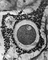
FIG. 1. Ovarian egg obtained by puncture of a follicle from the ovary of an unmated rabbit.
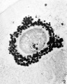
FIG. 2. Ovarian egg obtained by puncture of a follicle from the ovary of a doe mated 2 hours previously. The chromatin material condenses to tetrads. The vesicular membrane is still present.
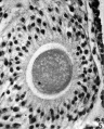
FIG. 5. Ovarian egg from a doe mated 6 hours previously. The first polar spindle begins to form.
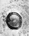
FIG. 6. Ovarian egg from a doe mated 8 hours previously. The first polar body has been given off.
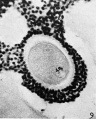
FIG. 9. Ovarian egg from a doe which had received 2 cc. thyroxin intravenously. Tetrads have formed in a vesicular nucleus.
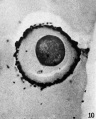
FIG. 10. Ovarian egg from unmated doe, inseminated with normal sperm in vitro. Sperm penetration has occurred. Note sperm head at lower right periphery.


Plate 2

FIG. 3. Ovarian egg from a doe mated 4 hours previously. Tetrads fully formed and vesicular nucleus dissolved.
FIG. 4. Ovarian egg from a doe mated 5 hours previously. The tetrads have become smaller and arranged themselves in a plate. All traces of the vesicular membrane have disappeared.
FIG. 7. Ovarian egg from a doe mated 9 hours previously. First polar body and second polar spindle.
FIG. 8. Ovarian egg cultured for 24 hours in Ringer-Locke solution containing maturity hormone. Note apparent fusion nuclei.
FIG. 11. Ovarian egg from a doe mated 6 hours previously and inseminated in vitro with normal sperm. Male and female pronuclei present side by side.
FIG. 12. Ovarian egg from a doe mated 8 hours previously and inseminated with normal sperm. The first polar body has formed and sperm penetration occurred. The entering spermatozoan has formed a male pronucleus (centre left).
Pincus G. and Enzmann EV. The comparative behavior of mammalian eggs in vivo and in vitro. (1935) J Exp Med. 62(5): 665-75. PMID 19870440
Cite this page: Hill, M.A. (2024, April 16) Embryology Paper - The Comparative Behavior of Mammalian Eggs in Vivo and in Vitro. Retrieved from https://embryology.med.unsw.edu.au/embryology/index.php/Paper_-_The_Comparative_Behavior_of_Mammalian_Eggs_in_Vivo_and_in_Vitro
- © Dr Mark Hill 2024, UNSW Embryology ISBN: 978 0 7334 2609 4 - UNSW CRICOS Provider Code No. 00098G