Paper - Preliminary note on the differentiation of angioblasts and the method by which they produce blood-vessels, blood-plasma, and red blood-cells as seen in the living chick: Difference between revisions
(Created page with "{{Ref-Sabin1917b}}") |
mNo edit summary |
||
| (One intermediate revision by the same user not shown) | |||
| Line 1: | Line 1: | ||
{{Header}} | |||
{{Ref-Sabin1917b}} | {{Ref-Sabin1917b}} | ||
{| class="wikitable mw-collapsible mw-collapsed" | |||
! Online Editor | |||
|- | |||
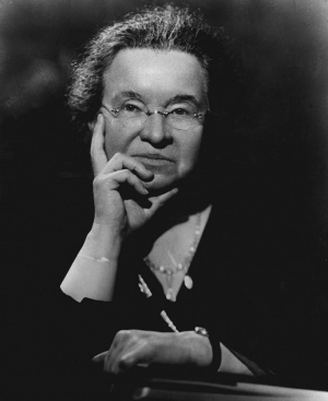
| [[File:Mark_Hill.jpg|90px|left]] This 1917 paper by [[Embryology History - Florence Sabin|Florence Rena Sabin]] (1871 - 1953) describes early vascular development in {{chicken}}. [[Embryology History - Florence Sabin|Florence Sabin]] was a key historic researcher in early 1900's establishing our early understanding of both vascular and lymphatic development. | |||
<br><br> | |||
'''Modern Notes:''' {{chicken}} | |||
<br><br> | |||
{{Heart Links}} | |||
<br> | |||
{{Immune Links}} | |||
<br> | |||
{{Chicken links}} | |||
<br> | |||
|- | |||
|} | |||
{{Historic Disclaimer}} | |||
=Preliminary note on the differentiation of angioblasts and the method by which they produce blood-vessels, blood-plasma, and red blood-cells as seen in the living chick= | |||
[[File:Florence Sabin 1938.jpg|thumb|alt=Florence Rena Sabin (1871 - 1953)|link=Embryology History - Florence Sabin|Florence Rena Sabin (1871-1953)]] | |||
[[Embryology History - Florence Sabin|Florence R. Sabin]] | |||
Anatomical Laboratory, Johns Hopkins University | |||
The question of the origin of the vascular system can be solved by the method of studying the living blastoderm of the chick in hanging-drop preparations. | |||
By watching chicks of the second day of incubation in these prei)arations it is possible to see all the processes by which blood-vessels and later blood-cells form. These observations can be made best on the area pellucida. Blood-vessels begin by the differentiation from mesodenn of a new type of cells, angiol)lasts or vaso-formal ivc cells. They differ from mesodenn in ha\-ing a much more granular cjiioplasm and in being more refractile. They differ also in their beha^^o^ and in their potentiahties. When a cell of the mesoderm divides, the daughter cells separate at least enough so that they can be recogruzed as distinct cells but angioblasts give daughter cells that remain together to form dense sjiicytial masses. These small masses soon jt)in similar masses by means of tiny processes of cytojjlasm put out from them exactly like the sprouts by which blood-vessels are known to grow. In this way angioblasts form a plexxis of dense masses of cells in sharp contrast to the more delicate plexus of mesotlerm wliich represents the early stages of the development of the coelom. The jilexus of angioblasts increases both by the division and the gi-owth of its cells anil by the constant addition of new angioblasts which diffiM'cntiatc from the mesoderm. | |||
Within the plexus of anpiohhists, vacuoles appear which represent a li(iuefactinn of the central ])art of the cytoplasm to fonn bloo(l-pla.sina. The vacuoles begin aKainst the nuclei and may occur anywhere in the mass but are especially frequent under the edges. The vacuoles just under the margius leave the cells along the border of the mass to become an endothelial lining of a ea\ity. The complete li(iuefaction of the central part of the mass into plasn\a takes from one to two hours and can be seen with great clearness in the living chick. The endothelial cells on the border of these cavities become less granular than the original angioblasts. Since the fluid is formed from the liquefying of the center there is no sign of distention of the cavities and no flattening of the cells along the border. There is a destruction both of the cytoplasm and of the nuclei of the mass to form the plasma. | |||
This process of the liquefaction of the central part of the mass of angioblasts to form vessels takes place not only within the )>lexus but in masses of angioI)lasts which are still isolated and in this way small vesicles are formed which then join the main plexus by processes of cyoplasm. I have seen such a tiny vesicle fonn from a single angiobhist proving that the lumen of a blood-vessel is intracellular. | |||
There are two processes from which the formation of bloodvessels must be sharply distinguished in watching these living specimens. The first is the fonnation of the coelom. The mesoderm of the chick is originally a continuous sheet of cells as is shown in Lilly's figm-e 40, A and B, on page 70' and in Duval's figures 18-i to 188 on i)late 12. As the chick grows larger this double sheet of cells forms a plexus with wide interspaces in the network where the mesoderm is entirelj' lacking lea\'ing nothing but ectoderm and endodenn. The coelom now begins as clefts full of fluid within the soUd bands, often at the nodes of the network of mesoderm. These spaces separate and, as it were, split the two layers of the mesodenn apart | |||
' Lilly, F. U. lOaS The development of the chicle. Henry Holt and Comp.'iny. New York. | |||
'Duval, .\I. 1S89 Atlas d'Embr>'ologie. G. Masson, Paris. | |||
and gradually flow together to iiuikc the ca\'ity of the coelom. The mesodenn on the Ijorders of these spaces gradually flattens into a mesothehmn. The process involves but httle destruction of tissue. At the stage of about six or seven somites a Hving specimen thus shows a double plexus over the area jiellucida, a dorsal plexus which is the developing coelom with large interspaces in the net and many tiny caxities representing the exo-coelom; and a ventral, more massive plexus of angioblasts with much more granular and more refractile c\-toplasm than the mesoderm. The granules of the angioblasts are strongly basophilic and stain intensely with haematox>'lin and with azur. The second structure from which developing vessels must be distinguished I shall call endodermal blebs or bhsters. They are collections of fluid beneath the endoderm, that is between endodenn and mesodenn, which are very frequent in the normal chick. They can be seen in any collection of mounted blastoderms. In the living chick they vary in appearance according to their size and shape. Their margins simulate endothelium to a marked degree. If they are distended and hence round, their margins will be thin, sharp and highly refractile; if they are flatter their walls will be still refractile but thicker. Often their nuclei seem to project into a ca\ity exactly like those of endothelial cells. They may be numerous, small and isolated or large and nmltiple reminding one of midtilocular cysts. .\s can be readily imagined from their structure they may change in shape rapidly, far more rapiiUy than true vessels change. They can be analyzed with the focusing screw both by following their margins over onto the endoderm and by noting their very superficial position. They occur under the ectoderm as well as under the endoderm but I think less freriuently. Xo one can follow the development of blood-vessels in the living chick without becoming thoroughly familiar with the appearances of these blisters. They have nothing to do with blood-vessels occurring both before the blood-vessels begin and afterward. They are however important physiological structures representing the method by which fluid is absorbed through tiie endoderm and ectoderm for the young chick. | |||
Red blnod-corpu8clc8 can be seen to grow from the endothelial lininR of hloiMJ-vossi'ls. Tlioy may develop from little masses of the oriRinal anpiulilasts which l)ecomc i)artially separated by the liquefaction of cytoplasm around them. Such a mass of eells becomes a blood-island by ha\inK haemoglobin develop within the cells. The color of haemoglobin can be detected in the li\ing cells earlier than I have been able to fix a,nd stain it. Again an endothelial cell of a blood-vessel will divide so that one daughter cell jirnjects into the lumen. This cell becomes filled with bivsojihilic granules and develops haemoglobin. It is then a imicelluhir blood-island. It divndes and the mass is increased also by the addition of other cells which differentiate from the endothelium in the neighborhood and creep ahmg the wall to join the first cell. These cells .soon form a yellow sjnicytial mass projecting into the lumen of the vessel. \i this stage the islands have a smooth, sharji contour. .Vs they develop, cells begin to rountl up on their siufacc until the whole mass comes to look Uke a mulberry and then the red cells break free from the mass and float away in the blood-plasma. | |||
These blood-islands I have seen develop in all the vessels of the area vasculosa, in the omphalo-mesenteric vein anil arteries and in the dorsal aorta. In the area pellucida they are most abundant in the i)l('xus of vessels just posterior to the area in which the omi)hal()-mesenteric arteries develop. In this area abnost any chick of fom-teen somites will show a plexus of angioblasts fuU of \acuoles and one of seventeen somites will show blood-islands. The circulation does not interfere with the development of the islands. If they clog the lumen the free cori)uscle8 either lodge behind them for a time or pass on thi'ough other channels in the jilexus. | |||
One of the most interesting points al)Out the nui.sses of angioblasts and of the islands is that all of the cells in them divide at about the same time. Moreover all of the separate islands of a given area seem to divide at once. In these total preparations, all one can .see of the ju-ocess of mitosis is the nucleus in the stage of the metaphase and the actual tlivision of the cj^toplasm. When the syncytial mass is about to divide it becomes intensely refractile and its cells become outlined; then one sees one nucleus after another pass into the nietaphase and finally all of the cells appear to be half as large as the mature cells. The island then becomes large by the giowth of all the cells to their original size. The blood-cells keep on dividing after they are free from the islands and after they have begim to circulate. | |||
These studies have been made with chicks which were grown in the mixture of Locke's solution and chicken bouillon develoj)ed by Margaret Reed Lewis for tissue-cultures.' For the young blastoderms it is better to increase the amount of the sodiiun chloride in the solution to L0(3 per cent. A weaker solution lakes the haemoglobin and is also less favorable for all the other cells. | |||
From these studies which are here reported briefly, certain jH-inciplos are established. Blood-\essels do not arise from the dilatation of tissue-spaces, but by the differentiation of angioblasts. They cannot be spoken of as arising from spaces because they develop within the bodies of these angioblasts; that is, they arise within cells, not between them. The processes by which they form are not in any way siimlar to the processes by which either the coelom or great systems of tissue-spaces like the cerebro-spinal spaces develop. The coelom forms by the splitting apart of two layers of mesoderm with Uttlc de.-^truction of tissue; the cerebro-spinal spaces form in a mass of typical mesenchjme with considerable destruction of tissue. Angioblasts differentiate tlu'oughout the wall of the yolk sac and in the embryo as well. I have seen the tlorsal aorta tlifferentiate in situ in the li\-ing chick even to some extent that part within the head. Angioblasts produce blood-plasma, endothelium and red blood-cells. .Vngioblasts and later endotheUal cells give rise to red blood-cells by develojiing haemoglol)in. | |||
The term blood-island has been used since the time of the early embryologists, notably Wolff and Pander, for the masses of vaso-formative cells which can be seen in the lU'ca opaque of the chick even before the first somite. I propose however to restrict the tenii hlood-islaiul to thoso masses of colls which can 1)0 shown to produco ha(>iiH)|a;lol)in and to boconio froo red blood-colls. Those masses are attached to the wall and hence are not strictly speaking islands. The more primitive masses which do boffin as isolated masses or islands of colls hut nmst lu"st i)roduce enilot helium and plasma, 1 shall call angioblasts. The retl blood-cells tlevelop after some plasma has been formed. .Ml of the blood-cells of the chick of the second day of incubation can be seen to have haemoglobin in the living chick and hence they cannot be consiilered as forerunners of white bloodcells. | |||
•Lewis. M. . :in<l \\. II. lOU The growth of embryonic chick tissues in artificial media, agar and liouiiion. Johns Hopkins Hospital Bulletin, 2°J. | |||
{{Footer}} | |||
[[Category:Chicken]][[Category:Blood]][[Category:Historic Embryology]][[Category:1910's]][[Category:Florence Sabin]] | |||
Revision as of 03:23, 9 September 2019
| Embryology - 19 Apr 2024 |
|---|
| Google Translate - select your language from the list shown below (this will open a new external page) |
|
العربية | català | 中文 | 中國傳統的 | français | Deutsche | עִברִית | हिंदी | bahasa Indonesia | italiano | 日本語 | 한국어 | မြန်မာ | Pilipino | Polskie | português | ਪੰਜਾਬੀ ਦੇ | Română | русский | Español | Swahili | Svensk | ไทย | Türkçe | اردو | ייִדיש | Tiếng Việt These external translations are automated and may not be accurate. (More? About Translations) |
Sabin FR. Preliminary note on the differentiation of angioblasts and the method by which they produce blood-vessels, blood-plasma, and red blood-cells as seen in the living chick. (1917) Anat. Rec. 13: 199-204
| Historic Disclaimer - information about historic embryology pages |
|---|
 |
Preliminary note on the differentiation of angioblasts and the method by which they produce blood-vessels, blood-plasma, and red blood-cells as seen in the living chick
Anatomical Laboratory, Johns Hopkins University
The question of the origin of the vascular system can be solved by the method of studying the living blastoderm of the chick in hanging-drop preparations.
By watching chicks of the second day of incubation in these prei)arations it is possible to see all the processes by which blood-vessels and later blood-cells form. These observations can be made best on the area pellucida. Blood-vessels begin by the differentiation from mesodenn of a new type of cells, angiol)lasts or vaso-formal ivc cells. They differ from mesodenn in ha\-ing a much more granular cjiioplasm and in being more refractile. They differ also in their beha^^o^ and in their potentiahties. When a cell of the mesoderm divides, the daughter cells separate at least enough so that they can be recogruzed as distinct cells but angioblasts give daughter cells that remain together to form dense sjiicytial masses. These small masses soon jt)in similar masses by means of tiny processes of cytojjlasm put out from them exactly like the sprouts by which blood-vessels are known to grow. In this way angioblasts form a plexxis of dense masses of cells in sharp contrast to the more delicate plexus of mesotlerm wliich represents the early stages of the development of the coelom. The jilexus of angioblasts increases both by the division and the gi-owth of its cells anil by the constant addition of new angioblasts which diffiM'cntiatc from the mesoderm.
Within the plexus of anpiohhists, vacuoles appear which represent a li(iuefactinn of the central ])art of the cytoplasm to fonn bloo(l-pla.sina. The vacuoles begin aKainst the nuclei and may occur anywhere in the mass but are especially frequent under the edges. The vacuoles just under the margius leave the cells along the border of the mass to become an endothelial lining of a ea\ity. The complete li(iuefaction of the central part of the mass into plasn\a takes from one to two hours and can be seen with great clearness in the living chick. The endothelial cells on the border of these cavities become less granular than the original angioblasts. Since the fluid is formed from the liquefying of the center there is no sign of distention of the cavities and no flattening of the cells along the border. There is a destruction both of the cytoplasm and of the nuclei of the mass to form the plasma.
This process of the liquefaction of the central part of the mass of angioblasts to form vessels takes place not only within the )>lexus but in masses of angioI)lasts which are still isolated and in this way small vesicles are formed which then join the main plexus by processes of cyoplasm. I have seen such a tiny vesicle fonn from a single angiobhist proving that the lumen of a blood-vessel is intracellular.
There are two processes from which the formation of bloodvessels must be sharply distinguished in watching these living specimens. The first is the fonnation of the coelom. The mesoderm of the chick is originally a continuous sheet of cells as is shown in Lilly's figm-e 40, A and B, on page 70' and in Duval's figures 18-i to 188 on i)late 12. As the chick grows larger this double sheet of cells forms a plexus with wide interspaces in the network where the mesoderm is entirelj' lacking lea\'ing nothing but ectoderm and endodenn. The coelom now begins as clefts full of fluid within the soUd bands, often at the nodes of the network of mesoderm. These spaces separate and, as it were, split the two layers of the mesodenn apart
' Lilly, F. U. lOaS The development of the chicle. Henry Holt and Comp.'iny. New York.
'Duval, .\I. 1S89 Atlas d'Embr>'ologie. G. Masson, Paris.
and gradually flow together to iiuikc the ca\'ity of the coelom. The mesodenn on the Ijorders of these spaces gradually flattens into a mesothehmn. The process involves but httle destruction of tissue. At the stage of about six or seven somites a Hving specimen thus shows a double plexus over the area jiellucida, a dorsal plexus which is the developing coelom with large interspaces in the net and many tiny caxities representing the exo-coelom; and a ventral, more massive plexus of angioblasts with much more granular and more refractile c\-toplasm than the mesoderm. The granules of the angioblasts are strongly basophilic and stain intensely with haematox>'lin and with azur. The second structure from which developing vessels must be distinguished I shall call endodermal blebs or bhsters. They are collections of fluid beneath the endoderm, that is between endodenn and mesodenn, which are very frequent in the normal chick. They can be seen in any collection of mounted blastoderms. In the living chick they vary in appearance according to their size and shape. Their margins simulate endothelium to a marked degree. If they are distended and hence round, their margins will be thin, sharp and highly refractile; if they are flatter their walls will be still refractile but thicker. Often their nuclei seem to project into a ca\ity exactly like those of endothelial cells. They may be numerous, small and isolated or large and nmltiple reminding one of midtilocular cysts. .\s can be readily imagined from their structure they may change in shape rapidly, far more rapiiUy than true vessels change. They can be analyzed with the focusing screw both by following their margins over onto the endoderm and by noting their very superficial position. They occur under the ectoderm as well as under the endoderm but I think less freriuently. Xo one can follow the development of blood-vessels in the living chick without becoming thoroughly familiar with the appearances of these blisters. They have nothing to do with blood-vessels occurring both before the blood-vessels begin and afterward. They are however important physiological structures representing the method by which fluid is absorbed through tiie endoderm and ectoderm for the young chick.
Red blnod-corpu8clc8 can be seen to grow from the endothelial lininR of hloiMJ-vossi'ls. Tlioy may develop from little masses of the oriRinal anpiulilasts which l)ecomc i)artially separated by the liquefaction of cytoplasm around them. Such a mass of eells becomes a blood-island by ha\inK haemoglobin develop within the cells. The color of haemoglobin can be detected in the li\ing cells earlier than I have been able to fix a,nd stain it. Again an endothelial cell of a blood-vessel will divide so that one daughter cell jirnjects into the lumen. This cell becomes filled with bivsojihilic granules and develops haemoglobin. It is then a imicelluhir blood-island. It divndes and the mass is increased also by the addition of other cells which differentiate from the endothelium in the neighborhood and creep ahmg the wall to join the first cell. These cells .soon form a yellow sjnicytial mass projecting into the lumen of the vessel. \i this stage the islands have a smooth, sharji contour. .Vs they develop, cells begin to rountl up on their siufacc until the whole mass comes to look Uke a mulberry and then the red cells break free from the mass and float away in the blood-plasma.
These blood-islands I have seen develop in all the vessels of the area vasculosa, in the omphalo-mesenteric vein anil arteries and in the dorsal aorta. In the area pellucida they are most abundant in the i)l('xus of vessels just posterior to the area in which the omi)hal()-mesenteric arteries develop. In this area abnost any chick of fom-teen somites will show a plexus of angioblasts fuU of \acuoles and one of seventeen somites will show blood-islands. The circulation does not interfere with the development of the islands. If they clog the lumen the free cori)uscle8 either lodge behind them for a time or pass on thi'ough other channels in the jilexus.
One of the most interesting points al)Out the nui.sses of angioblasts and of the islands is that all of the cells in them divide at about the same time. Moreover all of the separate islands of a given area seem to divide at once. In these total preparations, all one can .see of the ju-ocess of mitosis is the nucleus in the stage of the metaphase and the actual tlivision of the cj^toplasm. When the syncytial mass is about to divide it becomes intensely refractile and its cells become outlined; then one sees one nucleus after another pass into the nietaphase and finally all of the cells appear to be half as large as the mature cells. The island then becomes large by the giowth of all the cells to their original size. The blood-cells keep on dividing after they are free from the islands and after they have begim to circulate.
These studies have been made with chicks which were grown in the mixture of Locke's solution and chicken bouillon develoj)ed by Margaret Reed Lewis for tissue-cultures.' For the young blastoderms it is better to increase the amount of the sodiiun chloride in the solution to L0(3 per cent. A weaker solution lakes the haemoglobin and is also less favorable for all the other cells.
From these studies which are here reported briefly, certain jH-inciplos are established. Blood-\essels do not arise from the dilatation of tissue-spaces, but by the differentiation of angioblasts. They cannot be spoken of as arising from spaces because they develop within the bodies of these angioblasts; that is, they arise within cells, not between them. The processes by which they form are not in any way siimlar to the processes by which either the coelom or great systems of tissue-spaces like the cerebro-spinal spaces develop. The coelom forms by the splitting apart of two layers of mesoderm with Uttlc de.-^truction of tissue; the cerebro-spinal spaces form in a mass of typical mesenchjme with considerable destruction of tissue. Angioblasts differentiate tlu'oughout the wall of the yolk sac and in the embryo as well. I have seen the tlorsal aorta tlifferentiate in situ in the li\-ing chick even to some extent that part within the head. Angioblasts produce blood-plasma, endothelium and red blood-cells. .Vngioblasts and later endotheUal cells give rise to red blood-cells by develojiing haemoglol)in.
The term blood-island has been used since the time of the early embryologists, notably Wolff and Pander, for the masses of vaso-formative cells which can be seen in the lU'ca opaque of the chick even before the first somite. I propose however to restrict the tenii hlood-islaiul to thoso masses of colls which can 1)0 shown to produco ha(>iiH)|a;lol)in and to boconio froo red blood-colls. Those masses are attached to the wall and hence are not strictly speaking islands. The more primitive masses which do boffin as isolated masses or islands of colls hut nmst lu"st i)roduce enilot helium and plasma, 1 shall call angioblasts. The retl blood-cells tlevelop after some plasma has been formed. .Ml of the blood-cells of the chick of the second day of incubation can be seen to have haemoglobin in the living chick and hence they cannot be consiilered as forerunners of white bloodcells.
•Lewis. M. . :in<l \\. II. lOU The growth of embryonic chick tissues in artificial media, agar and liouiiion. Johns Hopkins Hospital Bulletin, 2°J.
Cite this page: Hill, M.A. (2024, April 19) Embryology Paper - Preliminary note on the differentiation of angioblasts and the method by which they produce blood-vessels, blood-plasma, and red blood-cells as seen in the living chick. Retrieved from https://embryology.med.unsw.edu.au/embryology/index.php/Paper_-_Preliminary_note_on_the_differentiation_of_angioblasts_and_the_method_by_which_they_produce_blood-vessels,_blood-plasma,_and_red_blood-cells_as_seen_in_the_living_chick
- © Dr Mark Hill 2024, UNSW Embryology ISBN: 978 0 7334 2609 4 - UNSW CRICOS Provider Code No. 00098G