Paper - Photographs of a Series of Sections of an Early Human Embryo
| Embryology - 24 Apr 2024 |
|---|
| Google Translate - select your language from the list shown below (this will open a new external page) |
|
العربية | català | 中文 | 中國傳統的 | français | Deutsche | עִברִית | हिंदी | bahasa Indonesia | italiano | 日本語 | 한국어 | မြန်မာ | Pilipino | Polskie | português | ਪੰਜਾਬੀ ਦੇ | Română | русский | Español | Swahili | Svensk | ไทย | Türkçe | اردو | ייִדיש | Tiếng Việt These external translations are automated and may not be accurate. (More? About Translations) |
Buxton BH. Photographs of a series of sections of an early human embryo. (1899) J Anat Physiol. 33(3): 381-384 PMID 17232381
| Online Editor |
|---|
| This 1899 historic paper by Buxton is one of the first photographic series of histological sections through an early human embryo. The practice of photomicroscopy is a key process in documenting these very small embryos. An estimated age of 25 days would make this a stage 10 or stage 11 embryo. |
| Historic Disclaimer - information about historic embryology pages |
|---|
| Pages where the terms "Historic" (textbooks, papers, people, recommendations) appear on this site, and sections within pages where this disclaimer appears, indicate that the content and scientific understanding are specific to the time of publication. This means that while some scientific descriptions are still accurate, the terminology and interpretation of the developmental mechanisms reflect the understanding at the time of original publication and those of the preceding periods, these terms, interpretations and recommendations may not reflect our current scientific understanding. (More? Embryology History | Historic Embryology Papers) |
Photographs of a Series of Sections of an Early Human Embryo
By B. H. Buxton (Plates XVI. - XXVII.)
- Estimated age - 25 days.
- Ovum - Size of pigeon's egg, covered with shaggy vili
- Embryo - Length, 5mm. Thickness, 2mm.
Taken fresh from uterus after hysterectomy, and kept for a year in 2 percent. formalin before hardening.
Hardened in absolute alcohol, imbedded in celloidin, and cut in112 serial sections, each 20 micro-millimetres thick. Stained with haematoxylin and eosin (Stain - Haematoxylin Eosin).
Photographs
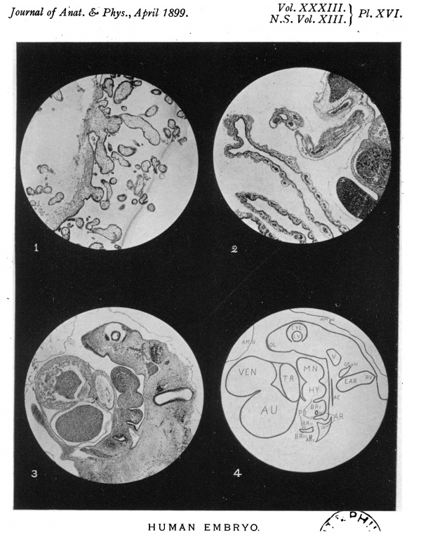
25 diameters, Spencer, 2in. Noocular.
100 diameters, Spencer, 1/2 in. Zeiss projection ocular.
180 diameters, Spencer, 1/4 in., Zeiss projection ocular.
500 diameters, Leitz, 1/12 oil. , Zeiss projection immersion.
The embryo is lying on its right side, and the sections are cut sagittally from left to right.
The first six plates are in regular order from left to right, and the numbers are those of the sections.
Special Points
Alltantois.-The cavity of the stalk can be traced a short dis- tancefromthecloaca. Itthenappearstocloseup. Thecavity of the yolk stalk is open, and continuous with that of the intes- tine.
Alimentary Canal.-The pituitary involution forms an un- branched tubule, extending upwards between the fore and hind brain from the roof of the mouth. The hyomandibular and three bronchial pouches can be clearly traced. A septum,containingnomesoblast,separatesthemfrom thecorrespondinggrooves. FromtheEsophagus,the laryngeal chamber branches of, and the trachea divides into two primary bronchi, but there are no secondary branches.
The stomach shows as an elongated dilatation.
The pancreas and bile-duct are present in early stages.
The liver is already of considerable size, and richly supplied with blood.
The vitelline loop extends outside the embryo, its cavity being continuous with that of the yolk stalk.
Beyond the vitelline loop the intestine dilates slightly, and then contracts again to form the rectum, which opens into the cloaca.
Beyond the cloaca is the narrow post-anal gut, which shows, however, a marked dilatation at its caudal extremity.
Circulatory
Heart
Single auricle and ventricle,from which latter arises the truncus arteriosus. Arches I. and IL. are obliterated, but I.,IV., and V. can be traced on either side to the anterior dorsal aorte, which join about opposite the lungs to form the single dorsal aorta, which, after giving of the vitelline artery, divides into the allantoic arteries a little anterior to the cloaca. The remnant of the dorsal aorta continues as the caudal aorta.
The blood is returned by the allantoic and vitelline veins, which unite, partly breaking up in the liver to form the portal system, and partly running directly into the sinus venosus by means of the ductus venosus.
The cardinal veins, running dorsally to the Wolffian bodies, return the blood from the embryo, the anterior and posterior on either side combining to form the Cuvierian veins, which empty intothesinusvenosus.
Nervous System
Cranial flexure is marked. Of the cranial nerves,-
- V. Supplies the mandibular arch.
- VII. Supplies the hyoid arch.
- VIII. Supplies the ear.
- IX. Supplies the first bronchial arch.
- X. Supplies the 2nd and 3rd bronchial arches.
The cervical nerves V.-VIII. and 1st dorsal join to form the brachial plexus.
There is no trace of the sacral plexus.
Skeletal
The notochord cannot be traced further forward than a point opposite the hyoid arch, but from that place to the extreme end of the tail is well marked. It exhibits several dilatations, one of which is shown on Plate XXVII., d. No head cavities or muscle plates anterior to those of the hypoglossal region can be found.
Muscle plates of hypoglossal region: Three distinct plates can be seen on either side; the anterior plate appears to split up, and may perhaps be considered to represent two plates,which would make up the usual number of four.
The dorsal muscle plates are differentiated into muscular and dermal layers, except in their more ventral portions.
The fore and hind limbs appear as buds, composed of undifferentiated cells.
Genito-Urinary
The Wolffian ducts commence just behind the lung, and run posteriorly outside the Wolffian bodies to the eloaca. Just before the ducts reach the cloaca the ureters appear as evaginations from them.
Special Sense Organs
Eye.-The optic vesicle buds off from the fore-brain, the lumen being still wide open. It rises up to form the optic cup, of which the retinal is already somewhat thicker than the pigmentary layer. The choroidal fissure is well marked, and the vesicle of the lens is still open to the exterior.
The Ear forms an elongated, roughly quadrangular sac, closed off from the exterior. Closely connected with its vestibular portion is the ganglion of the eighth nerve.
The Nose.- Olfactory plates are present as thickened ridges of the epiblast, but there are as yet no involutions to form the olfactory pits.
Description of Plates XVI - XXVII
The number in brackets is that of the section, the current numbers are those of the figures in the several plates.
Plate XVI
(25 diameters). 1, chorion; 2, yolk sac and stalk; 3, 4, (23), ear and bronchial arches.
Plate XVII
(25 diameters). 5, 6, (26), optic cup, bronchial arches,. heart; 7, 8, (34), truncus arteriosus, post-anal gut.
Plate XVIII
(25diameters). 9,10,(40),larynx-cesophagus; 11, 12,(43),uretersand alantois.
Plate XIX
(25diameters). 13, 14,(60),sinusvenosus,gal-blader, pituitaryinvolution; 15,16,(70),brain.
Plate XX
(25 diameters). 17, 18, (78), sixth cervical nerve, optic vesicle; 19, 20, (78), Wolffian bodies, transverse section.
Plate XXI
(25 diameters). 21, 22, (83), Wolffian bodies; 23, (92), Wolffian bodies on right and muscle plates on left of dorsal aorta. To extreme right is the optic vesicle and vesicle of lens; 24, (101), spinal cord-longitudinal section. The muscle plates on either side. On left-centraltransversesection. Onright-ventraltransversesection.
Plate XXII
(100diameters). 25, sinus venosus. The ductus venosus runs into it on the left, and the Cuvierian vein on the right. Above is the auricle, and below the lung-(see PL. XIX., 14); 26, allantoic arteries branching off on either side from the posterior end of the' dorsal aorta, and enclosing the Wolffian ducts and rectum; 27,spinal cord. On either side the ganglia, and further outthe muscle plates. Below is the notochord, and below this the dorsal aorta-(see P1. XX., 19); 28, ear and ganglion of eighth nerve-(see P1. XVI., 3).
Plate XXIII
(Eye series).29, 30, 31, 100 diameters-32, 180 diam- eters.-29, optic vesicle budding off from fore-brain-(see Pl. XX., 19); 30, optic vesicle rising up to form optic cup; 31, optic cup and choroidal fissure-(see P1. XVII., 5); 32, vesicle of lens-(see P1. XVI., 3).
Plate XXIV
(Ureter series) 100 diameters.- Ureters and cloaca. Serial sections.-On the left in 3.3 is the dilated posterior extremity of the Wolffian duct, and the following photographs show that the ureter isformed by evagination from this.
Plate XXV
(180diameters). 37, chorionic villus - (seeP1.XVI., 1); 38, sixth cervical nerve-(see P1. XX., 19); 39, caudal dilatation of' post-anal gut-(see P1. XVII., 7); 40, pituitary involution-(see Pl. XIX., 13).
Plate XXVI
(41,100diameters;42,43,44,180diameters). 41, larynx and aesophagus-(see P1. XVIII., 9); 42, muscle plates of hypoglossal region; 43,muscle plates, central transverse section (see P1. XXI., 24); 44, muscle plates, ventral transverse section (see, P1.XXI., 24).
Plate XXVII
(45, 46, 47, 180 diameters; 48, 500 diameters).-a, septum dividing first branchial pouch from first bronchial groove; b, Wolffian duct and tubules; c, Wolffian body, showing glomeruli; d,notochord.

Reference
Buxton BH. Photographs of a series of sections of an early human embryo. (1899) J Anat Physiol. 33(3): 381-384 PMID 17232381
Cite this page: Hill, M.A. (2024, April 24) Embryology Paper - Photographs of a Series of Sections of an Early Human Embryo. Retrieved from https://embryology.med.unsw.edu.au/embryology/index.php/Paper_-_Photographs_of_a_Series_of_Sections_of_an_Early_Human_Embryo
- © Dr Mark Hill 2024, UNSW Embryology ISBN: 978 0 7334 2609 4 - UNSW CRICOS Provider Code No. 00098G


