Paper - On the transitory or artificial fissures of the human cerebrum: Difference between revisions
mNo edit summary |
|||
| (3 intermediate revisions by the same user not shown) | |||
| Line 13: | Line 13: | ||
{{Historic Disclaimer}} | {{Historic Disclaimer}} | ||
=On the Transitory or Artificial Fissures of the Human Cerebrum= | =On the Transitory or Artificial Fissures of the Human Cerebrum= | ||
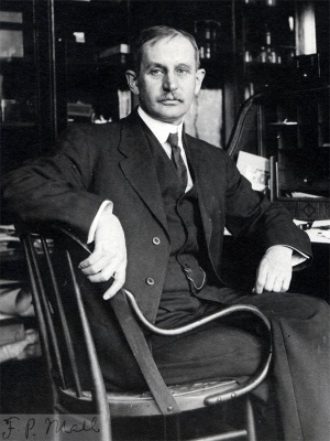
[[File:Franklin Mall 01.jpg|thumb|300px|link=Embryology History - Franklin Mall|Franklin Mall (1911)]] | |||
by | |||
Franklin P. Mall. | |||
Professor of Anatomy, Johns Hopkins University. | |||
With 1 Table. | |||
==Introduction== | |||
Nearly a century ago J. F. Meckel ‘ discovered the transitory fissures in the eerebrum of human embryos eight or nine Weeks old; these he believed to be normal and in no way connected with the permanent fissures. The presence of the transitory fissures was confined by numerous competent anatomists and from time to time their relation to the permanent fissures was discussed. According to Tiedemann they represent the earlier stages of the permanent fissures, and Cunningham states that most of them are obliterated while several “occupy positions which later on are occupied by permanent fissures, and either show direct continuity of existence with these, or at least act as their precursors.” | |||
It was gradually shown that the transitory fissures are usually present in embryos of the third and fourth months. To be sure suitable material for study is diificult to obtain and usually the specimens studied were those that had been preserved in alcohol for a considerable time. The influence of alcohol, especially weak alcohol, upon tissues is well known and this naturally led Bischoff in 1868 to suspect that the transitory fissures were artificial, having been produced by the macerating influence of weak alcohol. Furthermore, he found that the fissures were not present in the brains of embryos which had been hardened in chloride of zinc. This view is accepted by Marchand (1891) in his paper on the corpus callosum. | |||
Were it not so difficult to obtain fresh human embryos, this question would have been settled long ago. That these fissures are normal seems to be proved by Ecker, in 1869, who observed their presence in the fresh brain of an embryo three months old. Further observation by Hochstetter (1898) shows that they are not present in the fresh brain. Hochstetter examined the brains of two very fresh human embryos and in neither of them was there a trace of a transitory fissure. In other specimens which were well preserved the transitory fissures were only slightly marked, or were not present at all. The observations of Hochstetter are confirmed by Retzius, who had an opportunity to examine the fresh brain of an embryo of the third month. The membranous skull was removed and the specimen hardened in Zenker’s solution. After this treatment, it was found that the lateral and mesial surfaces of the brain were perfectly smooth with the exception of a slight depression on the mesial side. | |||
1 References to the literature upon this subject will be found in the following papers: Cunningham, Jour. of Aunt. and Phys., XXIV, 1890; Hochstetter, Bibliotheca medics, Stuttgart, 1898; and Retzius, Biol. Untersucl1., X, 1902. 334 Transitory fissures of the Human Cerebrum | |||
It appears then that when the brains of fresh embryos of the third and fourth months are examined no transitory fissures are found. Furthermore, when fresh specimens are carefully hardened the transitory fissures are insignificant and not numerous, or are not present at all. | |||
About five years ago I noticed that the cerebral vesicles of human embryos hardened in formalin are entirely different from those hardened in alcohol. Not only are the vesicles perfect in form with walls in apposition to the membranes of the skull, but the arrangement of the cells is definite and clear. Specimens hardened in alcohol are sometimes folded and usually macerated, the degree of maceration always being far in excess of that of the rest of the body. | |||
The recent publication of Retzius has induced me to tabulate the condition of the brains in my embryological collection to determine the frequency and degree of transitory fissures in brains hardened in formalin as well as in those hardened in alcohol. The table which I have constructed records the condition of the brains in over fifty specimens. There are about a dozen excellent formalin specimens in the collection not included in the table, for they have not been sectioned and I am unwilling to injure them before they are cut into serial sections. It appears to me that these specimens recorded in the table, together with the observations of Hochstetter and of Retzius, set the transitory fissures aside as artificial products of the effect of weak alcohol upon the brain. | |||
The appended table gives the numbers, the length and the condition of the cerebral vesicles of embryos in my collection in which there are any data relating to the transitory fissures. The specimens have been grouped in months, using for this purpose a rule which has been published recently? According to this rule the age of an embryo in days equals the square root of one hundred times its length from vertex to breech in millimeters. Thus an embryo 30 mm. long is \/ 30 )( 100, or 54 days old. Or, to determine the vertex-breech length of an embryo for a given number of days, square the nu_mber of days and divide by one hundred. Thus the vertex-breech length of an embryo 30 days old is 302 | |||
2 Mall, Johns Hopkins Hospital Bulletin, 1903. Franklin P. Mall 335 | |||
T0 or 9 millimeters. The data upon which this rule rests will be found in my paper on the pathology of early human embryos.’ This formula applies only to embryos up to 100 mm. in length. In embryos from 100 to 220 mm. long from vertex to breech the length in millimeters equals the age in days. | |||
In nearly all instances the embryos were hardened either in alcohol or in formalin. Not only is this recorded in my notes, but it is also indicated by the condition of the tissue in case the specimen has been cut. It is very apparent from all of my specimens, both normal and pathological, that when the embryo is macerated to the least degree the effect is much more marked in the ‘brain than elsewhere. It appears that any dissociating fluid effects the brain first. So in order to tabulate the specimens I have had to express the extent of maceration of the brain in degrees, which in general is two or three degrees more advanced than that of any other organ of the embryo. | |||
and | |||
The | The condition of the brain is marked 0 in the table in case its lateral mesial surfaces are perfectly smooth as pictured by Retzius for the fresh brain of a human embryo at the end of the third month. Those brains in which there are slight irregularities of the walls, as is the case when there is some shrinkage with separation of the vesicle, are marked 1. Some of these folds are certainly not true transitory fissures, for in the same embryos there is the same separation of the epithelial cells in the oesophagus and in the intestine. I have, however, included in this group those brains in which the transitory fissures are just beginning. The brain is marked 2 whenever it has the typical transitory fissures as usually described.‘ In case the infolding is more extended, showing signs of maceration and disintegration of the walls of the brain with loose cells within the ventricle, it is marked 3. When the maceration has gone so far that the vesicles are filled with cells and the brain is nearly solid, it is marked 4. In the specimens marked 1 to 4 the spinal cord is not macerated very much, but when the entire central nervous system is macerated and solid it is marked 5. So we have, in addition to the embryos in which the surfaces of the brain are smooth, those in which the cerebral vesicles are folded and macerated from the simple small fold up to a stage in which the entire central nervous system is converted into a pulpy mass. | ||
are | |||
to | |||
3 Mall, Johns Hopkins Hospital Reports, IX, 1900. | |||
equals the | 4 The condition of specimens marked 1 equals about those with the least number of fissures as pictured by Retzius on Plate 1 in his great monograph, Das Menschen him. Those marked 2 represent those figures on this same plate with the greatest number of fissures. | ||
It is seen from the table that the condition of the brain varies very much in embryos of the first month as well as in the later months. In four of the embryos the cerebral vesicles are perfect and these are from specimens which were carefully hardened. In the fifth, No. 80, there are no data except that the specimen was hardened in alcohol. One embryo, No. 164, is from an autopsy, and the uterus after it had been cut open was kept on ice for 24 hours before it came into my hands. The entire specimen was then placed in strong formalin. Since all of the sections show that the tissues of the body are macerated it is not diflicult to understand why the walls of the cerebral vesicles are also macerated and slightly folded. | |||
from | |||
The | The embryos of the second month also show a variety of conditions in the cerebral vesicles. There are six perfect ones and three of these were hardened in formalin. One formalin specimen, No. 106, is pretty well macerated, but the specimen had been in water 24 hours before it came into my hands. In it the brain and spinal cord are practically solid. | ||
In No. 86 there is one small fissure on the medial and one on the lateral side of the cerebral hemisphere. This embryo was brought to the laboratory with the amnion unbroken, and without opening it the entire specimen was placed in formalin. It may be that the slight amount of formalin which entered the embryo first acted as a dissociator, caused the cerebral vesicles to expand quicker than the membranes and these narrow transitory fissures followed. In this specimen the fissures are formed by the epithelial wall of the cerebral vesicle turning in without drawing the pia with it. The pia bridges straight over the transitory fissure and the capillaries to the cerebral vesicle are torn off. It is clearly a case of tearing the cerebral vesicle from the pia, which could only have taken place after the death of the embryo. | |||
During the third month it is said that the transitory fissures make their appearance. Among ten specimens there are two with perfect transitory fissures and three with well marked transitory fissures. There are five specimens without any fissures at all and four of them are formalin specimens. One specimen, No. 95, has well-marked total fissures all around the cerebral vesicle. This specimen came to the laboratory fresh and without opening the ovum it was placed in formalin. This was the first of the formalin specimens which was cut, and for a long time I considered it conclusive proof in favor of the transitory fissures being normal. Here also the slow penetration of the formalin may have acted more markedly as a dissociator which caused the cerebral vesicles to expand quicker than the membranous walls of the head and thereby produced the slight infolding. In this specimen, as in No. 86, the maceration has caused a separation of the cerebral walls from the pia over the transitory fissures. At other points the cerebral cells turn outward, forming small microscopic protuberances. In both these specimens the microscopic examination shows clearly that the transitory fissures are produced artificially by t.he unequal expansion of the cerebral vesicles and the membranous wall. ’l‘he walls of the cerebral vesicles naturally were torn away from the pia along the line of the transitory fissures. | |||
the | |||
I was fortunate enough to obtain a fresh embryo of the fourth month while tabulating the specimens of my collection. Although the abortion had taken place 21 hours previously, the brain showed no indications of fissures at all; in every respect the specimen was like that of Retzius. After the membranous wall had been removed the brain was placed in formalin, in which it retained its smooth form. | |||
all | |||
The specimens of the fourth and fifth months are very conclusive. There are nine specimens hardened in formalin and none of them have any transitory fissures. They are present in the four specimens which were hardened in alcohol. A single fresh specimen at the beginning of the fifth month was perfectly smooth on both mesial and lateral surfaces, although the embryo came into my possession 24 hours after the abortion. | |||
It is apparent from the specimens which have been described that | It is apparent from the specimens which have been described that fluids which dissociate tissues are more marked in their effect upon the walls of the cerebral vesicles than upon any of the other tissues of the embryo. .’\S the cells of the cerebral vesicles become thicker and the tissues firmer the brain substance is more resistant and does not macerate as easily as before, so that by the fifth month transitory fissures can be no longer produced artificially. Formalin, which in strong solutions causes the brain tissue to swell, is a dissociator in very weak solutions, and therefore occasionally produces transitory fissures. According to the experience of Hochstetter, Retzius and myself, the transitory fissures are not found in fresh brains. The transitory fissures are therefore artificial and are of no morphological significance. | ||
fluids which dissociate tissues are more marked in their effect upon the | |||
walls of the cerebral vesicles than upon any of the other tissues of the | |||
embryo. .’\S the cells of the cerebral vesicles become thicker and the | |||
tissues firmer the brain substance is more resistant and does not macerate | |||
as easily as before, so that by the fifth month transitory fissures can be | |||
no longer produced artificially. Formalin, which in strong solutions | |||
causes the brain tissue to swell, is a dissociator in very weak solutions, | |||
and therefore occasionally produces transitory fissures. According to | |||
the experience of Hochstetter, Retzius and myself, the transitory fissures | |||
are not found in fresh brains. The transitory fissures are therefore artificial and are of no morphological significance. | |||
Table of Embyros Giving the Common of The Brain When Hardened In Alcohol On In Formalin. | |||
0, indicates‘ that the surface of the brain is smooth; 1, indicates that there are | 0, indicates‘ that the surface of the brain is smooth; 1, indicates that there are small folds present; 2, typical transitory fissures; 3, folds very marked with the beginning of maceration; 4, maceration complete and cerebral vesicles nearly solid 5, entire central nervous system solid. | ||
small folds present; 2, typical transitory fissures; 3, folds very marked with the | |||
beginning of maceration; 4, maceration complete and cerebral vesicles nearly solid | |||
5, entire central nervous system solid. | |||
EMBRYOS OF THE fiRST MONTH. | EMBRYOS OF THE fiRST MONTH. | ||
Number 1e1‘1rg.tll31'of Hardening emggyo. emgrgg? in ‘l))£‘&tl):Je. fluid‘ Remarks. 12 2 0 Alcohol 60 75 164 3% 1 Formfilin On ice 24 hrs., then in formalin. 148 4;? 0 Alcohol 80 95 76 4 0 Alcohol Whole ovum in absolute alcohol. 1 4% 5 - - - - - - Salicylic acid. 80 5 0 Alcohol 116 5 2 Alcohol 19 5 x 5 Alcohol 2 7 0 Alcohol Ovum in strong alcohol. 4 7 1 Alcohol 18 7 2 Alcohol 113 8 4 Alcohol | |||
emggyo. emgrgg? in ‘l))£‘&tl):Je. fluid‘ Remarks. | EMBRYOS OF THE SECOND MONTH. 163 9 0 Formalin 88 10 0 Alcohol 114 .10 3 Alcohol 109 11 2 Alcohol 175 13 3 Alcohol 144 14 0 Formalin 43 16 0 Alcohol Embryo within amnion in strong alcohol 106 17 5 Formalin In water 24 hrs., then in formalin. 9 17% 0 Alcohol 5 18% 4 Alcohol 74 19 3 Alcohol 22 20 2 ‘ Alcohol Whole ovum placed in alcohol. 108 22 4 Alcohol 57 23 5 Alcohol 100 27 5 Alcohol Weak alcohol. 45 28 2 Alcohol 86 30 1 Formalin Embryo within amnion in formalin, two flssures. 75 30 3 Alcohol Franklin P. Mall 339 | ||
12 2 0 Alcohol 60 75 | |||
164 3% 1 Formfilin On ice 24 hrs., then in formalin. | |||
148 4;? 0 Alcohol 80 95 | |||
76 4 0 Alcohol Whole ovum in absolute alcohol. | |||
1 4% 5 - - - - - - Salicylic acid. | |||
80 5 0 Alcohol | |||
116 5 2 Alcohol | |||
19 5 x 5 Alcohol | |||
2 7 0 Alcohol Ovum in strong alcohol. | |||
4 7 1 Alcohol | |||
18 7 2 Alcohol | |||
113 8 4 Alcohol | |||
EMBRYOS OF THE SECOND MONTH. | |||
163 9 0 Formalin | |||
88 10 0 Alcohol | |||
114 .10 3 Alcohol | |||
109 11 2 Alcohol | |||
175 13 3 Alcohol | |||
144 14 0 Formalin | |||
43 16 0 Alcohol Embryo within amnion in strong alcohol | |||
106 17 5 Formalin In water 24 hrs., then in formalin. | |||
9 17% 0 Alcohol | |||
5 18% 4 Alcohol | |||
74 19 3 Alcohol | |||
22 20 2 ‘ Alcohol Whole ovum placed in alcohol. | |||
108 22 4 Alcohol | |||
57 23 5 Alcohol | |||
100 27 5 Alcohol Weak alcohol. | |||
45 28 2 Alcohol | |||
86 30 1 Formalin Embryo within amnion in formalin, | |||
two flssures. | |||
75 30 3 Alcohol | |||
Franklin P. Mall 339 | |||
EMBRYOS OF THE THIRD MONTH. | EMBRYOS OF THE THIRD MONTH. | ||
Alcohol | |||
206 40 2 218 42 0 i Fresh, 24 hrs. after the abortion. 96 44 0 - Formalln 95 46 1 Formalin Whole ovum hardened in formalin. 105 48 1 Alcohol 84 50 0 Alcohol 169 52 0 Formalin 151 52 1 Alcohol 139 55 0 Formalin Whole uterus with ovum hardened in formalin. 65 2 Alcohol 179 70 0 Formalin I EMBRYOS OF THE FOURTH MONTH. 80 2 Alcohol 90 0 Formalin 146 95 0 Formalin 100 0 Formalin 105 0 Formalin 110 0 Formalin 110 1 Alcohol 112 0 Fotmalin 138 112 0 Formalin EMBRYOS OF THE fiFTH MONTH. 219 115 0 Fresh, 24 hrs. after the abortion. 149 120 1 Alcohol 170 125 0 Formalin 48 130 1 Alcohol 150 0 Formalin | |||
{{Footer}} | {{Footer}} | ||
[[Category:Neural]][[Category:1900's]][[Category:Franklin Mall]] | |||
Latest revision as of 17:14, 4 November 2017
| Embryology - 16 Apr 2024 |
|---|
| Google Translate - select your language from the list shown below (this will open a new external page) |
|
العربية | català | 中文 | 中國傳統的 | français | Deutsche | עִברִית | हिंदी | bahasa Indonesia | italiano | 日本語 | 한국어 | မြန်မာ | Pilipino | Polskie | português | ਪੰਜਾਬੀ ਦੇ | Română | русский | Español | Swahili | Svensk | ไทย | Türkçe | اردو | ייִדיש | Tiếng Việt These external translations are automated and may not be accurate. (More? About Translations) |
Mall FP. On the transitory or artificial fissures of the human cerebrum. (1902) Amer. J Anat. 1(2): -259.
| Online Editor |
|---|

|
| Historic Disclaimer - information about historic embryology pages |
|---|
|
On the Transitory or Artificial Fissures of the Human Cerebrum
by
Franklin P. Mall.
Professor of Anatomy, Johns Hopkins University.
With 1 Table.
Introduction
Nearly a century ago J. F. Meckel ‘ discovered the transitory fissures in the eerebrum of human embryos eight or nine Weeks old; these he believed to be normal and in no way connected with the permanent fissures. The presence of the transitory fissures was confined by numerous competent anatomists and from time to time their relation to the permanent fissures was discussed. According to Tiedemann they represent the earlier stages of the permanent fissures, and Cunningham states that most of them are obliterated while several “occupy positions which later on are occupied by permanent fissures, and either show direct continuity of existence with these, or at least act as their precursors.”
It was gradually shown that the transitory fissures are usually present in embryos of the third and fourth months. To be sure suitable material for study is diificult to obtain and usually the specimens studied were those that had been preserved in alcohol for a considerable time. The influence of alcohol, especially weak alcohol, upon tissues is well known and this naturally led Bischoff in 1868 to suspect that the transitory fissures were artificial, having been produced by the macerating influence of weak alcohol. Furthermore, he found that the fissures were not present in the brains of embryos which had been hardened in chloride of zinc. This view is accepted by Marchand (1891) in his paper on the corpus callosum.
Were it not so difficult to obtain fresh human embryos, this question would have been settled long ago. That these fissures are normal seems to be proved by Ecker, in 1869, who observed their presence in the fresh brain of an embryo three months old. Further observation by Hochstetter (1898) shows that they are not present in the fresh brain. Hochstetter examined the brains of two very fresh human embryos and in neither of them was there a trace of a transitory fissure. In other specimens which were well preserved the transitory fissures were only slightly marked, or were not present at all. The observations of Hochstetter are confirmed by Retzius, who had an opportunity to examine the fresh brain of an embryo of the third month. The membranous skull was removed and the specimen hardened in Zenker’s solution. After this treatment, it was found that the lateral and mesial surfaces of the brain were perfectly smooth with the exception of a slight depression on the mesial side.
1 References to the literature upon this subject will be found in the following papers: Cunningham, Jour. of Aunt. and Phys., XXIV, 1890; Hochstetter, Bibliotheca medics, Stuttgart, 1898; and Retzius, Biol. Untersucl1., X, 1902. 334 Transitory fissures of the Human Cerebrum
It appears then that when the brains of fresh embryos of the third and fourth months are examined no transitory fissures are found. Furthermore, when fresh specimens are carefully hardened the transitory fissures are insignificant and not numerous, or are not present at all.
About five years ago I noticed that the cerebral vesicles of human embryos hardened in formalin are entirely different from those hardened in alcohol. Not only are the vesicles perfect in form with walls in apposition to the membranes of the skull, but the arrangement of the cells is definite and clear. Specimens hardened in alcohol are sometimes folded and usually macerated, the degree of maceration always being far in excess of that of the rest of the body.
The recent publication of Retzius has induced me to tabulate the condition of the brains in my embryological collection to determine the frequency and degree of transitory fissures in brains hardened in formalin as well as in those hardened in alcohol. The table which I have constructed records the condition of the brains in over fifty specimens. There are about a dozen excellent formalin specimens in the collection not included in the table, for they have not been sectioned and I am unwilling to injure them before they are cut into serial sections. It appears to me that these specimens recorded in the table, together with the observations of Hochstetter and of Retzius, set the transitory fissures aside as artificial products of the effect of weak alcohol upon the brain.
The appended table gives the numbers, the length and the condition of the cerebral vesicles of embryos in my collection in which there are any data relating to the transitory fissures. The specimens have been grouped in months, using for this purpose a rule which has been published recently? According to this rule the age of an embryo in days equals the square root of one hundred times its length from vertex to breech in millimeters. Thus an embryo 30 mm. long is \/ 30 )( 100, or 54 days old. Or, to determine the vertex-breech length of an embryo for a given number of days, square the nu_mber of days and divide by one hundred. Thus the vertex-breech length of an embryo 30 days old is 302
2 Mall, Johns Hopkins Hospital Bulletin, 1903. Franklin P. Mall 335
T0 or 9 millimeters. The data upon which this rule rests will be found in my paper on the pathology of early human embryos.’ This formula applies only to embryos up to 100 mm. in length. In embryos from 100 to 220 mm. long from vertex to breech the length in millimeters equals the age in days.
In nearly all instances the embryos were hardened either in alcohol or in formalin. Not only is this recorded in my notes, but it is also indicated by the condition of the tissue in case the specimen has been cut. It is very apparent from all of my specimens, both normal and pathological, that when the embryo is macerated to the least degree the effect is much more marked in the ‘brain than elsewhere. It appears that any dissociating fluid effects the brain first. So in order to tabulate the specimens I have had to express the extent of maceration of the brain in degrees, which in general is two or three degrees more advanced than that of any other organ of the embryo.
The condition of the brain is marked 0 in the table in case its lateral mesial surfaces are perfectly smooth as pictured by Retzius for the fresh brain of a human embryo at the end of the third month. Those brains in which there are slight irregularities of the walls, as is the case when there is some shrinkage with separation of the vesicle, are marked 1. Some of these folds are certainly not true transitory fissures, for in the same embryos there is the same separation of the epithelial cells in the oesophagus and in the intestine. I have, however, included in this group those brains in which the transitory fissures are just beginning. The brain is marked 2 whenever it has the typical transitory fissures as usually described.‘ In case the infolding is more extended, showing signs of maceration and disintegration of the walls of the brain with loose cells within the ventricle, it is marked 3. When the maceration has gone so far that the vesicles are filled with cells and the brain is nearly solid, it is marked 4. In the specimens marked 1 to 4 the spinal cord is not macerated very much, but when the entire central nervous system is macerated and solid it is marked 5. So we have, in addition to the embryos in which the surfaces of the brain are smooth, those in which the cerebral vesicles are folded and macerated from the simple small fold up to a stage in which the entire central nervous system is converted into a pulpy mass.
3 Mall, Johns Hopkins Hospital Reports, IX, 1900.
4 The condition of specimens marked 1 equals about those with the least number of fissures as pictured by Retzius on Plate 1 in his great monograph, Das Menschen him. Those marked 2 represent those figures on this same plate with the greatest number of fissures.
It is seen from the table that the condition of the brain varies very much in embryos of the first month as well as in the later months. In four of the embryos the cerebral vesicles are perfect and these are from specimens which were carefully hardened. In the fifth, No. 80, there are no data except that the specimen was hardened in alcohol. One embryo, No. 164, is from an autopsy, and the uterus after it had been cut open was kept on ice for 24 hours before it came into my hands. The entire specimen was then placed in strong formalin. Since all of the sections show that the tissues of the body are macerated it is not diflicult to understand why the walls of the cerebral vesicles are also macerated and slightly folded.
The embryos of the second month also show a variety of conditions in the cerebral vesicles. There are six perfect ones and three of these were hardened in formalin. One formalin specimen, No. 106, is pretty well macerated, but the specimen had been in water 24 hours before it came into my hands. In it the brain and spinal cord are practically solid.
In No. 86 there is one small fissure on the medial and one on the lateral side of the cerebral hemisphere. This embryo was brought to the laboratory with the amnion unbroken, and without opening it the entire specimen was placed in formalin. It may be that the slight amount of formalin which entered the embryo first acted as a dissociator, caused the cerebral vesicles to expand quicker than the membranes and these narrow transitory fissures followed. In this specimen the fissures are formed by the epithelial wall of the cerebral vesicle turning in without drawing the pia with it. The pia bridges straight over the transitory fissure and the capillaries to the cerebral vesicle are torn off. It is clearly a case of tearing the cerebral vesicle from the pia, which could only have taken place after the death of the embryo.
During the third month it is said that the transitory fissures make their appearance. Among ten specimens there are two with perfect transitory fissures and three with well marked transitory fissures. There are five specimens without any fissures at all and four of them are formalin specimens. One specimen, No. 95, has well-marked total fissures all around the cerebral vesicle. This specimen came to the laboratory fresh and without opening the ovum it was placed in formalin. This was the first of the formalin specimens which was cut, and for a long time I considered it conclusive proof in favor of the transitory fissures being normal. Here also the slow penetration of the formalin may have acted more markedly as a dissociator which caused the cerebral vesicles to expand quicker than the membranous walls of the head and thereby produced the slight infolding. In this specimen, as in No. 86, the maceration has caused a separation of the cerebral walls from the pia over the transitory fissures. At other points the cerebral cells turn outward, forming small microscopic protuberances. In both these specimens the microscopic examination shows clearly that the transitory fissures are produced artificially by t.he unequal expansion of the cerebral vesicles and the membranous wall. ’l‘he walls of the cerebral vesicles naturally were torn away from the pia along the line of the transitory fissures.
I was fortunate enough to obtain a fresh embryo of the fourth month while tabulating the specimens of my collection. Although the abortion had taken place 21 hours previously, the brain showed no indications of fissures at all; in every respect the specimen was like that of Retzius. After the membranous wall had been removed the brain was placed in formalin, in which it retained its smooth form.
The specimens of the fourth and fifth months are very conclusive. There are nine specimens hardened in formalin and none of them have any transitory fissures. They are present in the four specimens which were hardened in alcohol. A single fresh specimen at the beginning of the fifth month was perfectly smooth on both mesial and lateral surfaces, although the embryo came into my possession 24 hours after the abortion.
It is apparent from the specimens which have been described that fluids which dissociate tissues are more marked in their effect upon the walls of the cerebral vesicles than upon any of the other tissues of the embryo. .’\S the cells of the cerebral vesicles become thicker and the tissues firmer the brain substance is more resistant and does not macerate as easily as before, so that by the fifth month transitory fissures can be no longer produced artificially. Formalin, which in strong solutions causes the brain tissue to swell, is a dissociator in very weak solutions, and therefore occasionally produces transitory fissures. According to the experience of Hochstetter, Retzius and myself, the transitory fissures are not found in fresh brains. The transitory fissures are therefore artificial and are of no morphological significance.
Table of Embyros Giving the Common of The Brain When Hardened In Alcohol On In Formalin.
0, indicates‘ that the surface of the brain is smooth; 1, indicates that there are small folds present; 2, typical transitory fissures; 3, folds very marked with the beginning of maceration; 4, maceration complete and cerebral vesicles nearly solid 5, entire central nervous system solid.
EMBRYOS OF THE fiRST MONTH.
Number 1e1‘1rg.tll31'of Hardening emggyo. emgrgg? in ‘l))£‘&tl):Je. fluid‘ Remarks. 12 2 0 Alcohol 60 75 164 3% 1 Formfilin On ice 24 hrs., then in formalin. 148 4;? 0 Alcohol 80 95 76 4 0 Alcohol Whole ovum in absolute alcohol. 1 4% 5 - - - - - - Salicylic acid. 80 5 0 Alcohol 116 5 2 Alcohol 19 5 x 5 Alcohol 2 7 0 Alcohol Ovum in strong alcohol. 4 7 1 Alcohol 18 7 2 Alcohol 113 8 4 Alcohol EMBRYOS OF THE SECOND MONTH. 163 9 0 Formalin 88 10 0 Alcohol 114 .10 3 Alcohol 109 11 2 Alcohol 175 13 3 Alcohol 144 14 0 Formalin 43 16 0 Alcohol Embryo within amnion in strong alcohol 106 17 5 Formalin In water 24 hrs., then in formalin. 9 17% 0 Alcohol 5 18% 4 Alcohol 74 19 3 Alcohol 22 20 2 ‘ Alcohol Whole ovum placed in alcohol. 108 22 4 Alcohol 57 23 5 Alcohol 100 27 5 Alcohol Weak alcohol. 45 28 2 Alcohol 86 30 1 Formalin Embryo within amnion in formalin, two flssures. 75 30 3 Alcohol Franklin P. Mall 339
EMBRYOS OF THE THIRD MONTH.
Alcohol
206 40 2 218 42 0 i Fresh, 24 hrs. after the abortion. 96 44 0 - Formalln 95 46 1 Formalin Whole ovum hardened in formalin. 105 48 1 Alcohol 84 50 0 Alcohol 169 52 0 Formalin 151 52 1 Alcohol 139 55 0 Formalin Whole uterus with ovum hardened in formalin. 65 2 Alcohol 179 70 0 Formalin I EMBRYOS OF THE FOURTH MONTH. 80 2 Alcohol 90 0 Formalin 146 95 0 Formalin 100 0 Formalin 105 0 Formalin 110 0 Formalin 110 1 Alcohol 112 0 Fotmalin 138 112 0 Formalin EMBRYOS OF THE fiFTH MONTH. 219 115 0 Fresh, 24 hrs. after the abortion. 149 120 1 Alcohol 170 125 0 Formalin 48 130 1 Alcohol 150 0 Formalin
Cite this page: Hill, M.A. (2024, April 16) Embryology Paper - On the transitory or artificial fissures of the human cerebrum. Retrieved from https://embryology.med.unsw.edu.au/embryology/index.php/Paper_-_On_the_transitory_or_artificial_fissures_of_the_human_cerebrum
- © Dr Mark Hill 2024, UNSW Embryology ISBN: 978 0 7334 2609 4 - UNSW CRICOS Provider Code No. 00098G