Paper - On the role of the developing epidermis in forming sheaths and lumina to organs: Difference between revisions
(Created page with " {{Ref-Hart1907}} Hart DB. Paper - On the role of the developing epidermis in forming sheaths and lumina to organs|On the role of the developing epidermis in forming sheath...") |
|||
| (7 intermediate revisions by the same user not shown) | |||
| Line 1: | Line 1: | ||
{{Header}} | |||
{{Ref-Hart1907}} | |||
{| class="wikitable mw-collapsible mw-collapsed" | |||
! Online Editor | |||
|- | |||
| [[File:Mark_Hill.jpg|90px|left]] This 1914 historic paper describes sheaths and lumina to organs development in the embryo. See links below for the modern notes. | |||
<br> | |||
<br> | |||
{{Ref-Hart1896a}} | |||
'''Modern Notes:''' | |||
{{Genital Links}} | |||
{{Integumentary Links}} | |||
<br> | |||
|} | |||
{{Historic Disclaimer}} | |||
=On the Role of the Developing Epidermis in Forming Sheaths and Lumina to Organs, Illustrated Specially in the Development of the Prepuce and Urethra= | |||
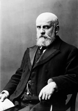
[[File:David Berry Hart.jpg|thumb|300px|alt=David Berry Hart|link=Embryology History - David Berry Hart|David Berry Hart (1851-1920)]] | |||
By D. Berry Hart, M.D.,F.R.C.P.E. & F.R.S.E., | |||
Lecturer on Midwifery, Edinburgh, ; C’u,rrr2/egiciRescarrcIz, Fellow. | |||
Both the clitoris and penis are protected by a sheath or prepuce of skin, tl1at of the penis being more complete than in the case of the clitoris, where it is deficient on its inferior aspect. The question arises as to how this prepuce is formed, and I purpose considering its method of formation on the basis of microscopical sections of the human preputium penis at the second and a half month and fourth and a half month; and of the preputium clitoridis at the fourth and seventh month respectively. | |||
As a rule, the preputium penis is well developed at full time, completely covering the glans and usually projecting beyond its tip, while it can in most infants be drawn back so as to uncover it. The same holds good for the clitoris, but opportunities for examining its conditions are not so frequent as in the male child. Occasionally, and more especially in the male child, the prepuce cannot be so drawn back, and is then said to be adherent. | |||
The usual description of the development of the prepuce is that a fold of skin grows forward and covers the glans, and this growth is generally considered to be of the nature of a free fold, so that the inner surfaces of the prepuce and that of the glans are not normally blended. | |||
If, however, We examine sections of the developing glans and prepuce, we find that the development of the prepuce is more complex than thus stated. In a coronal section of the prepuce and clitoris at the fourth month the glans is seen to be bounded on its outer surface by a horeshoe arrangement which is made up of the future preputial space filled with epidermal cells. The outer surface of the glans is covered with deeply stained columnar cells of a narrow type, while the inner surface of the prepuce is also lined with the more highly stained cells which lie at the deep boundary of the epidermis. The important point is that the preputial space does not as yet exist, but is represented by epidermal cells joining the glans and preputial cell boundaries already mentioned. | |||
If now we examine sagittal sections in a two and a half months male foetus we see an earlier stage of preputial development. Covering the exposed glans is a thick epidermal layer continuous with that of the body of the penis, while on the dorsum the epidermis has begun to dip down at the glans base, as if a dorsal fold were growing downwards and forwards (figs. 1 and 2). On the inferior aspect a very slight indication of a similar process is shown ; and as the same is going on laterally, it is evidently a dipping downwards and forwards, especially of the epidermis of the body of the penis all round the glans. I have not an intermediate stage between this and the fuller development a few months later, but this may be inferred with some safety from the early state. | |||
fiG, 1. Sagittal lateral section of penis in two and a. half months foetus, showing folding beginning to form prepuce above. | |||
If we look at the sagittal mesial section we see a similar arrangement (fig. 3); but as the urethral aperture is shown, the epidermis is seen passing into it and continuous with the covering of the free surface of the organ. The special point then is that the prepuce is not at this stage unattached to the glans, but forms continuous tissue with it. | |||
In the clitoris and prepuce of the seventh months foetus an arrangement similar to that of the fourth month is present, but the central cells in the epidermal ones joining the glans and inner preputial surface are distinctly flattened, ‘Le. approach to the condition of the superficial shedding cells of the ordinary surface epidermis (fig. 4). | |||
The prepuce does not, therefore, grow forward as a free unattached sheath, but develops somewhat as follows. | |||
as | |||
We begin with the two and a half months male, the earliest specimen examined. Here the glans has no prepuce, but is covered with a thick epidermal layer. At the glans root the epidermis dips down solidly and then grows forwards over the glans, the fold becoming inverted, so ‘that what was superficial is now deep, facing the glans swrface. The glans and the developing fold are blended and continuous as a tissue, and the future preputial space is thus filled with epidermal cells, the central ones corresponding to the superficialepidermal layers. This gives a fuller and more accurate account than one I had previously published, where I drew attention to the continuity of glans and preputial structure, but spoke less accurately of the epidermis passing in and back from the apex and mapping out the glans, without attaching suflicient importance to the important forward growth of solid folds from the glans base. We thus get a stage later in foetal development, where 1. The prepuce has formed. | |||
the | |||
fiG. 2. Fold beginning to form prepuce. | |||
2. The folds have not coalesced at the apex, a projecting epidermic plug preventing this. | |||
3. There is no separability of prepuce from glans, but epidermal cells, unbroken, join them. | |||
The question now arises as to how the prepuce becomes separable and free from the glans, as it does in the later months of foetal life,.apparently at or near full-time. | |||
The | The explanation is probably as follows. The surface epidermis is, when fully developed in the foetus, made up of multiple cells, with an active deep single layer of more columnar and deeply-staining cells. As one examines the cells nearer the surface We see them gradually assuming the transparent ordinary epidermal type, the nucleus becoming smaller and the cell substance less, until we get the flattened epidermal squames of the free surface. The epidermis has a power of desquamating at its free surface, and We get in this the explanation of the ultimate separation of prepuce and glans, so that the former becomes movable and can be drawn back, or the latter can project during erection. Thus in the seventh month foetus the central cells in the horseshoe arrangement are derived from the superficial epidermis and can be seen to have the flattened condition of superficial epidermal squames, i.e. are in the condition of desquamating cells. | ||
fiG. 3. Sagittal mesial section of penis in two and a. half months foetus, showing formation of prepuce and tunnelling of glans to form its urethra. | |||
It is this that frees the prepuce from the glans and gives its normal separable condition “ a desquamation of the central cells in the epidermal layer joining the inner surface of the prepuce and the outer surface of the glans.” | |||
of | We thus get the explanation of the power of the epidermis in forming lumina or bringing about mobility in apposed surfaces formed by its solid indipping, as it is evident that when epidermis dips in or grows in to the mesoblast so as to form a solid infolding, the superficial epidermal cells thus become central, and their ultimate desquamation will make lumina or movable folds, as the case may be. The deep epidermal layer is evidently the active one in the indipping, as it stains deeply and has a larger nucleus than the more superficial cells. It follows from this that the so-called adherent prepuce found in young children is really due to deficient breaking down of the preputial epidermic plug, and not to pathological adhesions, is due to a delay in a developmental process. | ||
fiG. 4. Preputial space filled with epidermis cells in seven months fcetus. Note central ones more squamous. 2%9. | |||
The Fomnation of the Urethra in the Male. | |||
The | The question I wish to discuss here is how the open part of the urethra in the early embryo (11-13 mm. of Nagel) has the urethral canal formed and the glans perforated to form the continuous tube of the male. In the female this part remains open and the glans imperforate, but the cause of this, as well as that of the opposite change in the male, is undetermined. In the 11-13 mm. embryo the condition in the urethral area below that part of the urethra derived from the urinogenital sinus is shown in Nagel’s diagram. | ||
In a male embryo of an age between the second and third month I was fortunate enough, in a sufficient number of sagittal mesial sections, to be able to study the process of “closure,” and now consider— | |||
1. The perforation of the glans penis. 2. The closure of the inferior portion of the urethra below that derived from the nrinogenital sinus. | |||
fiG. 5. | fiG. 5. Shows closure of inferior aspect of urethra in two and a. half months foetus. | ||
two and a. half months foetus. | |||
1. The Perforation of the Glans penis. | 1. The Perforation of the Glans penis. | ||
already described in the case of the prepuce. One can _see the deep | The glans clitoridis is imperforate, but in the male the glans becomes tunnelled by the epidermis at the apex passing in and back, and thus forming a solid plug, which by central desquamation forms a lumen, as already described in the case of the prepuce. One can _see the deep epidermal cell layer on the glans surface continuous with that in the urethra and the canal filled with epidermal cells, often in the form of cell nests. This tunnelling passes beyond the glans through the tissue corresponding to the future vestibule in the female, but an aperture is left immediately behind the fraenum, and also somewhat further back. The way in which these appear to be closed is interesting. | ||
epidermal cell layer on the glans surface continuous with that in the | |||
urethra and the canal filled with epidermal cells, often in the form | |||
of cell nests. This tunnelling passes beyond the glans through the | |||
tissue corresponding to the future vestibule in the female, but an | |||
aperture is left immediately behind the fraenum, and also somewhat | |||
further back. The way in which these appear to be closed is interesting. | |||
. the opening brought together as it were by means of the deep active | The superficial epidermis between these two apertures grows in two directions—superficially, and also with its deep layers streaming in at the apertures, 'i.e. passing in deeply. Fig. 5 shows well the deep margins of the opening brought together as it were by means of the deep active epidermal cell layer. Thus the epidermal layer does three things in closing the urethra below :— | ||
1. It proliferates in at the apertures solidly, carrying the superficial cells in its centre. | |||
2. The deep contiguous active cells unite to close any aperture; and | |||
3. The superficial epidermis proliferates over the closed apertures so as to make a surface. | |||
Finally, the central cells in the blocked urethra break down and form the lumen. | |||
Hypospadias, in its varying degrees, results from the failure of this closure. | |||
{{Footer}} | |||
[[Category:Historic Embryology]][[Category:1900's]][[Category:Genital]][[Category:Integumentary]] | |||
[[Category:Draft]] | |||
Latest revision as of 15:51, 29 January 2019
| Embryology - 19 Apr 2024 |
|---|
| Google Translate - select your language from the list shown below (this will open a new external page) |
|
العربية | català | 中文 | 中國傳統的 | français | Deutsche | עִברִית | हिंदी | bahasa Indonesia | italiano | 日本語 | 한국어 | မြန်မာ | Pilipino | Polskie | português | ਪੰਜਾਬੀ ਦੇ | Română | русский | Español | Swahili | Svensk | ไทย | Türkçe | اردو | ייִדיש | Tiếng Việt These external translations are automated and may not be accurate. (More? About Translations) |
Hart DB. On the role of the developing epidermis in forming sheaths and lumina to organs, illustrated specially in the development of the prepuce and urethra. (1907) J Anat Physiol. 42(1): 50–56.
| Historic Disclaimer - information about historic embryology pages |
|---|
 |
On the Role of the Developing Epidermis in Forming Sheaths and Lumina to Organs, Illustrated Specially in the Development of the Prepuce and Urethra
By D. Berry Hart, M.D.,F.R.C.P.E. & F.R.S.E.,
Lecturer on Midwifery, Edinburgh, ; C’u,rrr2/egiciRescarrcIz, Fellow.
Both the clitoris and penis are protected by a sheath or prepuce of skin, tl1at of the penis being more complete than in the case of the clitoris, where it is deficient on its inferior aspect. The question arises as to how this prepuce is formed, and I purpose considering its method of formation on the basis of microscopical sections of the human preputium penis at the second and a half month and fourth and a half month; and of the preputium clitoridis at the fourth and seventh month respectively.
As a rule, the preputium penis is well developed at full time, completely covering the glans and usually projecting beyond its tip, while it can in most infants be drawn back so as to uncover it. The same holds good for the clitoris, but opportunities for examining its conditions are not so frequent as in the male child. Occasionally, and more especially in the male child, the prepuce cannot be so drawn back, and is then said to be adherent.
The usual description of the development of the prepuce is that a fold of skin grows forward and covers the glans, and this growth is generally considered to be of the nature of a free fold, so that the inner surfaces of the prepuce and that of the glans are not normally blended.
If, however, We examine sections of the developing glans and prepuce, we find that the development of the prepuce is more complex than thus stated. In a coronal section of the prepuce and clitoris at the fourth month the glans is seen to be bounded on its outer surface by a horeshoe arrangement which is made up of the future preputial space filled with epidermal cells. The outer surface of the glans is covered with deeply stained columnar cells of a narrow type, while the inner surface of the prepuce is also lined with the more highly stained cells which lie at the deep boundary of the epidermis. The important point is that the preputial space does not as yet exist, but is represented by epidermal cells joining the glans and preputial cell boundaries already mentioned.
If now we examine sagittal sections in a two and a half months male foetus we see an earlier stage of preputial development. Covering the exposed glans is a thick epidermal layer continuous with that of the body of the penis, while on the dorsum the epidermis has begun to dip down at the glans base, as if a dorsal fold were growing downwards and forwards (figs. 1 and 2). On the inferior aspect a very slight indication of a similar process is shown ; and as the same is going on laterally, it is evidently a dipping downwards and forwards, especially of the epidermis of the body of the penis all round the glans. I have not an intermediate stage between this and the fuller development a few months later, but this may be inferred with some safety from the early state.
fiG, 1. Sagittal lateral section of penis in two and a. half months foetus, showing folding beginning to form prepuce above.
If we look at the sagittal mesial section we see a similar arrangement (fig. 3); but as the urethral aperture is shown, the epidermis is seen passing into it and continuous with the covering of the free surface of the organ. The special point then is that the prepuce is not at this stage unattached to the glans, but forms continuous tissue with it.
In the clitoris and prepuce of the seventh months foetus an arrangement similar to that of the fourth month is present, but the central cells in the epidermal ones joining the glans and inner preputial surface are distinctly flattened, ‘Le. approach to the condition of the superficial shedding cells of the ordinary surface epidermis (fig. 4).
The prepuce does not, therefore, grow forward as a free unattached sheath, but develops somewhat as follows.
We begin with the two and a half months male, the earliest specimen examined. Here the glans has no prepuce, but is covered with a thick epidermal layer. At the glans root the epidermis dips down solidly and then grows forwards over the glans, the fold becoming inverted, so ‘that what was superficial is now deep, facing the glans swrface. The glans and the developing fold are blended and continuous as a tissue, and the future preputial space is thus filled with epidermal cells, the central ones corresponding to the superficialepidermal layers. This gives a fuller and more accurate account than one I had previously published, where I drew attention to the continuity of glans and preputial structure, but spoke less accurately of the epidermis passing in and back from the apex and mapping out the glans, without attaching suflicient importance to the important forward growth of solid folds from the glans base. We thus get a stage later in foetal development, where 1. The prepuce has formed.
fiG. 2. Fold beginning to form prepuce.
2. The folds have not coalesced at the apex, a projecting epidermic plug preventing this.
3. There is no separability of prepuce from glans, but epidermal cells, unbroken, join them.
The question now arises as to how the prepuce becomes separable and free from the glans, as it does in the later months of foetal life,.apparently at or near full-time.
The explanation is probably as follows. The surface epidermis is, when fully developed in the foetus, made up of multiple cells, with an active deep single layer of more columnar and deeply-staining cells. As one examines the cells nearer the surface We see them gradually assuming the transparent ordinary epidermal type, the nucleus becoming smaller and the cell substance less, until we get the flattened epidermal squames of the free surface. The epidermis has a power of desquamating at its free surface, and We get in this the explanation of the ultimate separation of prepuce and glans, so that the former becomes movable and can be drawn back, or the latter can project during erection. Thus in the seventh month foetus the central cells in the horseshoe arrangement are derived from the superficial epidermis and can be seen to have the flattened condition of superficial epidermal squames, i.e. are in the condition of desquamating cells.
fiG. 3. Sagittal mesial section of penis in two and a. half months foetus, showing formation of prepuce and tunnelling of glans to form its urethra.
It is this that frees the prepuce from the glans and gives its normal separable condition “ a desquamation of the central cells in the epidermal layer joining the inner surface of the prepuce and the outer surface of the glans.”
We thus get the explanation of the power of the epidermis in forming lumina or bringing about mobility in apposed surfaces formed by its solid indipping, as it is evident that when epidermis dips in or grows in to the mesoblast so as to form a solid infolding, the superficial epidermal cells thus become central, and their ultimate desquamation will make lumina or movable folds, as the case may be. The deep epidermal layer is evidently the active one in the indipping, as it stains deeply and has a larger nucleus than the more superficial cells. It follows from this that the so-called adherent prepuce found in young children is really due to deficient breaking down of the preputial epidermic plug, and not to pathological adhesions, is due to a delay in a developmental process.
fiG. 4. Preputial space filled with epidermis cells in seven months fcetus. Note central ones more squamous. 2%9.
The Fomnation of the Urethra in the Male.
The question I wish to discuss here is how the open part of the urethra in the early embryo (11-13 mm. of Nagel) has the urethral canal formed and the glans perforated to form the continuous tube of the male. In the female this part remains open and the glans imperforate, but the cause of this, as well as that of the opposite change in the male, is undetermined. In the 11-13 mm. embryo the condition in the urethral area below that part of the urethra derived from the urinogenital sinus is shown in Nagel’s diagram.
In a male embryo of an age between the second and third month I was fortunate enough, in a sufficient number of sagittal mesial sections, to be able to study the process of “closure,” and now consider—
1. The perforation of the glans penis. 2. The closure of the inferior portion of the urethra below that derived from the nrinogenital sinus.
fiG. 5. Shows closure of inferior aspect of urethra in two and a. half months foetus.
1. The Perforation of the Glans penis.
The glans clitoridis is imperforate, but in the male the glans becomes tunnelled by the epidermis at the apex passing in and back, and thus forming a solid plug, which by central desquamation forms a lumen, as already described in the case of the prepuce. One can _see the deep epidermal cell layer on the glans surface continuous with that in the urethra and the canal filled with epidermal cells, often in the form of cell nests. This tunnelling passes beyond the glans through the tissue corresponding to the future vestibule in the female, but an aperture is left immediately behind the fraenum, and also somewhat further back. The way in which these appear to be closed is interesting.
The superficial epidermis between these two apertures grows in two directions—superficially, and also with its deep layers streaming in at the apertures, 'i.e. passing in deeply. Fig. 5 shows well the deep margins of the opening brought together as it were by means of the deep active epidermal cell layer. Thus the epidermal layer does three things in closing the urethra below :—
1. It proliferates in at the apertures solidly, carrying the superficial cells in its centre.
2. The deep contiguous active cells unite to close any aperture; and
3. The superficial epidermis proliferates over the closed apertures so as to make a surface.
Finally, the central cells in the blocked urethra break down and form the lumen.
Hypospadias, in its varying degrees, results from the failure of this closure.
Cite this page: Hill, M.A. (2024, April 19) Embryology Paper - On the role of the developing epidermis in forming sheaths and lumina to organs. Retrieved from https://embryology.med.unsw.edu.au/embryology/index.php/Paper_-_On_the_role_of_the_developing_epidermis_in_forming_sheaths_and_lumina_to_organs
- © Dr Mark Hill 2024, UNSW Embryology ISBN: 978 0 7334 2609 4 - UNSW CRICOS Provider Code No. 00098G