Paper - Methods used by C. H. Heuser in preparing and sectioning early embryos: Difference between revisions
mNo edit summary |
mNo edit summary |
||
| Line 1: | Line 1: | ||
{{Header}} | {{Header}} | ||
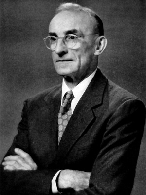
[[File:Chester Heuser.jpg|thumb|alt=Chester H. Heuser|link=Embryology History - Chester Heuser|Chester H. Heuser]] | |||
{{Ref-Heard1957}} | {{Ref-Heard1957}} | ||
| Line 6: | Line 7: | ||
=Methods Used by C. H. Heuser in Preparing and Sectioning Early Embryos= | =Methods Used by C. H. Heuser in Preparing and Sectioning Early Embryos= | ||
[[File: | [[File:Osborne Heard.jpg|thumb|Osborne Heard]] | ||
by [[Embryology History - Osborne Heard|Osborne O. Heard]] | |||
With 3 plates and 12 figures (1957) | With 3 plates and 12 figures (1957) | ||
Revision as of 17:04, 19 August 2016
| Embryology - 20 Apr 2024 |
|---|
| Google Translate - select your language from the list shown below (this will open a new external page) |
|
العربية | català | 中文 | 中國傳統的 | français | Deutsche | עִברִית | हिंदी | bahasa Indonesia | italiano | 日本語 | 한국어 | မြန်မာ | Pilipino | Polskie | português | ਪੰਜਾਬੀ ਦੇ | Română | русский | Español | Swahili | Svensk | ไทย | Türkçe | اردو | ייִדיש | Tiếng Việt These external translations are automated and may not be accurate. (More? About Translations) |
Heard OO. Methods used by C. H. Heuser in preparing and sectioning early embryos. (1957.) Carnegie Instn. Wash. Publ. 611, Contrib. Embryol., 36, 1-18.
| Historic Disclaimer - information about historic embryology pages |
|---|
 |
Methods Used by C. H. Heuser in Preparing and Sectioning Early Embryos

With 3 plates and 12 figures (1957)
Introduction
Readers of the Contributions to Embryology have long appreciated the remarkable technical ability of Chester H. Heuser 1 as shown not only in the preparations of early and monkey embryos, descriptions of which were published in collaboration with the late G. L. Streeter (:29, 1941), but also in those of human embryos, for example the perfect presomite specimen (Carnegie no. 30) described by Heuser (1932). When, owing to the enterprise and skill of A. T. Hertig and Iohn Rock, the Department of Embryology was entrusted with a unique studies of well preserved human embryos of the first three weeks, it was indeed fortunate that Dr. Heuser was at hand to prepare and section them and, with his unrivaled practical knowledge of very early mammalian embryology, to give advice and counsel on their study. Visiting colleagues who have seen Dr. Heuser working the microtome and have observed the many novel details of his technique have often urged that they be described in print. Dr. Heuser’s modesty and his desire for perfection, however, have deterred him from rendering this service to his colleagues. finally, with his consent, the present account was prepared in consultation with other members of the Department. The author, who was associated with Dr. Heuser for many years, has often assisted in the embedding and sectioning of the embryos. In collaboration with Dr. Heuser, and frequently also on his own initiative, he designed and constructed much of the special apparatus used in this work. Dr. Heuser has reviewed the draft of this paper and has given his approval.
While this article was being prepared for press Dr. Heuser reached his seventieth birthday. The Staff of the Department of Embryology of the Carnegie Institution of Washington takes pleasure in presenting it in the Contributions to Embryology, which Dr. Heuser's skill has so greatly enriched, as a tribute on this occasion to the lifelong scientific devotion of a beloved colleague.
Collection and Preservation of Embryonic Material
Sources and Types
Human embryonic material received at the Carnegie boratory is of three kinds. The first group comprises iecimens received from physicians outside of Baltimore, irgely in response to periodic appeals circulated by the iboratory. They consist almost entirely of aborted or irgically removed chorions and of Fallopian tubes exised because of tubal pregnancy. The instructions to -hysicians advise that such specimens be fixed in to ver cent aqueous formol. Although not the ideal fixative, ormol is easily obtained and diluted for use. Specimens .re safely kept and shipped in the same fluid without urther attention. Furthermore, formol makes the tissues ess opaque than do fixatives containing picric acid or nercurial salts, and thus leaves them in more favorable zondition for examination and exploration.
The second group comprises specimens obtained by the gynecologists and obstetricians of Baltimore hospitals with whom the laboratory is in close touch. These collaborators are requested to telephone when they have a fresh specimen so that a messenger may be sent to bring the material for immediate attention at the laboratory. Such specimens are fixed, usually in Bouin’s picroacetic formol, by the experienced personnel of the laboratory.
The third group comprises specimens obtained by Member of the Department of Embryology, Carnegie Institution of VVashing1on,. 1921-1950; Research Associate, 1950-: Professor of Microscopic Anatomy, Medical College of Georgia, 1950-. special collaborators thoroughly familiar with the procedures for fixation and subsequent dehydration, who carry out these steps on the fresh material in their own laboratories. In this category are the embryos obtained by Dr. Arthur T. Hertig and Dr. John Rock at the Free Hospital for Women, Brookline, Massachusetts, and sectioned by Dr. Heuser, which have so greatly contributed to our knowledge of human development during the first two weeks.
It will be convenient to discuss first the handling of material already fixed, and the larger specimens which arrive at the laboratory in the fresh condition. These materials include naked embryos and fetuses; chorions of various sizes and in various conditions—intact or tom open; clots and decidual fragments which may contain embryonic tissues; extirpated uteri containing established pregnancies; and Fallopian tubes extirpated because of ectopic pregnancy.
The preliminary exploration of these diverse materials differs so much from specimen to specimen that only general suggestions can be given. The first step is, of course, to free the gestation sac of obscuring tissues, by cautiously opening the uterus, or by dissecting away clots and debris as the case may require. Chorions less than 5 mm. in diameter must usually be studied and handled under the microscope, as will be described below. Larger gestation sacs may be handled by the usual methods employed with biological specimens of ordinary dimensions. Chorionic sacs found in aborted clots or in intra abdominal clots may simply be washed off with physiological salt solution, freed of adherent organized material as far as is safely possible, studied in a preliminary way, and, if small, fixed by immersion. Chorions 10 mm. or more in diameter should be opened in order to observe the condition of the embryo, and to decide whether to prepare it for sectioning. This statement applies whether the chorionic sac is obtained in an extirpated uterus or as an aborted ovum.
Tubal Pregnancies
An oviduct (Fallopian tube) containing a gestation sac should always be explored with care, for a good embryo may be present; indeed, it is the tubal pregnancies that offer the best chance of obtaining human embryos in the early somite stages.
A fresh pregnant tube should be dissected at once under physiological salt solution, since fixation adds greatly to the difficulty of exploration by forming adherent, stratified clots about the chorion. If, however, the specimen has been placed in fixing solution by the surgeon or pathologist, the embryologist must do the best he can. With either fresh or fixed material, working under the binocular microscope with finely painted forceps, needles, and scissors, he cautiously removes a portion of the wall of the oviduct so as to make a window into its lumen, and gently pulls away the clots or, when possible, dislodges them with a stream of salt solution. If chorionic villi are seen, he proceeds with great care to uncover the chorion or to locate the embryo if it has been extruded from the chorionic cavity. With a fixed specimen this operation will require patient, piecemeal removal of the clots, small bits being broken away one at a time with the needle.
The subsequent handling of the embryos will be de scribed below, after the recovery of ova and blastocysts has been discussed.
Recovery of Segmenting Oa and Unimplanted Blastocysts
The special methods used by Dr. Heuser are primarily applicable to the material of the second and third classes mentioned above, that is, specimens obtained from local sources in fresh condition and specimens representing very early pregnancies adequately fixed before shipment to the laboratory.
The procedures to be described are naturally not limited to use with human material, but are for the most part applicable to mammalian embryos in general. In fact, they were largely worked out, in so far as they are original with Dr. Heuser and his co—workers, on other species, as will be realized by those who are familiar with the published reports of his studies, partly in collaboration with the late G. L. Streeter, on the early development of the domestic pig and the rhesus monkey. The only technique specifically designed for human material is that for opening the uterus, and even this was developed by experience with the similar though much smaller uterus of the rhesus monkey.
Tubal Ova
The human ovum, like that of most other mammals, is in the oviduct (Fallopian tube) for about three days after ovulation, and may be recovered by washing out the lumen of the tube. The excised Fallopian tube should be brought from the operating room in physiological salt solution. The investigator holds it directly over a small dish (embryological watch glass, fig. 1) in which a small amount of
fig. 1. Method used for recovery of tubal ova.
the salt solution has been placed. The lumen is then irrigated with salt solution by means of a pipette with a large rubber bulb, inserted into the fimbriated end of the tube. Usually the uterine end is sufficiently contracted to cause distention of the ampulla, so that the spaces between the folds are irrigated; if the end of the tube is not contracted, it may be closed by compressing it between thumb and finger until the ampulla is slightly distended. The egg usually emerges with the first fluid that passes into the dish. The free end of the tube may whip about as the fluid is ejected; hence great care must be taken to keep it over the dish at all times in order not to lose even a drop.
If the egg is not recovered at the first washing, the process is repeated. The tube may be lightly “milked" downward by the fingers, and finally a fine-pointed
Cite this page: Hill, M.A. (2024, April 20) Embryology Paper - Methods used by C. H. Heuser in preparing and sectioning early embryos. Retrieved from https://embryology.med.unsw.edu.au/embryology/index.php/Paper_-_Methods_used_by_C._H._Heuser_in_preparing_and_sectioning_early_embryos
- © Dr Mark Hill 2024, UNSW Embryology ISBN: 978 0 7334 2609 4 - UNSW CRICOS Provider Code No. 00098G