Paper - Implantation of the human ovum in the uterus: Difference between revisions
mNo edit summary |
mNo edit summary |
||
| (13 intermediate revisions by the same user not shown) | |||
| Line 4: | Line 4: | ||
! Online Editor | ! Online Editor | ||
|- | |- | ||
| [[File:Mark_Hill.jpg|90px|left]] This historic 1904 paper by [[Embryology_History_-_Charles_Minot|Minot]] described the understanding of human implantation at that time. | | [[File:Mark_Hill.jpg|90px|left]] This historic 1904 paper by [[Embryology_History_-_Charles_Minot|Minot]] described the understanding of human implantation at that time. This was an invited lecture presented at the 1904 meeting of the American Gynecological Society. See also by [[Embryology_History_-_Charles_Minot|Charles Minot]]. | ||
<br> | |||
Note that all early human developmental stages were still described as the "ovum", today this would be described as the blastocyst implantation occurring in [[week 2]]. | |||
'''Modern Notes:''' {{Implantation}} | [[Week 2]] | [[Blastocyst]] | [[Embryology_History_-_Charles_Minot|Charles Minot]] | |||
<br> | <br> | ||
| Line 17: | Line 15: | ||
|} | |} | ||
{{Historic Disclaimer}} | {{Historic Disclaimer}} | ||
=Implantation of the Human Ovum in the Uterus= | |||
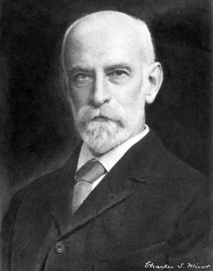
[[File:Charles minot.jpg|thumb|300px|link=Embryology History - Charles Minot|Charles Sedgwick Minot (1852–1914)]] | |||
By Charles Sedgwick Minot, LL.D., Sc.D., | |||
Professor In The Harvard Medical School, Boston. | |||
Read by invitation. | |||
The discovery of the fact that certain tissues can attack and destroy others is one of the most important discoveries of recent years. It is perhaps to be regarded as a process of digestion of one tissue by another. It is known that certain young tissues of the embryo are in part endowed with a capacity to destroy the tissues with which they come in contact. This phenomenon has an intimate relation to many diverse developmental processes. It appears that a tissue may produce a destructive agent, presumably chemical in character, from which the tissue itself under normal conditions does not suffer, but which will destroy other tissues with which it comes in contact. Our present knowledge leads us to believe that such a phenomenon occurs during the early stages of the human ovum and affords us an approximately correct conception of the most important steps in the earliest history of the ovum, by which its original relations to the uterus are established. Embryologists designate this early attachment, the insertion of ova upon the wall of the uterus, by the term implantation. In animals which have claws and are therefore called unguiculate, such as carnivora and primates, including man, the chorion is supplied with bloodvessels and enters into intimate relations with the wall of the uterus. It has been my privilege to investigate the changes involved in the establishment of such relations in various animals, and I find, as other observers also have found, that the ectoderm of the chorion undergoes a peculiar proliferation by which its cells become very much more numerous. Some of the cells rapidly assume a distinctive character, and are easily recognized by their large size. It further appears that wherever these modified ectodermal cells come in contact with the walls of the uterus, destructive changes go on in the uterine tissue. The facts indicated suggest that these cells produce a substance which is capable of digesting or dissolving the substance of the uterus. As the ectodermal cells in question are important and highly characteristic of many placental mammals, I propose for them a special name, trophoderm.<ref>In the address the term "trophoblast" was used in accordance with my understanding of [[Embryology History - Ambrosius Hubrecht|Professor Hubrecht's]] views and consent ; but as [[Embryology History - Ambrosius Hubrecht|Professor Hubrecht]] has objected to this application of his term, it has been necessary to propose a new one. I regret that so good a name as trophoblast has to be dropped.</ref> The function of the trophoderra is to corrode away a portion of the mucous membrane of the uterus. In man this corrosion takes place so as to make a cavity in which the ovum lodges itself. The trophoderm thereupon imdergoes a hypertrophic degeneration, which occurs so as to produce a series of irregular spaces, which persist and become the intervillous spaces of the placenta. Papillary outgrowths of the chorionic mesoderm meanwhile penetrate the trophoderm, initiating the formation of the chorionic villi. The trophodermic cells over each mesodermic outgrowth persist in two layers: the inner, cellular, and the outer, syncytial. These two layers represent the first stage of the ectoderm of the villi. | |||
In many animals the trophoderm does not cover the whole of the ovum as in man, but is spread only over a small portion of the chorion. This portion is sometimes designated as the placental area. A restricted placental area of this sort can be readily observed in the early stages of the rabbit, dog, or cat. On the other hand, there are a few animals, such as the hedgehog and guinea-pig, in which the trophoderm is very precociously developed, so as to be present at a time when the ovum has advanced but little beyond the first stages of segmentation. The trophodermic cells in these cases increase in number, form considerable layers, and where they come in contact with the walls of the uterus, attack and destroy part of it, so that a hole is made in its wall the exact size of the young ovum. The process in such cases is similar to that which has been observed in man, but with the difference that it occurs while the ovum is much less advanced in its development. | |||
Turn now to observations which concern the primates and man. Owing to the investigations of Selenka and of Kollmann, we know that a true trophoderm exists in the early stages in monkeys and apes. It has been actually observed in various old-world monkeys, in the gibbon, and chimpanzee. Ova of these animals have been also observed in which the trophoderm seems to have corroded away the uterine wall and made a place for the newly developing organism. The evidence in regard to implantation in man may be briefly indicated as follows: First, we have a number of ova in very early stages in which the trophoderm can be distinctly seen over the outside of the ova; it is most developed in the very earliest stages heretofore recorded, and it rapidly degenerates, but in these very earliest stages the implantation in the uterus has already taken place. The ovum is lodged in a cavity of the mucosa. Second, we have the numerous observations which show that the trophoderm has the power of destroying uterine tissue in various animals. The conclusion from this double evidence is obvious; that the trophoderm in man has for its special function the accomplisliment of the primitive insertion of the developing germ into the wall of the uterus. This conclusion is further corroborated by the evidence from apes and monkeys. In short, it seems scarcely too much too say positively, in the light of our present knowledge, that the function of the trophoderm as the means of implanting the human ovum is demonstrated. | |||
and | |||
the | |||
of the | |||
the insertion of | |||
[[File:Minot1904 fig01.jpg|600px]] | |||
'''Explanation of diagram, based on Peters' ovum.''' Ep, uterine epithelium; Ut, mucous membrane (decidua) of uterus; Tro, trophoderm ; Bl, spaces formed by the degeneration of the trophoderm ; maternal blood enters these spaces from the decidual bloodvessels ; Emb, embryonic shield; Am. cav, amniotic cavity; Yk.s, yolk sack, the entodermal lining of which is indicated by a heavy black line ; Mes, chorionic mesoderm ; Coe, extraembryonic celom. | |||
{| class="wikitable mw-collapsible mw-collapsed" | |||
! Online Editor - Peters Ovum | |||
|- | |||
| [[File:Mark_Hill.jpg|90px|left]] Thought to be classified as [[Carnegie stage 6]] in [[Week 2]] of development. | |||
<br> | |||
{{Ref-O'RahillyMüller1987}} | |||
:"Peters Ovum described in a monograph by Peters (1899). Autopsy. A famous embryo, for long the youngest known and the first to be described in detail. Photomicrographs have since been published (Rossenbeck, 1923, plate 42; Odgers, 1937, plate 2, fig. 2). The chorionic villi, some of which display a mesenchymal core, send cellular columns externally and these latter are beginning to form a cytotrophoblastic shell. Slight branching of villi (Krafka, 1941). Chorionic cavity contains magma réticulé of Velpeau (Mall, 1916). Blood islands on umbilical vesicle. Chorion, 1.5 x 2 mm. Chorionic cavity, 1.6 x 0.9 x 0.8 mm; capacity, 0.7 mm3 (Odgers, 1937). Embryonic disc, 0.18 x 0.24 mm (Krafka, 1941). The basement membrane (Hensen’s membrane prima) of the epiblast was noted by Graf Spee. Allantoic diverticulum and primitive streak uncertain. Presumed age, 13 days (Krafka,1941). A tabulation of normal human embryos compiled from the literature prior to 1900 and from Mall’s own collection was published by Mall (1900, pp. 38-46). The least advanced specimen was the Peters embryo, and included in the list were 92 embryos of 0.19-32 mm, as well as 17 fetuses of 33-210 mm." | |||
|} | |||
cellular | |||
of | |||
Peters' ovum still remains the earliest stage of man yet described. It has a small chorionic vesicle, on the exterior of which is a very considerable layer of trophodermic cells, which stretches completely around the vesicle, and which in thickness equals a quarter or a fifth of the diameter of the entire chorionic vesicle. This external layer is made up entirely of proliferated enlarged cells of the chorionic ectoderm. There is an embryonic shield present at this stage, but nothing which can properly be called an embryo. The total diameter of such a vesicle is 3 or 4 mm. Even in this the youngest stage, which has been actually observed, the trophoderm has undergone a certain amount of degeneration, and there are consequently already a series of cavities as represented in the accompanying diagram, which we identify as the beginnings of the intervillous spaces. The diagram also shows that there has been precociously developed a special layer of connective tissue lining the interior of the chorion and forming a series of projections or papillip. We are so fortunate as to have a series of specimens in the embryological collection at the Harvard Medical School sufBcient to enable us to gather a general notion of the origin and development of the chorionic villi. We find that the trophoderm undergoes its degeneration probably exclusively after it has performed its function of making a place for the ovum in the wall of the uterus. As it degenerates the trophoderm covering of the incipient villi becomes thinner, the mesoderm of the villi meanwhile continues to grow, and the original simple papilla? become branching, until finally we have the remnants of the trophoderm converted, as above stated, into the double layer of cells, the so-called chorionic ectoderm. For a considerable period, however, there are always accumulations of the original trophoblastic cells preserving something of their primitive character to be observed at the end of the villi. Thus, in an ovum of 8 mm. in diameter, we observed that the amount of the trophoblast has been greatly reduced as compared with Peters' ovum, and that the mesoderm had extended farther down, and hence the structure of the villus was more clearly defined. By the time the ovum is eleven or 12 mm. in diameter there is little trace of the trophoderm left, and it is about at this period of development that we begin to recognize the formation of embryonic structures, so that we may say that the entire history of the trophoderm is completed, and all its functions are accomplished before the embryonic structures are existent. Peters' observations indicate that the ovum becomes completely embedded in uterine mucous tissue and that the thickness of the mucous membrane at the time of implantation is a little greater than the total diameter of the ovum. The tissue of the uterus, which lies adjacent to the ovum with its trophoderm, has preserved its normal structure; it shows no change. The tissue in the space occupied by the ovum we might imagine to have been cut off as if one had taken a sharp instrument and made a spheric cavity of a few milimeters' diameter. We can explain this condition as indicated by assuming that the uterine tissue has been corroded out or dissolved away by the action of the trophoderm, and it is further to be presumed that the ovum has not only been thus providing a place for itself, but that also it has been actually digesting portions of the uterine tissue, growing, therefore, at the mother's expense. This involves the idea that the uterine tissue has been digested and absorbed by the trophoderm; hence the name which is given to this layer. The immediate consequence of the corrosion of the uterine tissue is the production of a wounded surface, and if there happens to be a bloodvessel which comes in the path of corrosion, it may have a portion of its wall corroded away, and then the blood, which would otherwise be restrained within the bloodvessel, can and does apparently escape into the spaces where the trophoderm has degenerated and disappeared. Thus, in this very early stage the maternal blood passes out from the maternal vessels into the trophodermic or later intervillous spaces. Therefore, there is an extravascular circulation established at the very start when the ovum first implants itself in the wall of the uterus. We must say, then, contrary to current descriptions, that the intervillous circulation is almost the first thing established in the uterus for the purpose of nourishing the embryo, and, indeed, we might almost say that the intervillous circulation begins before the villi have formed, for before there are villi we find maternal blood circulating in spaces bounded by fetal tissue. In brief, we may assert that the essential conditions for the development of the placenta are established during the implantation of the ovum and that the ultimate development of the placenta is brought about by a modification of this primitive disposition. | |||
It may be convenient to add here a few words as to further degenerative changes in the pregnant uterus. This is especially important as a means of illustrating the fate of the decidua reflexa. As is well known, this membrane is present during the first half of pregnancy and absent during the second. I am able to demonstrate by a series of specimens which I have in my laboratory that the disappearance of the decidua reflexa is accomphshed by degeneration, followed by absorption. As soon as the decidua reflexa has been thus removed, it becomes possible for the chorion of the embryo to come into immediate contact with the decidua- vera, and,, as is well known, this contact is characteristic of the second half of pregnancy. We do not yet know the exact date at which this condition is completely estabUshed. We know only that by the end of about the fifth month the decidua reflexua has usually entirely disappeared. So that even that portion of the chorion, which we can reach by passing up through the cervLx of the uterus, is in the later stages of pregnancy uncovered by any uterine membrane. If the decidua reflexa did not degenerate and disappear completely that bit of chorion would have a covering of decidual tissue, which, as you know, is not present at term. | |||
the | |||
into the | |||
as | |||
at | |||
the | |||
the | |||
Such are the main points, briefly outlined, relative to the implantation of the human ovum in the uterus. I have only to indicate, in conclusion, that the greater part of this investigation has been done by others, though I have, fortunately, some material in my own possession by which I have been enabled, to a considerable extent, to confirm these observations, and in some particulars to extend them. | |||
Before concluding, may I state that we are forming a collection of serial sections of human embryos in the Harvard | Before concluding, may I state that we are forming a collection of serial sections of human embryos in the Harvard Medical School. These sections are prepared by a skilled technician, who has been especially trained to the work. The collection is intended always to be open to competent investigators and students. May I not hope that we may have the co-operation of the members of this Society in securing specimens which would be available for the development of this collection? Those of the first and second months are especially desired. They may be easily preserved by placing them in a mixture of one part of formalin with nine parts of water, as soon as possible after they are obtained. | ||
Medical School. These sections are prepared by a skilled | |||
technician, who has been especially trained to the work. The | |||
collection is intended always to be open to competent investigators and students. May I not hope that we may have the | |||
co-operation of the members of this Society in securing specimens which would be available for the development of this | |||
in a mixture of one part of formalin with nine parts of water, | |||
as soon as possible after they are obtained. | |||
I beg to thank the Society for the courtesy shown me. | |||
<references/> | |||
{{Historic Disclaimer}} | {{Historic Disclaimer}} | ||
{{Footer}} | {{Footer}} | ||
][[Category:Charles Minot]] | |||
[Category:Historic Embryology]][[Category:Implantation]][[Category:1900's]] | [Category:Historic Embryology]][[Category:Implantation]][[Category:1900's]] | ||
Latest revision as of 10:06, 17 March 2020
| Embryology - 20 Apr 2024 |
|---|
| Google Translate - select your language from the list shown below (this will open a new external page) |
|
العربية | català | 中文 | 中國傳統的 | français | Deutsche | עִברִית | हिंदी | bahasa Indonesia | italiano | 日本語 | 한국어 | မြန်မာ | Pilipino | Polskie | português | ਪੰਜਾਬੀ ਦੇ | Română | русский | Español | Swahili | Svensk | ไทย | Türkçe | اردو | ייִדיש | Tiếng Việt These external translations are automated and may not be accurate. (More? About Translations) |
Minot CS. Implantation of the human ovum in the uterus. (1904) Trans. Am. Gynec. Soc., Philadelphia, 29: 395-402.
| Online Editor |
|---|

Note that all early human developmental stages were still described as the "ovum", today this would be described as the blastocyst implantation occurring in week 2. Modern Notes: implantation | Week 2 | Blastocyst | Charles Minot
|
| Historic Disclaimer - information about historic embryology pages |
|---|
|
Implantation of the Human Ovum in the Uterus
By Charles Sedgwick Minot, LL.D., Sc.D.,
Professor In The Harvard Medical School, Boston.
Read by invitation.
The discovery of the fact that certain tissues can attack and destroy others is one of the most important discoveries of recent years. It is perhaps to be regarded as a process of digestion of one tissue by another. It is known that certain young tissues of the embryo are in part endowed with a capacity to destroy the tissues with which they come in contact. This phenomenon has an intimate relation to many diverse developmental processes. It appears that a tissue may produce a destructive agent, presumably chemical in character, from which the tissue itself under normal conditions does not suffer, but which will destroy other tissues with which it comes in contact. Our present knowledge leads us to believe that such a phenomenon occurs during the early stages of the human ovum and affords us an approximately correct conception of the most important steps in the earliest history of the ovum, by which its original relations to the uterus are established. Embryologists designate this early attachment, the insertion of ova upon the wall of the uterus, by the term implantation. In animals which have claws and are therefore called unguiculate, such as carnivora and primates, including man, the chorion is supplied with bloodvessels and enters into intimate relations with the wall of the uterus. It has been my privilege to investigate the changes involved in the establishment of such relations in various animals, and I find, as other observers also have found, that the ectoderm of the chorion undergoes a peculiar proliferation by which its cells become very much more numerous. Some of the cells rapidly assume a distinctive character, and are easily recognized by their large size. It further appears that wherever these modified ectodermal cells come in contact with the walls of the uterus, destructive changes go on in the uterine tissue. The facts indicated suggest that these cells produce a substance which is capable of digesting or dissolving the substance of the uterus. As the ectodermal cells in question are important and highly characteristic of many placental mammals, I propose for them a special name, trophoderm.[1] The function of the trophoderra is to corrode away a portion of the mucous membrane of the uterus. In man this corrosion takes place so as to make a cavity in which the ovum lodges itself. The trophoderm thereupon imdergoes a hypertrophic degeneration, which occurs so as to produce a series of irregular spaces, which persist and become the intervillous spaces of the placenta. Papillary outgrowths of the chorionic mesoderm meanwhile penetrate the trophoderm, initiating the formation of the chorionic villi. The trophodermic cells over each mesodermic outgrowth persist in two layers: the inner, cellular, and the outer, syncytial. These two layers represent the first stage of the ectoderm of the villi.
In many animals the trophoderm does not cover the whole of the ovum as in man, but is spread only over a small portion of the chorion. This portion is sometimes designated as the placental area. A restricted placental area of this sort can be readily observed in the early stages of the rabbit, dog, or cat. On the other hand, there are a few animals, such as the hedgehog and guinea-pig, in which the trophoderm is very precociously developed, so as to be present at a time when the ovum has advanced but little beyond the first stages of segmentation. The trophodermic cells in these cases increase in number, form considerable layers, and where they come in contact with the walls of the uterus, attack and destroy part of it, so that a hole is made in its wall the exact size of the young ovum. The process in such cases is similar to that which has been observed in man, but with the difference that it occurs while the ovum is much less advanced in its development.
Turn now to observations which concern the primates and man. Owing to the investigations of Selenka and of Kollmann, we know that a true trophoderm exists in the early stages in monkeys and apes. It has been actually observed in various old-world monkeys, in the gibbon, and chimpanzee. Ova of these animals have been also observed in which the trophoderm seems to have corroded away the uterine wall and made a place for the newly developing organism. The evidence in regard to implantation in man may be briefly indicated as follows: First, we have a number of ova in very early stages in which the trophoderm can be distinctly seen over the outside of the ova; it is most developed in the very earliest stages heretofore recorded, and it rapidly degenerates, but in these very earliest stages the implantation in the uterus has already taken place. The ovum is lodged in a cavity of the mucosa. Second, we have the numerous observations which show that the trophoderm has the power of destroying uterine tissue in various animals. The conclusion from this double evidence is obvious; that the trophoderm in man has for its special function the accomplisliment of the primitive insertion of the developing germ into the wall of the uterus. This conclusion is further corroborated by the evidence from apes and monkeys. In short, it seems scarcely too much too say positively, in the light of our present knowledge, that the function of the trophoderm as the means of implanting the human ovum is demonstrated.

Explanation of diagram, based on Peters' ovum. Ep, uterine epithelium; Ut, mucous membrane (decidua) of uterus; Tro, trophoderm ; Bl, spaces formed by the degeneration of the trophoderm ; maternal blood enters these spaces from the decidual bloodvessels ; Emb, embryonic shield; Am. cav, amniotic cavity; Yk.s, yolk sack, the entodermal lining of which is indicated by a heavy black line ; Mes, chorionic mesoderm ; Coe, extraembryonic celom.
| Online Editor - Peters Ovum |
|---|
|
Peters' ovum still remains the earliest stage of man yet described. It has a small chorionic vesicle, on the exterior of which is a very considerable layer of trophodermic cells, which stretches completely around the vesicle, and which in thickness equals a quarter or a fifth of the diameter of the entire chorionic vesicle. This external layer is made up entirely of proliferated enlarged cells of the chorionic ectoderm. There is an embryonic shield present at this stage, but nothing which can properly be called an embryo. The total diameter of such a vesicle is 3 or 4 mm. Even in this the youngest stage, which has been actually observed, the trophoderm has undergone a certain amount of degeneration, and there are consequently already a series of cavities as represented in the accompanying diagram, which we identify as the beginnings of the intervillous spaces. The diagram also shows that there has been precociously developed a special layer of connective tissue lining the interior of the chorion and forming a series of projections or papillip. We are so fortunate as to have a series of specimens in the embryological collection at the Harvard Medical School sufBcient to enable us to gather a general notion of the origin and development of the chorionic villi. We find that the trophoderm undergoes its degeneration probably exclusively after it has performed its function of making a place for the ovum in the wall of the uterus. As it degenerates the trophoderm covering of the incipient villi becomes thinner, the mesoderm of the villi meanwhile continues to grow, and the original simple papilla? become branching, until finally we have the remnants of the trophoderm converted, as above stated, into the double layer of cells, the so-called chorionic ectoderm. For a considerable period, however, there are always accumulations of the original trophoblastic cells preserving something of their primitive character to be observed at the end of the villi. Thus, in an ovum of 8 mm. in diameter, we observed that the amount of the trophoblast has been greatly reduced as compared with Peters' ovum, and that the mesoderm had extended farther down, and hence the structure of the villus was more clearly defined. By the time the ovum is eleven or 12 mm. in diameter there is little trace of the trophoderm left, and it is about at this period of development that we begin to recognize the formation of embryonic structures, so that we may say that the entire history of the trophoderm is completed, and all its functions are accomplished before the embryonic structures are existent. Peters' observations indicate that the ovum becomes completely embedded in uterine mucous tissue and that the thickness of the mucous membrane at the time of implantation is a little greater than the total diameter of the ovum. The tissue of the uterus, which lies adjacent to the ovum with its trophoderm, has preserved its normal structure; it shows no change. The tissue in the space occupied by the ovum we might imagine to have been cut off as if one had taken a sharp instrument and made a spheric cavity of a few milimeters' diameter. We can explain this condition as indicated by assuming that the uterine tissue has been corroded out or dissolved away by the action of the trophoderm, and it is further to be presumed that the ovum has not only been thus providing a place for itself, but that also it has been actually digesting portions of the uterine tissue, growing, therefore, at the mother's expense. This involves the idea that the uterine tissue has been digested and absorbed by the trophoderm; hence the name which is given to this layer. The immediate consequence of the corrosion of the uterine tissue is the production of a wounded surface, and if there happens to be a bloodvessel which comes in the path of corrosion, it may have a portion of its wall corroded away, and then the blood, which would otherwise be restrained within the bloodvessel, can and does apparently escape into the spaces where the trophoderm has degenerated and disappeared. Thus, in this very early stage the maternal blood passes out from the maternal vessels into the trophodermic or later intervillous spaces. Therefore, there is an extravascular circulation established at the very start when the ovum first implants itself in the wall of the uterus. We must say, then, contrary to current descriptions, that the intervillous circulation is almost the first thing established in the uterus for the purpose of nourishing the embryo, and, indeed, we might almost say that the intervillous circulation begins before the villi have formed, for before there are villi we find maternal blood circulating in spaces bounded by fetal tissue. In brief, we may assert that the essential conditions for the development of the placenta are established during the implantation of the ovum and that the ultimate development of the placenta is brought about by a modification of this primitive disposition.
It may be convenient to add here a few words as to further degenerative changes in the pregnant uterus. This is especially important as a means of illustrating the fate of the decidua reflexa. As is well known, this membrane is present during the first half of pregnancy and absent during the second. I am able to demonstrate by a series of specimens which I have in my laboratory that the disappearance of the decidua reflexa is accomphshed by degeneration, followed by absorption. As soon as the decidua reflexa has been thus removed, it becomes possible for the chorion of the embryo to come into immediate contact with the decidua- vera, and,, as is well known, this contact is characteristic of the second half of pregnancy. We do not yet know the exact date at which this condition is completely estabUshed. We know only that by the end of about the fifth month the decidua reflexua has usually entirely disappeared. So that even that portion of the chorion, which we can reach by passing up through the cervLx of the uterus, is in the later stages of pregnancy uncovered by any uterine membrane. If the decidua reflexa did not degenerate and disappear completely that bit of chorion would have a covering of decidual tissue, which, as you know, is not present at term.
Such are the main points, briefly outlined, relative to the implantation of the human ovum in the uterus. I have only to indicate, in conclusion, that the greater part of this investigation has been done by others, though I have, fortunately, some material in my own possession by which I have been enabled, to a considerable extent, to confirm these observations, and in some particulars to extend them.
Before concluding, may I state that we are forming a collection of serial sections of human embryos in the Harvard Medical School. These sections are prepared by a skilled technician, who has been especially trained to the work. The collection is intended always to be open to competent investigators and students. May I not hope that we may have the co-operation of the members of this Society in securing specimens which would be available for the development of this collection? Those of the first and second months are especially desired. They may be easily preserved by placing them in a mixture of one part of formalin with nine parts of water, as soon as possible after they are obtained.
I beg to thank the Society for the courtesy shown me.
- ↑ In the address the term "trophoblast" was used in accordance with my understanding of Professor Hubrecht's views and consent ; but as Professor Hubrecht has objected to this application of his term, it has been necessary to propose a new one. I regret that so good a name as trophoblast has to be dropped.
| Historic Disclaimer - information about historic embryology pages |
|---|
|
Cite this page: Hill, M.A. (2024, April 20) Embryology Paper - Implantation of the human ovum in the uterus. Retrieved from https://embryology.med.unsw.edu.au/embryology/index.php/Paper_-_Implantation_of_the_human_ovum_in_the_uterus
- © Dr Mark Hill 2024, UNSW Embryology ISBN: 978 0 7334 2609 4 - UNSW CRICOS Provider Code No. 00098G
] [Category:Historic Embryology]]