Paper - Developmental Changes in the Pericardium, the Mesocardia, and the Pleural Sacs in the Human Embryo: Difference between revisions
mNo edit summary |
mNo edit summary |
||
| Line 16: | Line 16: | ||
# Embryo S3, 22 mm in length. | # Embryo S3, 22 mm in length. | ||
[[File:Waterston1915 fig01.jpg|600px]] | |||
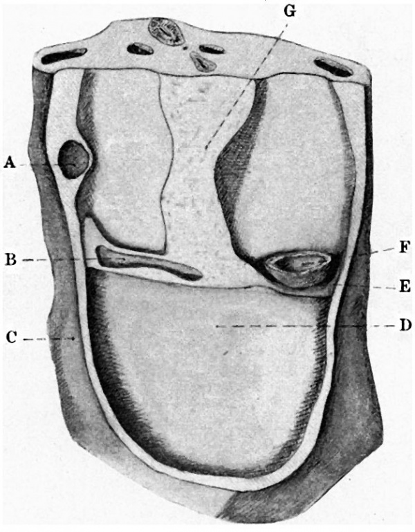
'''Fig. 1.''' — Dorsal wall of pericsrdial cavity of 3-mm. embryo. A, right duct of Cuvier; B, sinus venoaus; C, body-wall; D, floor of pericardium (septum transversum); E, left duct of Cuvier; F, left portion of atrium ; G, dorsal meaocardium. | '''Fig. 1.''' — Dorsal wall of pericsrdial cavity of 3-mm. embryo. A, right duct of Cuvier; B, sinus venoaus; C, body-wall; D, floor of pericardium (septum transversum); E, left duct of Cuvier; F, left portion of atrium ; G, dorsal meaocardium. | ||
| Line 50: | Line 50: | ||
The fold F, originally the dorsal margin of the septum transversum, has, however, undergone a considerable modification, and is now continued into a prominent fold on the caudal surface of the septum transversum which extends in a ventral direction to the liver, which now projects from the caudal surface of the septum, forming the ventral pillar of the diaphragm, and dorsally to the cephalic end of the Wolflian body — a fold which forms the dorsal and ventral pillars of the diaphragm. | The fold F, originally the dorsal margin of the septum transversum, has, however, undergone a considerable modification, and is now continued into a prominent fold on the caudal surface of the septum transversum which extends in a ventral direction to the liver, which now projects from the caudal surface of the septum, forming the ventral pillar of the diaphragm, and dorsally to the cephalic end of the Wolflian body — a fold which forms the dorsal and ventral pillars of the diaphragm. | ||
[[File:Waterston1915 fig02.jpg|600px]] | |||
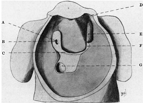
'''Fig. 2.'''— Dorsal wall and floor of pericardial cavity of 6-mm. embryo. A, right duct of Cuvier; B, mesocardlum ; C, dorsal margin of septum trausversum; D, upper remains of dorsal mesocardium; E, left duct. of Cuvier ; Iv‘, mesocardial lold. | '''Fig. 2.'''— Dorsal wall and floor of pericardial cavity of 6-mm. embryo. A, right duct of Cuvier; B, mesocardlum ; C, dorsal margin of septum trausversum; D, upper remains of dorsal mesocardium; E, left duct. of Cuvier ; Iv‘, mesocardial lold. | ||
| Line 59: | Line 60: | ||
The remains of the cephalic portion of the original dorsal mesocardium are seen connecting the aorta and the pulmonary artery to the dorsal wall and constituting the arterial mesocardium. The caudal (venous) portion of the mesocardium has no longer the U—shaped appearance of the second stage, for the limbs of the U have become shortened by the approximation of the ducts of Cuvier, and the base of the former U has drawn away from the orifice of the vena cava inferior. There is now a Wide mesocardium showing: (1) the orifice of the vena cava inferior caudally; (2) immediately cephalic to this point the mesocardium is widened transversely for the pulmonary veins of the two sides which are drawing apart; and (3) cephalic to these is a narrower vertical portion which bifurcates at the top to receive at each lateral extremity the duct of Cuvier. | The remains of the cephalic portion of the original dorsal mesocardium are seen connecting the aorta and the pulmonary artery to the dorsal wall and constituting the arterial mesocardium. The caudal (venous) portion of the mesocardium has no longer the U—shaped appearance of the second stage, for the limbs of the U have become shortened by the approximation of the ducts of Cuvier, and the base of the former U has drawn away from the orifice of the vena cava inferior. There is now a Wide mesocardium showing: (1) the orifice of the vena cava inferior caudally; (2) immediately cephalic to this point the mesocardium is widened transversely for the pulmonary veins of the two sides which are drawing apart; and (3) cephalic to these is a narrower vertical portion which bifurcates at the top to receive at each lateral extremity the duct of Cuvier. | ||
[[File:Waterston1915 fig03.jpg|600px]] | |||
'''Fig. 3.''' — Dorsal wall and floor of pericardial sac of 22-mm. embryo. A, right duct of Cuvier; B, aacendin aorta: C, dorsal arterial mesocardium ; D, arterial | '''Fig. 3.''' — Dorsal wall and floor of pericardial sac of 22-mm. embryo. A, right duct of Cuvier; B, aacendin aorta: C, dorsal arterial mesocardium ; D, arterial | ||
| Line 74: | Line 75: | ||
The changes thus indicated are more readily followed by reference to figs. 4: and 5, of which fig. 4 shows the portion of the dorsal wall of the coelomic cavity of embryo 1 as seen after removal of the septum transversum by the dissection described earlier in this paper. The upper portion of the stippled area in that figure (4) indicates the attachment of the dorsal mesocardium, and the lower part the dorsal attachment of the ventral mesentery of the stomach. The level of the pointers B indicates the level at which lay the dorsal margin of the septum transversum and the cephalic extremity of the pericardio-peritoneal passage, and in these passages on each side is the prominence formed by the mesodermal lung. | The changes thus indicated are more readily followed by reference to figs. 4: and 5, of which fig. 4 shows the portion of the dorsal wall of the coelomic cavity of embryo 1 as seen after removal of the septum transversum by the dissection described earlier in this paper. The upper portion of the stippled area in that figure (4) indicates the attachment of the dorsal mesocardium, and the lower part the dorsal attachment of the ventral mesentery of the stomach. The level of the pointers B indicates the level at which lay the dorsal margin of the septum transversum and the cephalic extremity of the pericardio-peritoneal passage, and in these passages on each side is the prominence formed by the mesodermal lung. | ||
[[File:Waterston1915 fig04.jpg|600px]] | |||
'''Fig. 4.''' — Dorsal wall of the pericardial and upper peritoneal portions of coelom with pericardio-peritoneal passages and commencing lung buds. A, ducts of Cuvier in bod -wall; B, mesodermal lung swelling: ; 0, D, pneumato-ante c recesses; E, dorsal mesocnrdlum. | '''Fig. 4.''' — Dorsal wall of the pericardial and upper peritoneal portions of coelom with pericardio-peritoneal passages and commencing lung buds. A, ducts of Cuvier in bod -wall; B, mesodermal lung swelling: ; 0, D, pneumato-ante c recesses; E, dorsal mesocnrdlum. | ||
Revision as of 16:48, 24 August 2015
| Embryology - 16 Apr 2024 |
|---|
| Google Translate - select your language from the list shown below (this will open a new external page) |
|
العربية | català | 中文 | 中國傳統的 | français | Deutsche | עִברִית | हिंदी | bahasa Indonesia | italiano | 日本語 | 한국어 | မြန်မာ | Pilipino | Polskie | português | ਪੰਜਾਬੀ ਦੇ | Română | русский | Español | Swahili | Svensk | ไทย | Türkçe | اردو | ייִדיש | Tiếng Việt These external translations are automated and may not be accurate. (More? About Translations) |
Waterston D. Developmental Changes in the Pericardium, the Mesocardia, and the Pleural Sacs in the Human Embryo. J Anat Physiol. 1915 Oct;50(Pt 1):24-9. PMID 17233049
| Historic Disclaimer - information about historic embryology pages |
|---|
| Pages where the terms "Historic" (textbooks, papers, people, recommendations) appear on this site, and sections within pages where this disclaimer appears, indicate that the content and scientific understanding are specific to the time of publication. This means that while some scientific descriptions are still accurate, the terminology and interpretation of the developmental mechanisms reflect the understanding at the time of original publication and those of the preceding periods, these terms, interpretations and recommendations may not reflect our current scientific understanding. (More? Embryology History | Historic Embryology Papers) |
Developmental Changes in the Pericardium, the Mesocardia, and the Pleural Sacs in the Human Embryo
By David Waterston, M.D.,
University of St Andrews.
The descriptions and the figures in this communication have been taken from wax-plate reconstructions which I have prepared from the following embryos :—
- Embryo 2W1, approximately 3 mm in length
- Embryo S1, 6 mm in length
- Embryo S3, 22 mm in length.
Fig. 1. — Dorsal wall of pericsrdial cavity of 3-mm. embryo. A, right duct of Cuvier; B, sinus venoaus; C, body-wall; D, floor of pericardium (septum transversum); E, left duct of Cuvier; F, left portion of atrium ; G, dorsal meaocardium.
The first of these embryos was described in the October number of this Journal. The other embryos have not yet been described, and for the present I need only say that they were both obtained from operation cases and were in excellent condition.
The completed models, which included the heart and adjacent portion of the trunk, were dissected so as to give corresponding views in the various embryos: viz., after removal of the ventral wall of the pericardium the heart was removed from that cavity by the division of the great vessels and of the mesocardia, and at a later stage, in the two earlier specimens, the septum transversum was removed in a piece by making a vertical cut through the lateral body—wall dorsal to the attachment of the septum so as to give a View of the thoracic and upper abdominal portions of the dorsal wall of the body cavity.
The first figure shows the appearances found in the model of the 3-mm embryo after the removal of the heart from the pericardial cavity.
The dorsal mesocardium forms at this stage a continuous fold lying vertically and extending from the truncus arteriosus (just above the upper margin in the figure) to the cephalic (dorsal) margin of the septum transversum below. The two, layers forming the mesocardium spread out dorsally on to the ridge formed by the mesoderm surrounding the trachea, and caudally on to the margin of the septum transversum. Two pulmonary veins lay in this mesocardium, but are not shown in the figure. The margins of the septum transversum passed laterally to the body-wall and there received the duct of Cuvier on each side. The mesocardium at this stage resembles an inverted T, the stem of the T being the dorsal mesocardium and the transverse piece corresponding to the lateral portions of the margin of the septum transversum. The vitelline and umbilical veins opened into the portion of the sinus venosus which is still embedded in the septum transversum.
The second figure shows the corresponding parts in the embryo 6 mm in length. The vertical portion of the dorsal mesocardium has almost entirely disappeared, only a small portion persisting at the cranial end, transmitting the truncus arteriosus to the dorsal wall. The disappearance of the central portion of the dorsal mesocardium gives origin to the transverse sinus of the pericardium.
The ducts of Cuvier now pass to the heart from the dorsal and not from the lateral wall of the trunk, and the left duct opens at a higher level than does the right, in contrast to the condition in the first specimen. The vena cava inferior opens into the heart through a prolongation of the mesocardial “bare area” on to the floor of the pericardium ventral to the dorsal edge of the septum transversum.
The attachment of the lower “venous” portion of the dorsal mesocardium now forms a U-shaped figure, one duct of Cuvier opening at the extremity of each of the limbs of the U, and the base of the U corresponds to the dorsal margin of the septum transversum.
The mesial portion of this septum retains its attachment to the dorsal body-wall, but laterally—e.g. dorsal to the level indicated by C in the figure—it is not so attached, and at this point on each side a pointer can be passed dorsal to the edge of the septum transversum caudally into the abdominal division of the ctrzlom, traversing the pericardio-peritoneal passage.
The ducts of Cuvier have, as it were, been drawn towards the head from the septum transversum, and the letter F in the figure indicates a prolongation of the thin margin of the septum, which is continued to the duct of Cuvier and which is most distinct on the left side. The pleuro-pericardial passage lies on each side between this fold and the ridge forming the dorsal wall of the pericardium.
The fold F, originally the dorsal margin of the septum transversum, has, however, undergone a considerable modification, and is now continued into a prominent fold on the caudal surface of the septum transversum which extends in a ventral direction to the liver, which now projects from the caudal surface of the septum, forming the ventral pillar of the diaphragm, and dorsally to the cephalic end of the Wolflian body — a fold which forms the dorsal and ventral pillars of the diaphragm.
Fig. 2.— Dorsal wall and floor of pericardial cavity of 6-mm. embryo. A, right duct of Cuvier; B, mesocardlum ; C, dorsal margin of septum trausversum; D, upper remains of dorsal mesocardium; E, left duct. of Cuvier ; Iv‘, mesocardial lold.
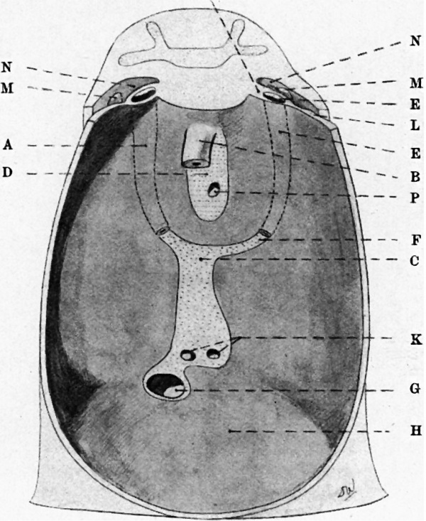
Fig. 3 shows the pericardial sac in the third embryo, in which the sac is completely closed.
The remains of the cephalic portion of the original dorsal mesocardium are seen connecting the aorta and the pulmonary artery to the dorsal wall and constituting the arterial mesocardium. The caudal (venous) portion of the mesocardium has no longer the U—shaped appearance of the second stage, for the limbs of the U have become shortened by the approximation of the ducts of Cuvier, and the base of the former U has drawn away from the orifice of the vena cava inferior. There is now a Wide mesocardium showing: (1) the orifice of the vena cava inferior caudally; (2) immediately cephalic to this point the mesocardium is widened transversely for the pulmonary veins of the two sides which are drawing apart; and (3) cephalic to these is a narrower vertical portion which bifurcates at the top to receive at each lateral extremity the duct of Cuvier.
Fig. 3. — Dorsal wall and floor of pericardial sac of 22-mm. embryo. A, right duct of Cuvier; B, aacendin aorta: C, dorsal arterial mesocardium ; D, arterial mesocardium; E, left duct of uvier, divided; F, meuocardial told; G vena cava Interior; H, floor of pericardium; K. pulmonary veins; L, pleura and pericardial wall; M, apex of lung; N, pleural cavity: P, pulmonary artery.
The left lateral prolongation forms the ligamentum venae cavae sinistrae of the adult, While the remaining portion of the mesocardium forms the definite adult venous mesocardium.
The pleuro-pericardial orifice was situated dorso-medial to the fold indicated by the letter F in this figure and in fig. 2. That orifice is now closed, and it is clear that the closure has been effected by the fusion of the fold F with the dorsal wall of the pericardium along the line extending from the duct of Cuvier to the point at which the central portion of the septum transversum is attached to that dorsal wall.
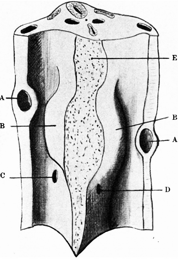
The changes thus indicated are more readily followed by reference to figs. 4: and 5, of which fig. 4 shows the portion of the dorsal wall of the coelomic cavity of embryo 1 as seen after removal of the septum transversum by the dissection described earlier in this paper. The upper portion of the stippled area in that figure (4) indicates the attachment of the dorsal mesocardium, and the lower part the dorsal attachment of the ventral mesentery of the stomach. The level of the pointers B indicates the level at which lay the dorsal margin of the septum transversum and the cephalic extremity of the pericardio-peritoneal passage, and in these passages on each side is the prominence formed by the mesodermal lung.
Fig. 4. — Dorsal wall of the pericardial and upper peritoneal portions of coelom with pericardio-peritoneal passages and commencing lung buds. A, ducts of Cuvier in bod -wall; B, mesodermal lung swelling: ; 0, D, pneumato-ante c recesses; E, dorsal mesocnrdlum.
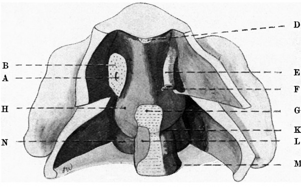
Fig. 5 shows the corresponding parts in the second embryo. In the middle line of the dorsal wall are shown: (1) the upper remains of the dorsal mesocardium, (2) below that the region of the transverse sinus of the pericardium, and (3) the upper margin of the ventral mesentery of the intestine.
Laterally are shown the right and left ducts of Cuvier, divided, and on the left side a small portion of the fold F, from which a fold is continued dorsally around the lateral surface of the prominence of the lung.
The thickened margin of the fold F, forms the pleuro-pericardial membrane, and the fold prolonged from F on the dorsal surface of the septum transversum forms the pleuro-peritoneal membrane. The folds or membranes which effect the closure of the pericardial sac and the separation of the pleural from the peritoneal divisions of the coelom are present, but have not as yet united to the walls opposed to them so as to effect that closure. Comparison of figs. 3 and 5 shows the great growth changes which occur, and which alter profoundly the relative positions of the lungs and pleural sacs to the pericardial cavity.
Fig. 5. — Dorsal wall of pericardial sac, pericsrdio-peritoneal passages, and upper part of peritoneal coelom in 6-mm embryo. A, duct of Cuvier; B, mesocardjsl fold; D. dorsal piesocardium, arterial; E, duct of Cuvier; F, mesocsx-dial fold, now forming pleuro-perlcardial and oval margin of pleuro-peritoneal membrane; G, ventral mesogastrium; H, lung bud in pleural division of caelom; K and L, right and left pnenmsto-enterlc recesses, the left very small; M, stomach; N, Wolffian ridge.
In fig. 5 the developing lungs lie dorsal and caudal to the pericardium and on the caudal and dorsal aspect of the original septum transversum.
Fig. 3 shows that the lung has grown towards the head behind the upper part of the pericardium, and has also extended ventrally so as to intervene between the pericardium and the lateral body-wall. With the growing lung, the pleural cavity has expanded in the loose mesenchyme tissue which occupied these regions. The illustrations forming figs. 3 and 5 were made by Mr W. Champneys, and the models of embryos 1 and 2 have been reproduced very successfully by Mr S. Boyes, 63 Hafton Road, Catford, S.E., who is prepared to supply copies of these models.
Part of the expense of the preparation of the original wax-plate models was defrayed by a grant from the Royal Society.
Cite this page: Hill, M.A. (2024, April 16) Embryology Paper - Developmental Changes in the Pericardium, the Mesocardia, and the Pleural Sacs in the Human Embryo. Retrieved from https://embryology.med.unsw.edu.au/embryology/index.php/Paper_-_Developmental_Changes_in_the_Pericardium,_the_Mesocardia,_and_the_Pleural_Sacs_in_the_Human_Embryo
- © Dr Mark Hill 2024, UNSW Embryology ISBN: 978 0 7334 2609 4 - UNSW CRICOS Provider Code No. 00098G

