Paper - Development of the ventral abdominal walls in man: Difference between revisions
(Created page with "{{Header}} {{Ref-Mall1898}} {| class="wikitable mw-collapsible mw-collapsed" ! Online Editor |- | 90px|left This historic 1998 paper by Mall desc...") |
mNo edit summary |
||
| (6 intermediate revisions by the same user not shown) | |||
| Line 4: | Line 4: | ||
! Online Editor | ! Online Editor | ||
|- | |- | ||
| [[File:Mark_Hill.jpg|90px|left]] This historic 1998 paper by Mall describes the early | | [[File:Mark_Hill.jpg|90px|left]] This historic 1998 paper by Mall describes the early development of the abdominal wall. | ||
<br> | <br> | ||
{{Mall Links}} | {{Mall Links}} | ||
| Line 10: | Line 10: | ||
'''Modern Notes:''' | '''Modern Notes:''' | ||
<br> | <br> | ||
{{ | {{Musculoskeletal Links}} | ||
|} | |} | ||
{{Historic Disclaimer}} | {{Historic Disclaimer}} | ||
=Development of the Ventral Abdominal Walls in Man= | |||
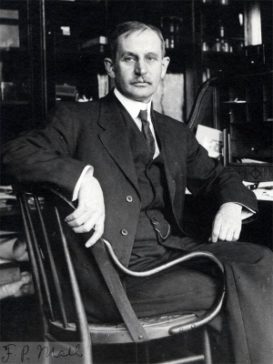
[[File:Franklin Mall 01.jpg|thumb|link=Embryology_History_-_Franklin_Mall|Franklin Mall (1911)]] | |||
[[Embryology_History_-_Franklin_Mall|Franklin P. Mall]] | |||
([[:Category:1800's|1898]]) | |||
* The present paper is to be considered a continuation of two papers published recently; one upon the development of the human coelom (_/our». qf [Harp/1., vol. xii), and the other upon the development of the human intestine (His’s Archiv, Supplement Band, 1897). | |||
==Introduction== | |||
After the neural plate is formed within the amniotic cavity, the tail end of the embryo remains attached to the chorion by means of the allantoic stalk (Bauchstiel). The amnion surrounds the embryo only on its dorsal side, as has been shown recently by the study of a number of young human embryos. The ventral side of the embryo, however, hangs free in the coelomic cavity, as the body walls have not been formed by the extension of the amnion. The extension of the amnion proceeds hand in hand with the flexion of the embryo. It first extends over the face of the embryo, then tucks under the tail end, and at the same time it encroaches from both sides of the body. In covering the head it first crosses the mouth and then the heart, this movement being accelerated by the growth of the amnion over the head end of the body from left to right.<ref>Mall, Journ. of Morph., vol. xii, p. 430, Figs. 27, 28 .</ref> While this is taking place, the stem of the umbilical vesicle elongates to produce the general condition, as represented in embryos four weeks old. Soon, however, the amnion covers the whole allantoic stalk, including within its folds the stem of the umbilical vesicle, thus forming the true umbilical cord. At this stage the intestine begins to enter the cord. | |||
After we have reached the stage in which the umbilical cord is formed we find that in transverse sections of human embryos, as well as in embryos of other mammals of the same stage, the ventral abdominal walls are composed wholly of a membrane of connective tissue, without any ribs, muscles, or blood vessels as they are found in the adult. | |||
It is now generally believed, and to a great extent proved, that the ribs and muscles wander into the ventral walls of the embryo from the sclerosomes and myomeres on the dorsal side of the embryo. It is easy to demonstrate the growth of the ribs from the dorsal to the ventral side of the embryo, but it is not so easy to demonstrate the growth of many muscles from the muscle plates to their final position in mammalian embryos. With the growth of the ribs and the wandering of the muscles from the myotomes into the ventral wall of the embryo we have associated with them their nerves and blood vessels, which ultimately form the intercostal nerves and blood vessels respectively. The segmental arteries fuse at their tips early in their development immediately below the rectus abdominis to form the internal mammary and deep epigastric arteries. This chain of anastomosis is formed in a manner with that seen in the formation of the vertebral artery, as shown by His, Froriep, and Hochstetter. The primitive internal mammary and deep epigastric arteries, together with the rectus, at first lie immediately below the mammary line in pigs’ embryos, and all three, artery, muscle, and line, wander together towards the ventral middle line of the body. No doubt the same process takes place in the human embryo, but the mammary line lacks prominence here and cannot be used as a landmark. | |||
As the segmental nerves appear, each is immediately connected will: its corresponding myotome, and all of the muscles arz'sz'¢zg from a myolome are always z'mzer72az'ed by branches of tile nerve w/ziclz orzigzhzally belonged to it. | |||
This generalization isdifiicult to prove in all of its details, but all of the work in embryology, as well as a tabulation of our knowledge of motor nerve distribution, indicates that it must be true. The simpler muscles of the back, as well as the intercostal, are innervated by their corresponding nerves. The muscles of the eye, of mastication, and of the face can be correspondingly grouped. The trapezius, latissimus dorsi, serrates anticis, and rectus abdominis wander great distances and carry with them their original nerves. Moreover, it would be practically impossible to explain why each muscle has its proper nerve supply, which is so evenly distributed in it, were not this original relation between muscle and nerve retained. With this view we have no grave mechanical difficulties to overcome in conceiving how it is possible for the simple peripheral nervous system of the embryo to be evolved into that of the adult. It is simply that each segmental nerve grows to its myotome, or corresponding group of tissue or branchial arch, and with the further growth of muscles from myotomes the original nervous connections are retained. The required free growth of a nerve is then extremely short, as the myotomes receive them as soon as they leave the spinal cord, and then as the different muscles or parts of muscles arise from each myotome the nerve bundles split to correspond. The primitive muscle, then, is from its earliest appearance connected with the cord, and, as its muscle fibers appear and increase in number, this first connection guides the new nerve fibers to their destination. | |||
The studies in comparative anatomy by Gegenbaur<ref>Gegenbaur, Grundriss d. vergleich. Anatomic. </ref> and by Huxley<ref>Huxley, Comparative Anatomy of Vertebrates. </ref> all lead to the same general conclusion that nerve and muscle are associated with each other in their earliest stages of development. This idea has been the foundation for a number of studies in comparative anatomy, and the more it is investigated the stronger the foundation becomes, as may be seen in the critical review by Kollmann.<ref>Kollmann, His’s Archiv, 1891, p. 76.</ref> In a discussion of Testut’s “Anomalies musculaires chez l’Homme,” Gegenbaur<ref>Gegenbaur, Marplz. fa/E-rZ:., Bd. 1:, p. 331.</ref> shows clearly that the study of variations of muscles would have great meaning if their nerve supply were included, and that all the reports of muscle anomalies for a period of more than one hundred years cannot be used at present, due to this omission. The great value of the comparative study of the muscles in connection with their nerve supply has recently been shown again by Ruge.<ref>Ruge, Morph. _/sz/ir5., Bd. xix, p. 376.</ref> His study shows that comparison of muscles can be made satisfactorily only when their nerve supply is included. | |||
All this indicates that the history of a muscle is indicated by its nerve, and that in studying the development of a muscle our main guide is its nerve. This has been plainly indicated recently by Nussbaum<ref>Nussbaum, Verhatzdl. d. Rnaf. Ges. I894-I896. </ref> and in my studies in embryology and anatomy I cannot find a single instance to contradict this idea. | |||
The same generalization, no doubt, applies also to the sensory nerves, which are distributed in a segmental way to the integument. Their first distribution would then be to the regions immediately over the spinal ganglia, and as the skin shifts in its development it carries with it its original nervous supply. | |||
That the nerves cannot be distributed to any great extent without some guide is further demonstrated by all experiments on regeneration, which show that they have great powers of growth if they are guided in their distribution by a canal or by the connective tissue sheath of some degenerated nerve. | |||
Although it is impossible to prove at present that all of the skeletal muscles arise from muscle plates or their corresponding coelomic diverticula into the branchial arches, the generalization that they do arise from the mesoderm segments is based upon sufficient observation to make it highly probable. In sharks the coelom extends into the branchial arches and into the myotomes ; in man the cavity in the muscle plates is never connected with the coelomic. cavity, while in the branchial arches no such cavity exists. Yet we do not hesitate to assert that the condition found in sharks is the primitive, while that in man is due to secondary changes. Of course the problem is more difficult when the fate of the plates is to be studied. In the sharks, where they are definite, it is easy to show that all of the skeletal muscles of the body arise from them, while in man it is extremely difficult to show from what myotome any given muscle arises. The first difficulty encountered is that in the head (with the exception of in the occipital region) there is no grouping of the mesoderm in the branchial arches, while in the extremities the myotomes lose their sharp outline and appear to blend with the surrounding mesenchym. This fact has been sufficiently emphasized by Paterson<ref>Paterson, Qmzr. journ. Micro. .Sci., N.S., vol. xxviii, p. 109. </ref> a number of years ago, and recently has been used by Fischel<ref>Fischel, Morph. Jalzrln, Bd. xxiii, p. 544.</ref> and by Harrison<ref>Harrison, Archiv mzvér. Amzt., Bd. xlvi,-p. 5oo.</ref> in combating the notion that all muscles arise directly from the myotomes. Harrison studied the development of the fin muscles in the trout, and finds that, with the exception of those in the pectoral fin, they arise directly through buds from the myotomes. In this one fin the muscle bud is small, and is so closely blended with the mesenchym that it is impossible to show whether or not the muscles of this fin arise from the muscle bud or mesenchym or from both. It appears to me that if all the body muscles of sharks and all of them in the trout arise directly from the myotomes, with the exception of those of the pectoral fin, it is better for the present to ascribe the muscles of this fin to the myotomes rather than to mesenchym, when mesenchym and buds are so completely blended. | |||
Not only does Harrison<ref>Harrison, Johns Hopkins University Circuiar, No. 3, 1894.</ref> deny that all the skeletal muscles A arise from the myotomes, but he also attempts to prove that the segments of a muscle, like the rectus, do not indicate that it has arisen from a corresponding number of muscle plates. He has shown that in teleosts the rectus is first split from the muscle plates as a band of tissue, and then undergoes secondary segmentation. After the muscle is laid down its front end remains connected with the myotomes until it has segmented, and hence these segments of the rectus correspond with the myotomes, while behind they do not. That the segments in the posterior end are formed after the muscle is separated from the myotomes indicates that there are mechanical causes which favor segmentation, but it is not proved that these segments do not correspond to the myotomes until it is shown that the nerve supply of the two are different. Even if it were shown that the succession of segments in the rectus did not correspond fully with the segmental nerves it would not prove anything special other than the history of this muscle. We must only recall the serratus anticus, which is composed of beautiful segments, all of which are of secondary formation. In its development the muscle wanders down from the neck, attaches itself to the scapula, and then successively to the ribs. During all this process of growth it retains its original nerve connection, which shows its origin; that it is segmented is due to the ribs, a mechanical element. | |||
While in the sharks it is comparatively easy to follow the development of the eye muscles with their respective nerve connections, it is impossible at present to follow them in higher animals. The same applies to the muscles arising from the hyoid arch and innervated by the seventh nerve. It is a remarkable fact, however, as emphasized by Rab], that just those muscles which belong to the hyoid arch are innervated in man by the seventh nerve. Even where we can see no "sharp line whatever in the embryo, there is one which has been indicated to us by the study of sharks. Rabl<ref>Rabl, Anat. Ans, Bd. ii, p. 226.</ref> states, after speaking of the muscles of mastication: “ Die iibrige Gesichtsmuskulatur dagegen wird vom Facialis innerviert und sie ist, wie Gegenbaur und Ruge auf vergleichend-anatomischem Wege gezeigt haben, aus einer Differenzierung des Platysma hervorgegangen; das Platysma selbst aber gehiirt genetisch dem Hyoidbogen an und wird daher, wie seine Differenzierungsprodukte, vom Facialis versorgt. Ebenso scharf sondem sich auch die beiden Innervationsgebiete in der Paukenhohle. Hammer und Amboss entwickeln sich aus dem ersten, der Steigbiigel, wie ich auseinandergesetzt habe, aus dem zweiten Kiemenbogen; der Muskel des Steigbiigels vom Facialis innerviert. Der Musc. tensor tymp. gehort mit dem Tensor veli palatini genetisch und anatomisch zu einer Gruppe (Schwalbe) ; der Musc. stapedius bildet in iihnlicher Weise aller Wahrscheinlichkeit nach mit dem Stylohyoideus und hinteren Biventerbauch eine Gruppe."<ref>There is a very extensive literature to show that the muscles of the limbs arise from the myotomes. The systematic work of Mol1ier(/brat. Hafiz, No. viii) is one of the best of this group of papers. In my discussion above I have only brought forth some general objections to the views of Paterson and the much stronger and better arguments of Harrison.</ref> | |||
The short muscles of the trunk are innervated by single nerves, and in them the embryonic condition is retained. But in the case of the shifting of a muscle its nerve supply is not so simple as in case of the diaphragm. In this extreme example the muscle wanders down with the development of the septum transversum<ref>Mall, Journal. of Morphol., vol. xii, p. 395, figs. 30, 41. </ref> and its nerve supply indicates that the muscle must have arisen from the fourth or fifth cervical myotome or both. After the septum transversum has reached its final location, to form the diaphragm, muscles arising from the lower dorsal myotomes wander into its periphery and these are innervated by the lower intercostal nerves. In the same way the sterno~mastoid and trapezius grow over from the head to the shoulder, while the latissimus dorsi and serratus anticus grow from the shoulder to the ribs and pelvis. I have already spoken of the serratus anticus above, and wish to add a few words about the latissimus dorsi. It undoubtedly arises from the seventh and eighth cervical myotomes, is soon separated, and free to move. It attaches itself first to the humerus and then gradually spreads out over the back, attaching itself first to the vertebrae, then to the ribs, and finally to the ilium. As the growth takes place in this order the shoulder end is the older, and the pelvic end the younger, thus accounting for the nerve distribution as shown recently by Nussbaum.<ref>Nussbaum,‘ Varhandl. J. Anat. Gas., 1894, p. 179; 1895, p. 26; 1896, p. 64. </ref> In spreading, its fasciculi cross and encircle branches from the intercostal and lumbar nerves, thus making them perforate this muscle. Other muscles that shift, like the deltoid and pectoralis major, are likewise perforated but not innervated by nerves foreign to them. | |||
Sometimes muscles which remain segmental cover metameres beyond their nervous supply. Quain’s<ref>Quain’s Anatomy, vol. ii, p. 3 54.</ref> Anatomy, in discussing the quadratus lumborum and complexus muscles, asserts that this indicates a reduction of the number of nerves, while from the above discussion it appears to me that it is not a reduction of nerves but an extension of the muscle beyond its original bounds. | |||
Likewise a complete absence of an important muscle (trapezius), or a group of muscles (facial), is not necessarily to be accounted for by a degeneration of the nerve. Anything which arrests the development of the muscle in its very earliest stage will accomplish absence of the nerve, while a nerve destroyed at so early a stage might be replaced by a neighboring nerve through anastomoses. | |||
One of the most complicating elements in the study of the development of a muscle is the formation of nerve plexuses. In those instances in which there is but little cross shifting in the movement of a muscle, its nerve remains single or nearly so. But when there is considerable cross shifting very early in the development we have the formation of a plexus. Furbringer<ref>Furbringer, Morph. Jahrb, Bd. v, p. 324. </ref> has shown from comparative anatomical studies that there is a plexus formation in the nerves supplying the limbs which shift in development, and that the plexus formation indicates that the limb is wandering. In the development of the extremities in man it has been clearly pointed out by the method of reconstruction that the arm bud makes a rapid excursion from the head backward in the early embryo.<ref>His, Abhandhl d. .K s.Ges. d. Wiss , Bd. xiv, p. 34!, Taf. II; and Mall, Journ. qf 11407395., vol. v, p. 4 59, Pl. XXX. </ref> In so doing it appears to drag the nerves together, thus fonning a plexus. The upper intercostal nerves do not shift and, therefore, do not form a plexus, while the lower intercostal nerves passing to the anterior abdominal wall shift in their development, and thus favor the plexus formation. | |||
In this shifting and mixing of nerves the portions of the muscle plates which they supply are also hopelessly mixed, and this I think accounts, in part at least, for the fact that the muscle buds are no longer defined in the arm and leg by buds. But after the muscles are fairly well outlined they move on to their destination without any more mixing of nerve fibers further than additions which may grow along the existing trunks from the spinal cord. The differentiation of the muscles in the extremities takes place after the plexus nearest the spinal cord is formed, thus permitting of a second group of nerve anastomoses in the extremity. This process in the forearm, for instance, accounts for the double nerve supply of some of the muscles in it. | |||
Physiologists have studied the distribution of nerves in muscles much more carefully than anatomists, and we have to thank Mays<ref>Mays, Zeit. fur Biol., vol. xx, p. 449.</ref> for very careful description of nerve plexuses in muscles. In a segmental muscle like the frog’s rectus abdominis, irritation of the nerve supplying one segment not only makes this contract but the rest of the muscle also, showing that there must be an anastomosis between the nerves supplying the different segments.<ref>Mays, Ueber die Nervatur d. Musculus rectus abdominis d. Frosches, Heidelberg, 1886.</ref> Therefore, the irritation of the nerve passing to a single segment sets the whole muscle to contracting. The heart is affected in a similar way, for irritation of the minutest sympathetic twig has the same effect upon the heart beat as the irritation of its main trunk.<ref>Johansson, Archiv. fur Physiol., 1897, p. 103.</ref> And the same is true regarding the influence of the splanchnic nerve upon the muscle Walls of the vena portae.<ref>Mall, Archiv. fur Physiol., 1892, p. 423; and Johns Hopkins Hospital Reports, vol. i, p. 148. </ref> | |||
In addition to the physiological verification of these nerve plexuses we have ample confirmation of them by Nussbaum, and very recently again by von Bardeleben and Frohse <ref>von Bardeleben and Frohse, Verfzandl. a. A max. Gun, 1897, p. 38.</ref> for a number of muscles in man. So every bundle of nerves which leaves the spinal cord to pass to a muscle has a chance to mix its fibers once, twice, or three times, as the case may be, before it reaches the muscle fibers, and a nerve stimulus may pass to a given muscle in a number of ways and from a number of sources. T | |||
The plexus within the muscle will, no doubt, prove to be a valuable object for investigation, but it does not concern us much when we consider the origin of a given muscle. I have given it for the sake of completion and have discussed the subject somewhat extensively in order to show a number of difficulties in my way, as well as my standpoint in the present problem. | |||
===Development of the Rectus=== | |||
In the earliest human embryos which have been studied it is observed that the allantoic stalk and the umbilical vesicle are located much nearer the head than in the adult. The relation of these organs to the body of the embryo is shown in the accompanying figure, which is taken from an embryo not over two weeks old.<ref>The anatomy of this embryo is fully described by me in the four». of Morph., vol. xii, p. 417, and in His’s Archiv, Supplement Band, 1897.</ref> It will be noticed from the figure that the allantoic stalk and the umbilical vesicle must shift their position through a distance of at least twelve segments in order to gain the position they will hold in older embryos. The possibility of this shifting is all easily understood when we consider how soft the tissues are at this time, and the lack of, any framework to hold the different parts of the embryo together. Later on, when nerve bundles and skeleton appear, the shifting still takes place, but by no means to as great an extent, and its course is now marked by the arrangement of the nerves as well as other tissues. At this stage, however, the most fixed points appear to be the myotomes, and from them I have made all of my measurements. For reasons given in the earlier descriptions of this embryo I have ascribed three of the muscle plates to the occipital region, making the first dorsal myotome immediately over therneurenteric canal. In later stages each myotome lies immediately over“ a spinal ganglion, and in referring to the myotomes I give them the numbers corresponding to the spinal nerves. This causes less confusion than by numbering them from the auditory vesicle backward, for by so doing we do not convey any idea of adult anatomy. | |||
In embryos slightly older than the one figured above, the spinal ganglia and vertebral column are outlined, thus aiding us easily to locate the position of any of the organs. With the descent of the heart the communication of the umbilical vesicle with the midgut is rapidly cut off, and the stems connecting them, together with the allantoic stalk are then encircled with the amnion. All these intermediate stages are sufliciently well shown in His’s Atlas, and I need not describe them. | |||
At the end of the fourth week the umbilical cord is fully formed, as is shown by a number of specimens in my possession, two of which are especially valuable because there is no doubt whatever as to their being normal. One is the specimen I have studied a great deal, which was obtained from a criminal abortion, and the other was gotten from a murdered woman and sent me by the coroner’s physician of Chicago. The first specimen (No. II) is somewhat older than the second (No. . LXXVI), and for that reason is more valuable. | |||
Fig. 2 is a profile of this embryo<ref>Mall, Journ. of.Morph, vol. v, p. 459. Reference Handbook of the Medical Sciences, New York, Supplement, pp. 184, 391, 87 5. jaurn. ry‘ Mar;/z., vol. xii, p. '43:. His’s Ardiizr, Supplement Band, 1897.</ref> drawn from the specimen before it was cut, with the outlines of the myotomes of the body added from a reconstruction. It shows the relation of the cord to the rest of the body and the extent of the myotomes have grown towards it. In the body itself the myotomes have distinct buds which are growing into the membrana reuniens, and each of these is intimately associated with its nerve. Where buds from the myotomes enter the extremities, their ventral tips appear to be fully blended to correspond with the blending of the nerves which enter the limbs. | |||
The extent of the growth of the myotome into the membrana reuniens is shown in fig. 3. It shows the marked bud from the myotome. Earlier stages need not be given, and a later stage has already been described and pictured by Kollmann. | |||
There is, however, a time when the buds from certain of the dorsal myotomes blend to form the rectus, and I have not been able to obtain a good specimen to demonstrate this. A stage somewhat older than one pictured by Kollmann<ref>Kollmann, His’s Archie, 1891, P1. III, fig. 1.</ref> will no doubt give the rectus arising from the myotomes. A good specimen of this desired stage is not at my disposal.<ref>His’s embryo Ko (Aqilzandz. a’. lie. .9. Gas. d. I/Vz'.s'.s*., Bd. xiv) and one described by Fraser (Tram. Ray. Acad. Med., Ireland, vol. xi, Pl. VI, fig. 1) represent the missing stage between embryos II and XLIII. Since writing the above I have obtained an excellent human embryo from an operation for tubal pregnancy. The specimen, No. CIX, was hardened immediately in graded alcohol, and measured 11 mm. Sections show that the ribs are just beginning, and between them the intercostal muscles are sharply outlined. Projecting from them there are spouts which at points appear to be blended to make the rectus abdominis rectus is innervated by a single intercostal nerve and, therefore, must be viewed as belonging to the same myotome.</ref> | |||
fig. 4 is from a reconstruction of the body of embryo XLIII, to which has been added the head taken from a photograph of the embryo before it was cut into sections. In the drawing only the superficial layer of abdominal muscles is shown. The form and extent of the rectus, as well as its relation to the external oblique, is indicated. The transverse lines in the rectus correspond with the digitations in the oblique, and each of these digitations with its corresponding segment in the | |||
The lateral position of the rectus, which has already been noted by Kolliker <ref>Kolliker, Grundriss, 1884, p. 340.</ref> is also easily seen in transverse section, as shown in fig. 5. Unfortunately, the stages between XLIII and II are missing, but the lateral position of the rectus does show that a finished rectus is formed at a very early date, and that it must still shift one-half the distance around the body in order to obtain its final position. The muscle is nearer the middle line at its upper end than at its lower, due no doubt to the more rapid growth of the ribs than of the pubis, to which it is now attached. | |||
In a later stage, fig. 6, we have the same condition of things as in No. XLIII, only more advanced. In this reconstruction it was impossible to make out definitely all the serration of the external oblique andthe segments in the rectus, because the sections were transverse to the embryo, while in XLIII they were sagittal. The transverse sections (figs. 7, 8) show the exact position of the rectus at its different levels. | |||
The two recti reach the middle line above the umbilical cord about the eighth week, while the main bulk of the intestine is still in the cord. At this time the ventral abdominal walls above the umbilicus are completed, and their further growth is simply a matter of expansion. About the end of the ninth week the intestine is withdrawn from the cord, the opening, soon becoming closed by an adhesion of the edges of the periph ery of the communication of the coelom of the cord and the abdominal cavity. As soon as this is accomplished the stem of the cord is surrounded by a membrane about 5 mm. in diameter, which in turn is encircled by the two recti. It is very easy with a probe to reestablish the old communication, and in so doing the membrane again returns to its former position around the end of the cord, showing by this reverse process its origin. Very soon after this membrane is formed the recti invade it also, thus completing the abdominal walls at this point. | |||
Relmrion of Ike Segment: of the Ream: to Z/ze Umbz'lz'cu.s‘. — Judging by the illustrations in the various atlases and text-books on anatomy there is considerable difference of opinion regarding the positions of the transverse lines in the rectus abdominis muscle. No doubt this lack of agreement is due to the variations in the arrangement of the muscle segments, for we know that in some instances all of them, as indicated by their innervation, may be present, while on the other hand a number of them may be but slightly marked or even wholly missing. As a rule a number of well-marked cross striations are present, but their position and number is differently pictured by different authors. Two of the newer atlases express very well the two views which are usually entertained. In Toldt’s<ref>Toldt, Anat. Atlas, Wien u. Leipzig, 1896. </ref> atlas a muscle segment is immediately opposite the umbilicus, while in Spalteholz’s<ref>Spalteholz, Hand-Atlas d. Anat. d. Muscle, Leipzig, 1396. </ref> atlas this muscle segment is wholly above the umbilicus, placing the lowest transverse line immediately opposite the umbilicus. With the exception of the insertion of the muscle, the illustration given by Spalteholz corresponds with that given by Quain, Henle, Testut, as well as by our more popular English text-books, Morris and Gray. In them the insertion, as pictured correctly by Spalteholz, is given only in a diagrammatic way. I have examined a great number of cadavers with special reference to this muscle and find that the great majority of specimens correspond in every detail with Spalteholz’s illustration. | |||
The muscle segment immediately above the umbilicus then is the rule, but whether it arises from the same myotome in all instances is not proved until it is shown that it is always innervated by the same nerve. This question I have investigated carefully in a number of cadavers and find that in nearly all instances the muscle segment immediately above the umbilicus is innervated by the ninth intercostal nerve. All atlases in which the exact innervation of the rectus is given confirm the above observation.<ref>See in the magnificent Atlas of Hirschfeld and Léveillé, Pl. LIII. This figure is copied extensively; for instance, Quain, vol. iii, p. 309.</ref> The skin immediately over this segment is also supplied by the ninth intercostal nerve.<ref>Head, Brain, 1893, 1894.</ref> This then gives us a very fixed point to work from in the further study of the walls of the abdomen, and from it we can make all of our counts. The segment above the ninth is usually the eighth, but occasionally the eighth and seventh combined, while the one lying upon the ribs belongs to the fifth and sixth, or sixth and seventh, or fifth, sixth, and seventh segments, as the case may be. The portion of the muscle below the umbilicus usually represents three segments, the tenth, eleventh, and twelfth, but occasionally a portion of the first lumbar may be included. It is of course understood that the numbers of the segments indicate from which myotomes they arose. | |||
The comparative studies of Ruge<ref>Ruge, Morph. Jahrd., Bd. xix, p. 376.</ref> show that the number of segments in the rectus increase as we pass down the scale of mammals, and the relation of the umbilicus to the segments changes in proportion to the number. As the number of segments increases, the umbilicus is pushed more and more away from the head. In man it is nearest the head, but when there is a variation in its position it is in the form of a reversion. Ruge’s figs. 12, 13, and 14 illustrate this well, showing that the umbilicus may be below the tenth segment. | |||
I have shown in an earlier communication that in the shifting of the viscera during development there is a fixed point opposite the first sacral segment, beyond which they do not pass. Everything descends towards this point, and those which go farther protrude into the umbilical cord. With the extreme shifting of the viscera there is a partial shifting of the abdominal walls, which seems to be partly controlled by the fixed point opposite the sacrum. This lower and more fundamental fixed point appears to be the meeting point of two forces, one the movement from the head towards the tail, and the other in the opposite direction. The point is well established in embryo XXII, and may control to a great extent the further shifting of the abdominal muscles. If there is any meaning to this fixed point opposite the sacrum, it will be necessary to find that it lies farther and farther away from the head as we pass down the scale of mammals. | |||
After the ninth segment has become definitely located just above the umbilicus the portion of the muscle between the umbilicus and pubis is relatively much shorter than it is in the adult. The distance between the end of the sternum and the umbilicus is twice the distance between the umbilicus and the pubis in embryos of about eight weeks, while in the adult these two measurements are equal.<ref>See also the figures given by Merkel, Aéfiandl. d. &. Gee. J. WzZr:., Giittingen, Bd. xi.</ref> Before the two pubic bones have met to form the symphysis, this difference is still greater. This narrowed area between the pubis and umbilicus in the early embryos may possibly account for the blending of the three muscle segments of the rectus in this region. The appearance in embryos XLIII and XXII, figs. 4 and 6, also indicate this. | |||
===Shifting of the Milk Lines in Pig Embryos=== | |||
The invasion of the membrana reuniens by the rectus is also indicated by the shifting of the milk line. O. Schultze<ref>O. Schultze, Anat. Anz., Bd. vii, and Verizaudl. a’. Pkys.-med. G-‘e.s'., Wiirzburg, Bd. xxvi. </ref> has shown that the mammary glands in pigs arise as a ridge of ectoderm on either side of the body immediately to the ventral side of the Wolffian ridge. In a short time the milk ridge breaks up into islands, each of which marks a later mammary gland. | |||
Figs. 9, 11, and 13 show the position of the milk line in pigs’ embryos, giving the relative positions of the line in the three different stages. Fresh specimens show clearly that the abdominal walls are extremely thin on the ventral side of the line, while on the dorsal side they are thicker and apparently finished. Sections of the three embryos pictured in figs. 9, 11,and 13 are shown in figs. 10, 12, and 14. The sections show that the line lies immediately over the rectus, and from this we can locate the position of the rectus in any embryo before it is cut. The wandering of the rectus is indicated then in pigs’ embryo by the shifting of the milk line. | |||
===Development of the Internal Mammary and Deep Epigastric Arteries=== | |||
Before the intercostal vessels are formed there appears to be a circulation through the membrana reuniens, as is indicated to us by a figure given by Kolliker,<ref>Kolliker, Grundriss, p. 103, fig. 58.</ref> as well as by the description of His.<ref>His, Anat. Mensch. Embryonem, Bd. iii, p. 206, also fig. 130.</ref> Kolliker pictures a cow’s embryo with the whole membrana reuniens filled with a minute plexus of veins which radiate from the myotomes towards the umbilical cord, while His describes this same region in the human embryo as filled with branches of the umbilical vein which empty into the sinus reuniens above, and into the umbilical vein below. According to His’s description they arise when the communica tion between the umbilical veins and the sinus reuniens is severed. Although the picture given by Kolliker does not correspond with His’s description it does not contradict it, nor is it peculiar to the cow's embryo. I have in my collection a well—preserved human embryo (No. LXXVI), in which the membrana reuniens is filled with a plexus of veins much like that in the cow’s embryo. The specimen was taken from the uterus seven hours after the death of the woman, and without opening the ovum was hardened in absolute alcohol. All the vessels down to the capillaries are filled with blood, thus making it an excellent specimen for the study of the blood vessels. It seems to represent a stage somewhat more advanced than the one pictured by Kolliker, as the plexus of veins does not cover the whole membrana reunieus. The ventral wall of the heart near the liver contains no vessels, while the membrana reuniens covering the upper end of the heart is filled with a plexus of vessels which communicate with the capillaries of the mandibular arch. There is an extensive plexus through the arm and lateral body walls which extends through the membrana reuniens covering the liver, and finally encircles the cord and communicates with the umbilical veins. | |||
The specimen just described must be about 22 days old and, although I have six other good embryos between 14 and 28 days old, I find no such plexus in the membrana reuniens, although in all but one of them (14 days) the arm shows a rich plexus of capillaries filled with blood. In stages older than four weeks I find no blood vessels in the membrana reuniens with the exception of that portion encircling the cord where there is a rich network of veins. Although I have a number of excellent specimens of five and six weeks, the membrana reuniens over the heart and liver contains no blood vessels until it is invaded by the ventral plate, which is accompanied by the development of the intercostal vessels. In pigs’ embryos the extensive membrane is also free from veins, with the exception of the zone encircling the cord, which again has a venous plexus more marked, however, than in the human embryo. | |||
It appears, then, that during the third week of development, while the umbilical veins still empty into the sinus reuniens, an extensive plexus is formed throughout the greater extent of the membrana reuniens, which receives blood from the aorta on its dorsal side, and empties it into the umbilical vein on its ventral side. As the umbilical vein changes its position to enter the liver, this circulation through the membrana reuniens is broken up as a much earlier circulation though the umbilical vesicle was broken up.<ref>In Journ. of Morph., vol. xii, fig. 16, it will be seen that the aorta sends a number of segmental arteries to the umbilical vesicle. In a short time these all disappear, only to repeat themselves over the body of the embryo. In turn this second set disappears as soon as the umbilical vein enters the liver.</ref> | |||
The earliest collecting vein for the descending aorta is the omphalomesenteric vein; next it is the umbilical vein, and finally, when the abdominal walls are completed, it is the cardinal. This in turn is partly converted into the vena cava inferior. | |||
The arterial system is already well outlined in embryo II. The aortic arches and segmental arteries are sufliciently well marked to number them. The vertebral artery is in process of development; it being formed by a union of a number of segmental arteries, as shown by His,<ref>His, Amzt. .Z|Iemv:}z. Embrjzone, vol. iii, I88 5. </ref> by Froriep,<ref>Froriep, His’s Archiv J, 1886, p. 69.</ref> and by Hochstetter<ref>Hochstetter, Morph. fa/zrb., Bd. xvi.</ref> The sixth cervical segmental artery gives rise to the subclavian artery. The lower cervical, all the dorsal and lumbar segmental arteries, are concerned in the development of the thoracic and abdominal walls. | |||
Fig. 2 shows the general extent of the dorsal myotomes, while fig. 15 gives the extent of the arteries. It shows a. simple arrangement from the vertebral to the hypogastric artery. The lower lumbar arteries are not shown. A section of this embryo is shown in fig. 3. It shows the relation of the segmental arteries to the myotome. The segmental arteries supply primarily the spinal cord and ganglia in two groups of branches, one near the middle line and one more lateral. A more ventral group of segmental arteries supplies the Wolffian body. The blood from all these groups of arteries appears to be collected by the cardinal veins; certainly that from the ventral and lateral group is. | |||
The lateral group gives rise to the intercostal arteries by first supplying the myotomes, and, as these grow into the membrana reuniens by a process of budding, the vascular loop follows it. In so doing the loop is first on the dorsal side of the sympathetic and finally on its lateral side, thus making the sympathetic cord cross the intercostal arteries and veins on their ventral side, as is the case in the adult. | |||
No sooner is the vascular loop extended to the lateral side of the sympathetic than it begins to anastomose with neighboring segmental loops, as single vessels near the subclavian and hypogastric arteries, and as a plexus midway between these two. This gives us at this early period a complete anastomosis from the subclavian to the femoral artery, as fig. 16 shows. It remains only for this system to shift around towards the median line with the muscle, nerves, and ribs to form the condition of things as found in the adult. | |||
Fig. 16 shows that the upper and lower segmental arteries of the series do not correspond with the same in the adult. Above the superior intercostal is missing, while below there is only a small fourth lumbar present, and it arises from the middle sacral. Ilio-lumbar and circumflex-iliac arteries are altogether wanting, and I should judge from the relation of the arteries in embryo XLIII that the arch formed by the ilio-lumbar and circumflex-iliac is of secondary origin and has nothing to do with the segmental arteries. That they form anastomoses with the lower lumbar arteries in the adult can be explained in other ways. | |||
The hypogastric artery is present long before the segmental arteries are formed near its junction with the aorta, and on that account we can no more call the trunk of the common iliac artery segmental than we can apply the same term to the descending aorta. We can only locate its origin in the neighborhood of the fourth lumbar artery. 7 | |||
Hochstetter has settled definitely that the subclavian is a branch from the seventh cervical segmental artery.<ref>Hochstetter numbers the segmental arteries to correspond with the vertebrae above them. Throughout I give the arteries the numbers of their accompanying nerves. He states that the subclavian arises from the sixth segmental, this being what I call the seventh cervical segmental.</ref> Between the seventh cervical (if we number the arteries with the nerves) and the third dorsal we have three segmental arteries. In fig. 15 the segmental arteries in the region are all shown to be simple with the exception of the seventh, which sends a marked branch into the arm. From this stage to the one pictured in fig. 16 there is a marked jump, but in it we see the intermediate stage between fig. I 5 and the adult. | |||
All of the arteries below the vertebral are destined to go behind the sympathetic, and it.is only excluded on account of its direction. In embryo XLIII (fig. 16) the eighth cervical segmental passes on the ventral side of the nerve, which shows conclusively that it must either be a new artery or a secondary connection between the eighth segmental and subclavian. Since the first and second intercostal arteries pass behind the sympathetic in this embryo and in front of it in the adult, we must accept Hochstetter’s opinion that the superior intercostal is formed by secondary connections between the upper intercostals and the subclavian. If the old connection remains, it forms the arteria aberrans. | |||
I stated above that the sympathetic lies in front of the subclavian, while in the adult it liesin great part behind it. Hochstetter explains this change of position by a wandering of the trunk of the artery through the group of embryonic nerve cells. In the early embryos the sympathetic system is as a group of sprouts from the segmental nerves which cross the segmental arteries, and the sympathetic cord is of secondary formation. This cord grows very rapidly during the fifth and sixth weeks, and makes a great mass of cells extending from the vagus ganglion to the adrenals, connecting all of the branches with the segmental nerves to make of them rami communicantes- This all goes on hand in hand with the descent of the heart into the thorax. At the same time the arm is rotating towards the ventral median line, and drags with it the subclavian artery. In so doing the subclavian is caught in the sympathetic cordnin its earliest‘ stage, while the greater portion of the cord is developed on the dorsal side of the artery. The portion of the sympathetic which from the first lies on the ventral side of the subclavian becomes the ansa subclavia. | |||
The descent of the heart into the thorax on the inside with the descent of the arm over the clavicle on the outside causes great tension on the upper intercostal arteries, and favors the new formation of blood vessels in a more direct line. This is the reason why the main branch of superior intercostal is a secondary and direct artery from the subclavian. | |||
===Résumé=== | |||
The permanent abdominal walls, then, are formed by its various structures growing from the ventral plate into the membrana reuniens. The muscles grow as buds from the myotomes, and their original segmental nature is retained in great part by the intercostal and abdominal muscles. Each segment in shifting retains its original nervous connection, so that the origin of each segmental muscle is indicated by its nervous supply. | |||
The segmental arteries soon unite at their tips, much after the fashion of the formation of the vertebral, to form the deep epigastric and internal mammary arteries. This arterial bridge is complete while it still lies on the lateral side of the body, and then obtains its definite position by shifting around to the ventral middle line of the body hand in hand with the ribs and rectus abdominis muscle. | |||
In pigs’ embryos the milk line lies immediately over the rectus and the arterial arch, and it shifts towards the middle line with them. Not only can this be determined by sections but also by observing live embryos. In the latter instance a cutaneous vessel is seen along the mammary, which belongs to the internal mammary and deep epigastric arteries. So the rectus, milk line, and the arterial arch with its cutaneous branches mark the border of the ventral plate as it invades the membrana reuniens. | |||
JOHNS HOPKINS UNIVERSITY, | |||
September 28, I897. | |||
==Footnotes== | |||
<references/> | |||
==Figures== | |||
fiG. 1. -— Profile view of embryo Xll two weeks old. Enlarged 38 times. A .m.. amnion; U. V ., umbilical vesicle ; (Z-.2!., auditory vesicle ; 0.2)., optic vesicle; 0.3, third occipital myotome ; C .8, eighth cervical myotome; HZ, heart; V .0. m., omphalomesenteric vein; /P[.R., membrana reuniens. | |||
fiG. z.—— Profile view of embryo II four weeks old. Enlarged 10 times. The myotomes of the dorsal region have been added from the sections. 4, 12, fourth and twelfth dorsal myotomes. | |||
Ernr 4:! and Mid! del. fozzriz. _/W07]i)'2., PI. 15’. | |||
fiG. 3.—Section through the fourth dorsal myotome of embryo II. 11‘.u1arged 40 times. On one side the muscle bud, ;1f.1>‘., is shown. A., aorta; 1*‘. W ., furamen of Winslow; 12., liver; H., heart. | |||
fiG. 4. — Embryo XLIII. Enlarged 5 times. The position of the rcctus is well shown. Srk m :21’! a’.-..’/. 1’/. C. | |||
F IG. 5. -—Transverse section through embryo LXXIV, being of the same stage as embryo XLIII. Enlarged :2 times. 18., rectus; !.M., internal mammary artery. | |||
.S'c/zmz}z’t cfel. fiG. 6. — Embryo XXI 1, six weeks old. Enlarged r, limes. ,/omvz. Q’ Jlfeirp/3., V02. XIV. Pl. D. | |||
fiG. 7. — Section through lower part of the body of embryo XXII. Enlarged to times. 117, kidney; 0.15., external oblique; R., origin of the rectus. | |||
fiG. 8. l--- Section through the middle of the body of embryo XXII. Enlarged 10 times. 8 and 9, eighth and ninth ribs; 0.E., external oblique ; Tr-., transversalis; R., reetus. | |||
F105. 9 AND no. — Outline and section of a. pig's embryo :6 mm. long. I?., rectus still attached to the myotome; f.M., internal mammary artery. | |||
Sc/amid! dd. _/cum. (J jlforp/2., I/0!. X] I7. jJ[_ [51 | |||
fiGS. : I AND :2. Outline and section of a pig's embryo 23 mm. long. 1?., rectus;1.M., internal mammary artery. | |||
fiGS. 13 AND 14. Outline and section of a pig's embyro 33 mm. long. 13., rectus; I.M’., initenml mammary artery. A superficial vessel S., which runs along the milk line, is shown in sections. This vessel is a vein,aud can be seen beautifully in the living specimen. | |||
Sc}: 2;: Fa’! del. _/(mm. qf Mo2pk., Vol. XIV. 1'7» F fiG. x 5. —- Arterial system of embryo II. 4, fourth dorsal segmental artery; V ., vertebra! P., pulmonary; .S‘., subclavian; C., coeliac axis. | |||
fiG. 16. Arterial system of embryo XLIII. Sr:hmz'd't and Mall def. | |||
{{Footer}} | {{Footer}} | ||
[[Category:Historic Embryology]][[Category:1800's]] | [[Category:Historic Embryology]][[Category:1800's]] | ||
[[Category: | [[Category:Franklin Mall]][[Category:Draft]] | ||
Latest revision as of 13:49, 12 September 2017
| Embryology - 19 Apr 2024 |
|---|
| Google Translate - select your language from the list shown below (this will open a new external page) |
|
العربية | català | 中文 | 中國傳統的 | français | Deutsche | עִברִית | हिंदी | bahasa Indonesia | italiano | 日本語 | 한국어 | မြန်မာ | Pilipino | Polskie | português | ਪੰਜਾਬੀ ਦੇ | Română | русский | Español | Swahili | Svensk | ไทย | Türkçe | اردو | ייִדיש | Tiếng Việt These external translations are automated and may not be accurate. (More? About Translations) |
Mall FP. Development of the ventral abdominal walls in man. (1898) Jour. Morph. 14: 347-366.
| Historic Disclaimer - information about historic embryology pages |
|---|
 |
Development of the Ventral Abdominal Walls in Man
(1898)
- The present paper is to be considered a continuation of two papers published recently; one upon the development of the human coelom (_/our». qf [Harp/1., vol. xii), and the other upon the development of the human intestine (His’s Archiv, Supplement Band, 1897).
Introduction
After the neural plate is formed within the amniotic cavity, the tail end of the embryo remains attached to the chorion by means of the allantoic stalk (Bauchstiel). The amnion surrounds the embryo only on its dorsal side, as has been shown recently by the study of a number of young human embryos. The ventral side of the embryo, however, hangs free in the coelomic cavity, as the body walls have not been formed by the extension of the amnion. The extension of the amnion proceeds hand in hand with the flexion of the embryo. It first extends over the face of the embryo, then tucks under the tail end, and at the same time it encroaches from both sides of the body. In covering the head it first crosses the mouth and then the heart, this movement being accelerated by the growth of the amnion over the head end of the body from left to right.[1] While this is taking place, the stem of the umbilical vesicle elongates to produce the general condition, as represented in embryos four weeks old. Soon, however, the amnion covers the whole allantoic stalk, including within its folds the stem of the umbilical vesicle, thus forming the true umbilical cord. At this stage the intestine begins to enter the cord.
After we have reached the stage in which the umbilical cord is formed we find that in transverse sections of human embryos, as well as in embryos of other mammals of the same stage, the ventral abdominal walls are composed wholly of a membrane of connective tissue, without any ribs, muscles, or blood vessels as they are found in the adult.
It is now generally believed, and to a great extent proved, that the ribs and muscles wander into the ventral walls of the embryo from the sclerosomes and myomeres on the dorsal side of the embryo. It is easy to demonstrate the growth of the ribs from the dorsal to the ventral side of the embryo, but it is not so easy to demonstrate the growth of many muscles from the muscle plates to their final position in mammalian embryos. With the growth of the ribs and the wandering of the muscles from the myotomes into the ventral wall of the embryo we have associated with them their nerves and blood vessels, which ultimately form the intercostal nerves and blood vessels respectively. The segmental arteries fuse at their tips early in their development immediately below the rectus abdominis to form the internal mammary and deep epigastric arteries. This chain of anastomosis is formed in a manner with that seen in the formation of the vertebral artery, as shown by His, Froriep, and Hochstetter. The primitive internal mammary and deep epigastric arteries, together with the rectus, at first lie immediately below the mammary line in pigs’ embryos, and all three, artery, muscle, and line, wander together towards the ventral middle line of the body. No doubt the same process takes place in the human embryo, but the mammary line lacks prominence here and cannot be used as a landmark.
As the segmental nerves appear, each is immediately connected will: its corresponding myotome, and all of the muscles arz'sz'¢zg from a myolome are always z'mzer72az'ed by branches of tile nerve w/ziclz orzigzhzally belonged to it.
This generalization isdifiicult to prove in all of its details, but all of the work in embryology, as well as a tabulation of our knowledge of motor nerve distribution, indicates that it must be true. The simpler muscles of the back, as well as the intercostal, are innervated by their corresponding nerves. The muscles of the eye, of mastication, and of the face can be correspondingly grouped. The trapezius, latissimus dorsi, serrates anticis, and rectus abdominis wander great distances and carry with them their original nerves. Moreover, it would be practically impossible to explain why each muscle has its proper nerve supply, which is so evenly distributed in it, were not this original relation between muscle and nerve retained. With this view we have no grave mechanical difficulties to overcome in conceiving how it is possible for the simple peripheral nervous system of the embryo to be evolved into that of the adult. It is simply that each segmental nerve grows to its myotome, or corresponding group of tissue or branchial arch, and with the further growth of muscles from myotomes the original nervous connections are retained. The required free growth of a nerve is then extremely short, as the myotomes receive them as soon as they leave the spinal cord, and then as the different muscles or parts of muscles arise from each myotome the nerve bundles split to correspond. The primitive muscle, then, is from its earliest appearance connected with the cord, and, as its muscle fibers appear and increase in number, this first connection guides the new nerve fibers to their destination.
The studies in comparative anatomy by Gegenbaur[2] and by Huxley[3] all lead to the same general conclusion that nerve and muscle are associated with each other in their earliest stages of development. This idea has been the foundation for a number of studies in comparative anatomy, and the more it is investigated the stronger the foundation becomes, as may be seen in the critical review by Kollmann.[4] In a discussion of Testut’s “Anomalies musculaires chez l’Homme,” Gegenbaur[5] shows clearly that the study of variations of muscles would have great meaning if their nerve supply were included, and that all the reports of muscle anomalies for a period of more than one hundred years cannot be used at present, due to this omission. The great value of the comparative study of the muscles in connection with their nerve supply has recently been shown again by Ruge.[6] His study shows that comparison of muscles can be made satisfactorily only when their nerve supply is included.
All this indicates that the history of a muscle is indicated by its nerve, and that in studying the development of a muscle our main guide is its nerve. This has been plainly indicated recently by Nussbaum[7] and in my studies in embryology and anatomy I cannot find a single instance to contradict this idea.
The same generalization, no doubt, applies also to the sensory nerves, which are distributed in a segmental way to the integument. Their first distribution would then be to the regions immediately over the spinal ganglia, and as the skin shifts in its development it carries with it its original nervous supply.
That the nerves cannot be distributed to any great extent without some guide is further demonstrated by all experiments on regeneration, which show that they have great powers of growth if they are guided in their distribution by a canal or by the connective tissue sheath of some degenerated nerve.
Although it is impossible to prove at present that all of the skeletal muscles arise from muscle plates or their corresponding coelomic diverticula into the branchial arches, the generalization that they do arise from the mesoderm segments is based upon sufficient observation to make it highly probable. In sharks the coelom extends into the branchial arches and into the myotomes ; in man the cavity in the muscle plates is never connected with the coelomic. cavity, while in the branchial arches no such cavity exists. Yet we do not hesitate to assert that the condition found in sharks is the primitive, while that in man is due to secondary changes. Of course the problem is more difficult when the fate of the plates is to be studied. In the sharks, where they are definite, it is easy to show that all of the skeletal muscles of the body arise from them, while in man it is extremely difficult to show from what myotome any given muscle arises. The first difficulty encountered is that in the head (with the exception of in the occipital region) there is no grouping of the mesoderm in the branchial arches, while in the extremities the myotomes lose their sharp outline and appear to blend with the surrounding mesenchym. This fact has been sufficiently emphasized by Paterson[8] a number of years ago, and recently has been used by Fischel[9] and by Harrison[10] in combating the notion that all muscles arise directly from the myotomes. Harrison studied the development of the fin muscles in the trout, and finds that, with the exception of those in the pectoral fin, they arise directly through buds from the myotomes. In this one fin the muscle bud is small, and is so closely blended with the mesenchym that it is impossible to show whether or not the muscles of this fin arise from the muscle bud or mesenchym or from both. It appears to me that if all the body muscles of sharks and all of them in the trout arise directly from the myotomes, with the exception of those of the pectoral fin, it is better for the present to ascribe the muscles of this fin to the myotomes rather than to mesenchym, when mesenchym and buds are so completely blended.
Not only does Harrison[11] deny that all the skeletal muscles A arise from the myotomes, but he also attempts to prove that the segments of a muscle, like the rectus, do not indicate that it has arisen from a corresponding number of muscle plates. He has shown that in teleosts the rectus is first split from the muscle plates as a band of tissue, and then undergoes secondary segmentation. After the muscle is laid down its front end remains connected with the myotomes until it has segmented, and hence these segments of the rectus correspond with the myotomes, while behind they do not. That the segments in the posterior end are formed after the muscle is separated from the myotomes indicates that there are mechanical causes which favor segmentation, but it is not proved that these segments do not correspond to the myotomes until it is shown that the nerve supply of the two are different. Even if it were shown that the succession of segments in the rectus did not correspond fully with the segmental nerves it would not prove anything special other than the history of this muscle. We must only recall the serratus anticus, which is composed of beautiful segments, all of which are of secondary formation. In its development the muscle wanders down from the neck, attaches itself to the scapula, and then successively to the ribs. During all this process of growth it retains its original nerve connection, which shows its origin; that it is segmented is due to the ribs, a mechanical element.
While in the sharks it is comparatively easy to follow the development of the eye muscles with their respective nerve connections, it is impossible at present to follow them in higher animals. The same applies to the muscles arising from the hyoid arch and innervated by the seventh nerve. It is a remarkable fact, however, as emphasized by Rab], that just those muscles which belong to the hyoid arch are innervated in man by the seventh nerve. Even where we can see no "sharp line whatever in the embryo, there is one which has been indicated to us by the study of sharks. Rabl[12] states, after speaking of the muscles of mastication: “ Die iibrige Gesichtsmuskulatur dagegen wird vom Facialis innerviert und sie ist, wie Gegenbaur und Ruge auf vergleichend-anatomischem Wege gezeigt haben, aus einer Differenzierung des Platysma hervorgegangen; das Platysma selbst aber gehiirt genetisch dem Hyoidbogen an und wird daher, wie seine Differenzierungsprodukte, vom Facialis versorgt. Ebenso scharf sondem sich auch die beiden Innervationsgebiete in der Paukenhohle. Hammer und Amboss entwickeln sich aus dem ersten, der Steigbiigel, wie ich auseinandergesetzt habe, aus dem zweiten Kiemenbogen; der Muskel des Steigbiigels vom Facialis innerviert. Der Musc. tensor tymp. gehort mit dem Tensor veli palatini genetisch und anatomisch zu einer Gruppe (Schwalbe) ; der Musc. stapedius bildet in iihnlicher Weise aller Wahrscheinlichkeit nach mit dem Stylohyoideus und hinteren Biventerbauch eine Gruppe."[13]
The short muscles of the trunk are innervated by single nerves, and in them the embryonic condition is retained. But in the case of the shifting of a muscle its nerve supply is not so simple as in case of the diaphragm. In this extreme example the muscle wanders down with the development of the septum transversum[14] and its nerve supply indicates that the muscle must have arisen from the fourth or fifth cervical myotome or both. After the septum transversum has reached its final location, to form the diaphragm, muscles arising from the lower dorsal myotomes wander into its periphery and these are innervated by the lower intercostal nerves. In the same way the sterno~mastoid and trapezius grow over from the head to the shoulder, while the latissimus dorsi and serratus anticus grow from the shoulder to the ribs and pelvis. I have already spoken of the serratus anticus above, and wish to add a few words about the latissimus dorsi. It undoubtedly arises from the seventh and eighth cervical myotomes, is soon separated, and free to move. It attaches itself first to the humerus and then gradually spreads out over the back, attaching itself first to the vertebrae, then to the ribs, and finally to the ilium. As the growth takes place in this order the shoulder end is the older, and the pelvic end the younger, thus accounting for the nerve distribution as shown recently by Nussbaum.[15] In spreading, its fasciculi cross and encircle branches from the intercostal and lumbar nerves, thus making them perforate this muscle. Other muscles that shift, like the deltoid and pectoralis major, are likewise perforated but not innervated by nerves foreign to them.
Sometimes muscles which remain segmental cover metameres beyond their nervous supply. Quain’s[16] Anatomy, in discussing the quadratus lumborum and complexus muscles, asserts that this indicates a reduction of the number of nerves, while from the above discussion it appears to me that it is not a reduction of nerves but an extension of the muscle beyond its original bounds.
Likewise a complete absence of an important muscle (trapezius), or a group of muscles (facial), is not necessarily to be accounted for by a degeneration of the nerve. Anything which arrests the development of the muscle in its very earliest stage will accomplish absence of the nerve, while a nerve destroyed at so early a stage might be replaced by a neighboring nerve through anastomoses.
One of the most complicating elements in the study of the development of a muscle is the formation of nerve plexuses. In those instances in which there is but little cross shifting in the movement of a muscle, its nerve remains single or nearly so. But when there is considerable cross shifting very early in the development we have the formation of a plexus. Furbringer[17] has shown from comparative anatomical studies that there is a plexus formation in the nerves supplying the limbs which shift in development, and that the plexus formation indicates that the limb is wandering. In the development of the extremities in man it has been clearly pointed out by the method of reconstruction that the arm bud makes a rapid excursion from the head backward in the early embryo.[18] In so doing it appears to drag the nerves together, thus fonning a plexus. The upper intercostal nerves do not shift and, therefore, do not form a plexus, while the lower intercostal nerves passing to the anterior abdominal wall shift in their development, and thus favor the plexus formation.
In this shifting and mixing of nerves the portions of the muscle plates which they supply are also hopelessly mixed, and this I think accounts, in part at least, for the fact that the muscle buds are no longer defined in the arm and leg by buds. But after the muscles are fairly well outlined they move on to their destination without any more mixing of nerve fibers further than additions which may grow along the existing trunks from the spinal cord. The differentiation of the muscles in the extremities takes place after the plexus nearest the spinal cord is formed, thus permitting of a second group of nerve anastomoses in the extremity. This process in the forearm, for instance, accounts for the double nerve supply of some of the muscles in it.
Physiologists have studied the distribution of nerves in muscles much more carefully than anatomists, and we have to thank Mays[19] for very careful description of nerve plexuses in muscles. In a segmental muscle like the frog’s rectus abdominis, irritation of the nerve supplying one segment not only makes this contract but the rest of the muscle also, showing that there must be an anastomosis between the nerves supplying the different segments.[20] Therefore, the irritation of the nerve passing to a single segment sets the whole muscle to contracting. The heart is affected in a similar way, for irritation of the minutest sympathetic twig has the same effect upon the heart beat as the irritation of its main trunk.[21] And the same is true regarding the influence of the splanchnic nerve upon the muscle Walls of the vena portae.[22]
In addition to the physiological verification of these nerve plexuses we have ample confirmation of them by Nussbaum, and very recently again by von Bardeleben and Frohse [23] for a number of muscles in man. So every bundle of nerves which leaves the spinal cord to pass to a muscle has a chance to mix its fibers once, twice, or three times, as the case may be, before it reaches the muscle fibers, and a nerve stimulus may pass to a given muscle in a number of ways and from a number of sources. T
The plexus within the muscle will, no doubt, prove to be a valuable object for investigation, but it does not concern us much when we consider the origin of a given muscle. I have given it for the sake of completion and have discussed the subject somewhat extensively in order to show a number of difficulties in my way, as well as my standpoint in the present problem.
Development of the Rectus
In the earliest human embryos which have been studied it is observed that the allantoic stalk and the umbilical vesicle are located much nearer the head than in the adult. The relation of these organs to the body of the embryo is shown in the accompanying figure, which is taken from an embryo not over two weeks old.[24] It will be noticed from the figure that the allantoic stalk and the umbilical vesicle must shift their position through a distance of at least twelve segments in order to gain the position they will hold in older embryos. The possibility of this shifting is all easily understood when we consider how soft the tissues are at this time, and the lack of, any framework to hold the different parts of the embryo together. Later on, when nerve bundles and skeleton appear, the shifting still takes place, but by no means to as great an extent, and its course is now marked by the arrangement of the nerves as well as other tissues. At this stage, however, the most fixed points appear to be the myotomes, and from them I have made all of my measurements. For reasons given in the earlier descriptions of this embryo I have ascribed three of the muscle plates to the occipital region, making the first dorsal myotome immediately over therneurenteric canal. In later stages each myotome lies immediately over“ a spinal ganglion, and in referring to the myotomes I give them the numbers corresponding to the spinal nerves. This causes less confusion than by numbering them from the auditory vesicle backward, for by so doing we do not convey any idea of adult anatomy.
In embryos slightly older than the one figured above, the spinal ganglia and vertebral column are outlined, thus aiding us easily to locate the position of any of the organs. With the descent of the heart the communication of the umbilical vesicle with the midgut is rapidly cut off, and the stems connecting them, together with the allantoic stalk are then encircled with the amnion. All these intermediate stages are sufliciently well shown in His’s Atlas, and I need not describe them.
At the end of the fourth week the umbilical cord is fully formed, as is shown by a number of specimens in my possession, two of which are especially valuable because there is no doubt whatever as to their being normal. One is the specimen I have studied a great deal, which was obtained from a criminal abortion, and the other was gotten from a murdered woman and sent me by the coroner’s physician of Chicago. The first specimen (No. II) is somewhat older than the second (No. . LXXVI), and for that reason is more valuable.
Fig. 2 is a profile of this embryo[25] drawn from the specimen before it was cut, with the outlines of the myotomes of the body added from a reconstruction. It shows the relation of the cord to the rest of the body and the extent of the myotomes have grown towards it. In the body itself the myotomes have distinct buds which are growing into the membrana reuniens, and each of these is intimately associated with its nerve. Where buds from the myotomes enter the extremities, their ventral tips appear to be fully blended to correspond with the blending of the nerves which enter the limbs.
The extent of the growth of the myotome into the membrana reuniens is shown in fig. 3. It shows the marked bud from the myotome. Earlier stages need not be given, and a later stage has already been described and pictured by Kollmann.
There is, however, a time when the buds from certain of the dorsal myotomes blend to form the rectus, and I have not been able to obtain a good specimen to demonstrate this. A stage somewhat older than one pictured by Kollmann[26] will no doubt give the rectus arising from the myotomes. A good specimen of this desired stage is not at my disposal.[27]
fig. 4 is from a reconstruction of the body of embryo XLIII, to which has been added the head taken from a photograph of the embryo before it was cut into sections. In the drawing only the superficial layer of abdominal muscles is shown. The form and extent of the rectus, as well as its relation to the external oblique, is indicated. The transverse lines in the rectus correspond with the digitations in the oblique, and each of these digitations with its corresponding segment in the
The lateral position of the rectus, which has already been noted by Kolliker [28] is also easily seen in transverse section, as shown in fig. 5. Unfortunately, the stages between XLIII and II are missing, but the lateral position of the rectus does show that a finished rectus is formed at a very early date, and that it must still shift one-half the distance around the body in order to obtain its final position. The muscle is nearer the middle line at its upper end than at its lower, due no doubt to the more rapid growth of the ribs than of the pubis, to which it is now attached.
In a later stage, fig. 6, we have the same condition of things as in No. XLIII, only more advanced. In this reconstruction it was impossible to make out definitely all the serration of the external oblique andthe segments in the rectus, because the sections were transverse to the embryo, while in XLIII they were sagittal. The transverse sections (figs. 7, 8) show the exact position of the rectus at its different levels.
The two recti reach the middle line above the umbilical cord about the eighth week, while the main bulk of the intestine is still in the cord. At this time the ventral abdominal walls above the umbilicus are completed, and their further growth is simply a matter of expansion. About the end of the ninth week the intestine is withdrawn from the cord, the opening, soon becoming closed by an adhesion of the edges of the periph ery of the communication of the coelom of the cord and the abdominal cavity. As soon as this is accomplished the stem of the cord is surrounded by a membrane about 5 mm. in diameter, which in turn is encircled by the two recti. It is very easy with a probe to reestablish the old communication, and in so doing the membrane again returns to its former position around the end of the cord, showing by this reverse process its origin. Very soon after this membrane is formed the recti invade it also, thus completing the abdominal walls at this point.
Relmrion of Ike Segment: of the Ream: to Z/ze Umbz'lz'cu.s‘. — Judging by the illustrations in the various atlases and text-books on anatomy there is considerable difference of opinion regarding the positions of the transverse lines in the rectus abdominis muscle. No doubt this lack of agreement is due to the variations in the arrangement of the muscle segments, for we know that in some instances all of them, as indicated by their innervation, may be present, while on the other hand a number of them may be but slightly marked or even wholly missing. As a rule a number of well-marked cross striations are present, but their position and number is differently pictured by different authors. Two of the newer atlases express very well the two views which are usually entertained. In Toldt’s[29] atlas a muscle segment is immediately opposite the umbilicus, while in Spalteholz’s[30] atlas this muscle segment is wholly above the umbilicus, placing the lowest transverse line immediately opposite the umbilicus. With the exception of the insertion of the muscle, the illustration given by Spalteholz corresponds with that given by Quain, Henle, Testut, as well as by our more popular English text-books, Morris and Gray. In them the insertion, as pictured correctly by Spalteholz, is given only in a diagrammatic way. I have examined a great number of cadavers with special reference to this muscle and find that the great majority of specimens correspond in every detail with Spalteholz’s illustration.
The muscle segment immediately above the umbilicus then is the rule, but whether it arises from the same myotome in all instances is not proved until it is shown that it is always innervated by the same nerve. This question I have investigated carefully in a number of cadavers and find that in nearly all instances the muscle segment immediately above the umbilicus is innervated by the ninth intercostal nerve. All atlases in which the exact innervation of the rectus is given confirm the above observation.[31] The skin immediately over this segment is also supplied by the ninth intercostal nerve.[32] This then gives us a very fixed point to work from in the further study of the walls of the abdomen, and from it we can make all of our counts. The segment above the ninth is usually the eighth, but occasionally the eighth and seventh combined, while the one lying upon the ribs belongs to the fifth and sixth, or sixth and seventh, or fifth, sixth, and seventh segments, as the case may be. The portion of the muscle below the umbilicus usually represents three segments, the tenth, eleventh, and twelfth, but occasionally a portion of the first lumbar may be included. It is of course understood that the numbers of the segments indicate from which myotomes they arose.
The comparative studies of Ruge[33] show that the number of segments in the rectus increase as we pass down the scale of mammals, and the relation of the umbilicus to the segments changes in proportion to the number. As the number of segments increases, the umbilicus is pushed more and more away from the head. In man it is nearest the head, but when there is a variation in its position it is in the form of a reversion. Ruge’s figs. 12, 13, and 14 illustrate this well, showing that the umbilicus may be below the tenth segment.
I have shown in an earlier communication that in the shifting of the viscera during development there is a fixed point opposite the first sacral segment, beyond which they do not pass. Everything descends towards this point, and those which go farther protrude into the umbilical cord. With the extreme shifting of the viscera there is a partial shifting of the abdominal walls, which seems to be partly controlled by the fixed point opposite the sacrum. This lower and more fundamental fixed point appears to be the meeting point of two forces, one the movement from the head towards the tail, and the other in the opposite direction. The point is well established in embryo XXII, and may control to a great extent the further shifting of the abdominal muscles. If there is any meaning to this fixed point opposite the sacrum, it will be necessary to find that it lies farther and farther away from the head as we pass down the scale of mammals.
After the ninth segment has become definitely located just above the umbilicus the portion of the muscle between the umbilicus and pubis is relatively much shorter than it is in the adult. The distance between the end of the sternum and the umbilicus is twice the distance between the umbilicus and the pubis in embryos of about eight weeks, while in the adult these two measurements are equal.[34] Before the two pubic bones have met to form the symphysis, this difference is still greater. This narrowed area between the pubis and umbilicus in the early embryos may possibly account for the blending of the three muscle segments of the rectus in this region. The appearance in embryos XLIII and XXII, figs. 4 and 6, also indicate this.
Shifting of the Milk Lines in Pig Embryos
The invasion of the membrana reuniens by the rectus is also indicated by the shifting of the milk line. O. Schultze[35] has shown that the mammary glands in pigs arise as a ridge of ectoderm on either side of the body immediately to the ventral side of the Wolffian ridge. In a short time the milk ridge breaks up into islands, each of which marks a later mammary gland.
Figs. 9, 11, and 13 show the position of the milk line in pigs’ embryos, giving the relative positions of the line in the three different stages. Fresh specimens show clearly that the abdominal walls are extremely thin on the ventral side of the line, while on the dorsal side they are thicker and apparently finished. Sections of the three embryos pictured in figs. 9, 11,and 13 are shown in figs. 10, 12, and 14. The sections show that the line lies immediately over the rectus, and from this we can locate the position of the rectus in any embryo before it is cut. The wandering of the rectus is indicated then in pigs’ embryo by the shifting of the milk line.
Development of the Internal Mammary and Deep Epigastric Arteries
Before the intercostal vessels are formed there appears to be a circulation through the membrana reuniens, as is indicated to us by a figure given by Kolliker,[36] as well as by the description of His.[37] Kolliker pictures a cow’s embryo with the whole membrana reuniens filled with a minute plexus of veins which radiate from the myotomes towards the umbilical cord, while His describes this same region in the human embryo as filled with branches of the umbilical vein which empty into the sinus reuniens above, and into the umbilical vein below. According to His’s description they arise when the communica tion between the umbilical veins and the sinus reuniens is severed. Although the picture given by Kolliker does not correspond with His’s description it does not contradict it, nor is it peculiar to the cow's embryo. I have in my collection a well—preserved human embryo (No. LXXVI), in which the membrana reuniens is filled with a plexus of veins much like that in the cow’s embryo. The specimen was taken from the uterus seven hours after the death of the woman, and without opening the ovum was hardened in absolute alcohol. All the vessels down to the capillaries are filled with blood, thus making it an excellent specimen for the study of the blood vessels. It seems to represent a stage somewhat more advanced than the one pictured by Kolliker, as the plexus of veins does not cover the whole membrana reunieus. The ventral wall of the heart near the liver contains no vessels, while the membrana reuniens covering the upper end of the heart is filled with a plexus of vessels which communicate with the capillaries of the mandibular arch. There is an extensive plexus through the arm and lateral body walls which extends through the membrana reuniens covering the liver, and finally encircles the cord and communicates with the umbilical veins.
The specimen just described must be about 22 days old and, although I have six other good embryos between 14 and 28 days old, I find no such plexus in the membrana reuniens, although in all but one of them (14 days) the arm shows a rich plexus of capillaries filled with blood. In stages older than four weeks I find no blood vessels in the membrana reuniens with the exception of that portion encircling the cord where there is a rich network of veins. Although I have a number of excellent specimens of five and six weeks, the membrana reuniens over the heart and liver contains no blood vessels until it is invaded by the ventral plate, which is accompanied by the development of the intercostal vessels. In pigs’ embryos the extensive membrane is also free from veins, with the exception of the zone encircling the cord, which again has a venous plexus more marked, however, than in the human embryo.
It appears, then, that during the third week of development, while the umbilical veins still empty into the sinus reuniens, an extensive plexus is formed throughout the greater extent of the membrana reuniens, which receives blood from the aorta on its dorsal side, and empties it into the umbilical vein on its ventral side. As the umbilical vein changes its position to enter the liver, this circulation through the membrana reuniens is broken up as a much earlier circulation though the umbilical vesicle was broken up.[38]
The earliest collecting vein for the descending aorta is the omphalomesenteric vein; next it is the umbilical vein, and finally, when the abdominal walls are completed, it is the cardinal. This in turn is partly converted into the vena cava inferior.
The arterial system is already well outlined in embryo II. The aortic arches and segmental arteries are sufliciently well marked to number them. The vertebral artery is in process of development; it being formed by a union of a number of segmental arteries, as shown by His,[39] by Froriep,[40] and by Hochstetter[41] The sixth cervical segmental artery gives rise to the subclavian artery. The lower cervical, all the dorsal and lumbar segmental arteries, are concerned in the development of the thoracic and abdominal walls.
Fig. 2 shows the general extent of the dorsal myotomes, while fig. 15 gives the extent of the arteries. It shows a. simple arrangement from the vertebral to the hypogastric artery. The lower lumbar arteries are not shown. A section of this embryo is shown in fig. 3. It shows the relation of the segmental arteries to the myotome. The segmental arteries supply primarily the spinal cord and ganglia in two groups of branches, one near the middle line and one more lateral. A more ventral group of segmental arteries supplies the Wolffian body. The blood from all these groups of arteries appears to be collected by the cardinal veins; certainly that from the ventral and lateral group is.
The lateral group gives rise to the intercostal arteries by first supplying the myotomes, and, as these grow into the membrana reuniens by a process of budding, the vascular loop follows it. In so doing the loop is first on the dorsal side of the sympathetic and finally on its lateral side, thus making the sympathetic cord cross the intercostal arteries and veins on their ventral side, as is the case in the adult.
No sooner is the vascular loop extended to the lateral side of the sympathetic than it begins to anastomose with neighboring segmental loops, as single vessels near the subclavian and hypogastric arteries, and as a plexus midway between these two. This gives us at this early period a complete anastomosis from the subclavian to the femoral artery, as fig. 16 shows. It remains only for this system to shift around towards the median line with the muscle, nerves, and ribs to form the condition of things as found in the adult.
Fig. 16 shows that the upper and lower segmental arteries of the series do not correspond with the same in the adult. Above the superior intercostal is missing, while below there is only a small fourth lumbar present, and it arises from the middle sacral. Ilio-lumbar and circumflex-iliac arteries are altogether wanting, and I should judge from the relation of the arteries in embryo XLIII that the arch formed by the ilio-lumbar and circumflex-iliac is of secondary origin and has nothing to do with the segmental arteries. That they form anastomoses with the lower lumbar arteries in the adult can be explained in other ways.
The hypogastric artery is present long before the segmental arteries are formed near its junction with the aorta, and on that account we can no more call the trunk of the common iliac artery segmental than we can apply the same term to the descending aorta. We can only locate its origin in the neighborhood of the fourth lumbar artery. 7
Hochstetter has settled definitely that the subclavian is a branch from the seventh cervical segmental artery.[42] Between the seventh cervical (if we number the arteries with the nerves) and the third dorsal we have three segmental arteries. In fig. 15 the segmental arteries in the region are all shown to be simple with the exception of the seventh, which sends a marked branch into the arm. From this stage to the one pictured in fig. 16 there is a marked jump, but in it we see the intermediate stage between fig. I 5 and the adult.
All of the arteries below the vertebral are destined to go behind the sympathetic, and it.is only excluded on account of its direction. In embryo XLIII (fig. 16) the eighth cervical segmental passes on the ventral side of the nerve, which shows conclusively that it must either be a new artery or a secondary connection between the eighth segmental and subclavian. Since the first and second intercostal arteries pass behind the sympathetic in this embryo and in front of it in the adult, we must accept Hochstetter’s opinion that the superior intercostal is formed by secondary connections between the upper intercostals and the subclavian. If the old connection remains, it forms the arteria aberrans.
I stated above that the sympathetic lies in front of the subclavian, while in the adult it liesin great part behind it. Hochstetter explains this change of position by a wandering of the trunk of the artery through the group of embryonic nerve cells. In the early embryos the sympathetic system is as a group of sprouts from the segmental nerves which cross the segmental arteries, and the sympathetic cord is of secondary formation. This cord grows very rapidly during the fifth and sixth weeks, and makes a great mass of cells extending from the vagus ganglion to the adrenals, connecting all of the branches with the segmental nerves to make of them rami communicantes- This all goes on hand in hand with the descent of the heart into the thorax. At the same time the arm is rotating towards the ventral median line, and drags with it the subclavian artery. In so doing the subclavian is caught in the sympathetic cordnin its earliest‘ stage, while the greater portion of the cord is developed on the dorsal side of the artery. The portion of the sympathetic which from the first lies on the ventral side of the subclavian becomes the ansa subclavia.
The descent of the heart into the thorax on the inside with the descent of the arm over the clavicle on the outside causes great tension on the upper intercostal arteries, and favors the new formation of blood vessels in a more direct line. This is the reason why the main branch of superior intercostal is a secondary and direct artery from the subclavian.
Résumé
The permanent abdominal walls, then, are formed by its various structures growing from the ventral plate into the membrana reuniens. The muscles grow as buds from the myotomes, and their original segmental nature is retained in great part by the intercostal and abdominal muscles. Each segment in shifting retains its original nervous connection, so that the origin of each segmental muscle is indicated by its nervous supply.
The segmental arteries soon unite at their tips, much after the fashion of the formation of the vertebral, to form the deep epigastric and internal mammary arteries. This arterial bridge is complete while it still lies on the lateral side of the body, and then obtains its definite position by shifting around to the ventral middle line of the body hand in hand with the ribs and rectus abdominis muscle.
In pigs’ embryos the milk line lies immediately over the rectus and the arterial arch, and it shifts towards the middle line with them. Not only can this be determined by sections but also by observing live embryos. In the latter instance a cutaneous vessel is seen along the mammary, which belongs to the internal mammary and deep epigastric arteries. So the rectus, milk line, and the arterial arch with its cutaneous branches mark the border of the ventral plate as it invades the membrana reuniens.
JOHNS HOPKINS UNIVERSITY,
September 28, I897.
Footnotes
- ↑ Mall, Journ. of Morph., vol. xii, p. 430, Figs. 27, 28 .
- ↑ Gegenbaur, Grundriss d. vergleich. Anatomic.
- ↑ Huxley, Comparative Anatomy of Vertebrates.
- ↑ Kollmann, His’s Archiv, 1891, p. 76.
- ↑ Gegenbaur, Marplz. fa/E-rZ:., Bd. 1:, p. 331.
- ↑ Ruge, Morph. _/sz/ir5., Bd. xix, p. 376.
- ↑ Nussbaum, Verhatzdl. d. Rnaf. Ges. I894-I896.
- ↑ Paterson, Qmzr. journ. Micro. .Sci., N.S., vol. xxviii, p. 109.
- ↑ Fischel, Morph. Jalzrln, Bd. xxiii, p. 544.
- ↑ Harrison, Archiv mzvér. Amzt., Bd. xlvi,-p. 5oo.
- ↑ Harrison, Johns Hopkins University Circuiar, No. 3, 1894.
- ↑ Rabl, Anat. Ans, Bd. ii, p. 226.
- ↑ There is a very extensive literature to show that the muscles of the limbs arise from the myotomes. The systematic work of Mol1ier(/brat. Hafiz, No. viii) is one of the best of this group of papers. In my discussion above I have only brought forth some general objections to the views of Paterson and the much stronger and better arguments of Harrison.
- ↑ Mall, Journal. of Morphol., vol. xii, p. 395, figs. 30, 41.
- ↑ Nussbaum,‘ Varhandl. J. Anat. Gas., 1894, p. 179; 1895, p. 26; 1896, p. 64.
- ↑ Quain’s Anatomy, vol. ii, p. 3 54.
- ↑ Furbringer, Morph. Jahrb, Bd. v, p. 324.
- ↑ His, Abhandhl d. .K s.Ges. d. Wiss , Bd. xiv, p. 34!, Taf. II; and Mall, Journ. qf 11407395., vol. v, p. 4 59, Pl. XXX.
- ↑ Mays, Zeit. fur Biol., vol. xx, p. 449.
- ↑ Mays, Ueber die Nervatur d. Musculus rectus abdominis d. Frosches, Heidelberg, 1886.
- ↑ Johansson, Archiv. fur Physiol., 1897, p. 103.
- ↑ Mall, Archiv. fur Physiol., 1892, p. 423; and Johns Hopkins Hospital Reports, vol. i, p. 148.
- ↑ von Bardeleben and Frohse, Verfzandl. a. A max. Gun, 1897, p. 38.
- ↑ The anatomy of this embryo is fully described by me in the four». of Morph., vol. xii, p. 417, and in His’s Archiv, Supplement Band, 1897.
- ↑ Mall, Journ. of.Morph, vol. v, p. 459. Reference Handbook of the Medical Sciences, New York, Supplement, pp. 184, 391, 87 5. jaurn. ry‘ Mar;/z., vol. xii, p. '43:. His’s Ardiizr, Supplement Band, 1897.
- ↑ Kollmann, His’s Archie, 1891, P1. III, fig. 1.
- ↑ His’s embryo Ko (Aqilzandz. a’. lie. .9. Gas. d. I/Vz'.s'.s*., Bd. xiv) and one described by Fraser (Tram. Ray. Acad. Med., Ireland, vol. xi, Pl. VI, fig. 1) represent the missing stage between embryos II and XLIII. Since writing the above I have obtained an excellent human embryo from an operation for tubal pregnancy. The specimen, No. CIX, was hardened immediately in graded alcohol, and measured 11 mm. Sections show that the ribs are just beginning, and between them the intercostal muscles are sharply outlined. Projecting from them there are spouts which at points appear to be blended to make the rectus abdominis rectus is innervated by a single intercostal nerve and, therefore, must be viewed as belonging to the same myotome.
- ↑ Kolliker, Grundriss, 1884, p. 340.
- ↑ Toldt, Anat. Atlas, Wien u. Leipzig, 1896.
- ↑ Spalteholz, Hand-Atlas d. Anat. d. Muscle, Leipzig, 1396.
- ↑ See in the magnificent Atlas of Hirschfeld and Léveillé, Pl. LIII. This figure is copied extensively; for instance, Quain, vol. iii, p. 309.
- ↑ Head, Brain, 1893, 1894.
- ↑ Ruge, Morph. Jahrd., Bd. xix, p. 376.
- ↑ See also the figures given by Merkel, Aéfiandl. d. &. Gee. J. WzZr:., Giittingen, Bd. xi.
- ↑ O. Schultze, Anat. Anz., Bd. vii, and Verizaudl. a’. Pkys.-med. G-‘e.s'., Wiirzburg, Bd. xxvi.
- ↑ Kolliker, Grundriss, p. 103, fig. 58.
- ↑ His, Anat. Mensch. Embryonem, Bd. iii, p. 206, also fig. 130.
- ↑ In Journ. of Morph., vol. xii, fig. 16, it will be seen that the aorta sends a number of segmental arteries to the umbilical vesicle. In a short time these all disappear, only to repeat themselves over the body of the embryo. In turn this second set disappears as soon as the umbilical vein enters the liver.
- ↑ His, Amzt. .Z|Iemv:}z. Embrjzone, vol. iii, I88 5.
- ↑ Froriep, His’s Archiv J, 1886, p. 69.
- ↑ Hochstetter, Morph. fa/zrb., Bd. xvi.
- ↑ Hochstetter numbers the segmental arteries to correspond with the vertebrae above them. Throughout I give the arteries the numbers of their accompanying nerves. He states that the subclavian arises from the sixth segmental, this being what I call the seventh cervical segmental.
Figures
fiG. 1. -— Profile view of embryo Xll two weeks old. Enlarged 38 times. A .m.. amnion; U. V ., umbilical vesicle ; (Z-.2!., auditory vesicle ; 0.2)., optic vesicle; 0.3, third occipital myotome ; C .8, eighth cervical myotome; HZ, heart; V .0. m., omphalomesenteric vein; /P[.R., membrana reuniens.
fiG. z.—— Profile view of embryo II four weeks old. Enlarged 10 times. The myotomes of the dorsal region have been added from the sections. 4, 12, fourth and twelfth dorsal myotomes.
Ernr 4:! and Mid! del. fozzriz. _/W07]i)'2., PI. 15’.
fiG. 3.—Section through the fourth dorsal myotome of embryo II. 11‘.u1arged 40 times. On one side the muscle bud, ;1f.1>‘., is shown. A., aorta; 1*‘. W ., furamen of Winslow; 12., liver; H., heart.
fiG. 4. — Embryo XLIII. Enlarged 5 times. The position of the rcctus is well shown. Srk m :21’! a’.-..’/. 1’/. C.
F IG. 5. -—Transverse section through embryo LXXIV, being of the same stage as embryo XLIII. Enlarged :2 times. 18., rectus; !.M., internal mammary artery.
.S'c/zmz}z’t cfel. fiG. 6. — Embryo XXI 1, six weeks old. Enlarged r, limes. ,/omvz. Q’ Jlfeirp/3., V02. XIV. Pl. D.
fiG. 7. — Section through lower part of the body of embryo XXII. Enlarged to times. 117, kidney; 0.15., external oblique; R., origin of the rectus.
fiG. 8. l--- Section through the middle of the body of embryo XXII. Enlarged 10 times. 8 and 9, eighth and ninth ribs; 0.E., external oblique ; Tr-., transversalis; R., reetus.
F105. 9 AND no. — Outline and section of a. pig's embryo :6 mm. long. I?., rectus still attached to the myotome; f.M., internal mammary artery.
Sc/amid! dd. _/cum. (J jlforp/2., I/0!. X] I7. jJ[_ [51
fiGS. : I AND :2. Outline and section of a pig's embryo 23 mm. long. 1?., rectus;1.M., internal mammary artery.
fiGS. 13 AND 14. Outline and section of a pig's embyro 33 mm. long. 13., rectus; I.M’., initenml mammary artery. A superficial vessel S., which runs along the milk line, is shown in sections. This vessel is a vein,aud can be seen beautifully in the living specimen.
Sc}: 2;: Fa’! del. _/(mm. qf Mo2pk., Vol. XIV. 1'7» F fiG. x 5. —- Arterial system of embryo II. 4, fourth dorsal segmental artery; V ., vertebra! P., pulmonary; .S‘., subclavian; C., coeliac axis.
fiG. 16. Arterial system of embryo XLIII. Sr:hmz'd't and Mall def.
Cite this page: Hill, M.A. (2024, April 19) Embryology Paper - Development of the ventral abdominal walls in man. Retrieved from https://embryology.med.unsw.edu.au/embryology/index.php/Paper_-_Development_of_the_ventral_abdominal_walls_in_man
- © Dr Mark Hill 2024, UNSW Embryology ISBN: 978 0 7334 2609 4 - UNSW CRICOS Provider Code No. 00098G