Paper - Development of the interfore-brain commissures in the human embryo: Difference between revisions
(Created page with "Development of the interfore-brain commissures in the human embryo. By George L. Streeter. Wistar Institute of Anatomy, Philadelphia. A morphological study of the corpus...") |
mNo edit summary |
||
| Line 1: | Line 1: | ||
Development of the | {{Header}} | ||
{{Ref-Streeter1907}} | |||
{| class="wikitable mw-collapsible mw-collapsed" | |||
! Online Editor | |||
|- | |||
| [[File:Mark_Hill.jpg|90px|left]] This historic 1907 paper by Streeter described central nervous system neural development. The "papilla of Retzius" was first described by Gustaf Retzius (1842–1919) in ''Das Menschenhirn''. (1895) Stockholm, one of the first photographic atlases of the brain. Retzius was also a supporter of the "neuron theory" of [[Embryology History - Santiago Ramón y CajalRamón y Cajal]]. | |||
See also: | |||
{{#pmid:21109741}} | |||
{{#pmid:20979584}} | |||
:"The fetal cortex consists of a densely packed band of cells in which only the superficial marginal zone (layer 1) is readily distinguished. In fetuses of 16–24 weeks gestation it is not uncommon to see irregularity of the cells of layer 1 and 2 of the cortex with projections of layer 2 cells into the relatively acellular layer 1 or molecular layer (Fig. 1). These are called ‘Papillae of Retzius’ and are commonly seen in the absence of any other abnormality. " | |||
<br><br> | |||
{{Streeter Links}} | |||
<br><br> | |||
'''Modern Notes:''' | |||
{{Neural Links}} | |||
|} | |||
{{Historic Disclaimer}} | |||
=Development of the Interfore-brain Commissures in the Human Embryo= | |||
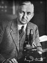
[[File:George_L._Streeter.jpg|thumb|200px|alt=Embryology History George Streeter|link=Embryology History - George Streeter|George Linius Streeter (1873-1948)]] | |||
By George L. Streeter. Wistar Institute of Anatomy, Philadelphia. | By George L. Streeter. Wistar Institute of Anatomy, Philadelphia. | ||
| Line 5: | Line 29: | ||
A morphological study of the corpus callosum and the commissure | A morphological study of the corpus callosum and the commissure | ||
of the hippocampus, based on a series of wax-plate reconstructions of | of the hippocampus, based on a series of wax-plate reconstructions of | ||
human embryos varying from 80 to 150 mm. in length. All three | human embryos varying from 80 to 150 mm. in length. All three structures cross the median line in that portion of the brain wall developed | ||
from the lamina terminalis. In 80 mm. embryos the corpus callosum | from the lamina terminalis. In 80 mm. embryos the corpus callosum | ||
consists of a round bundle of fibers lying directly on the commissure of | consists of a round bundle of fibers lying directly on the commissure of | ||
Revision as of 13:36, 6 November 2018
| Embryology - 18 Apr 2024 |
|---|
| Google Translate - select your language from the list shown below (this will open a new external page) |
|
العربية | català | 中文 | 中國傳統的 | français | Deutsche | עִברִית | हिंदी | bahasa Indonesia | italiano | 日本語 | 한국어 | မြန်မာ | Pilipino | Polskie | português | ਪੰਜਾਬੀ ਦੇ | Română | русский | Español | Swahili | Svensk | ไทย | Türkçe | اردو | ייִדיש | Tiếng Việt These external translations are automated and may not be accurate. (More? About Translations) |
Streeter GL. The cortex of the brain in the human embryo during the fourth month with special reference to the so-called Papilla of Retzius (1907) Amer. J Anat.
| Online Editor |
|---|
 See also: Triarhou LC. (2011). A review of Edward Flatau's 1894 Atlas of the Human Brain by the neurologist Sigmund Freud. Eur. Neurol. , 65, 10-5. PMID: 21109741 DOI. Squier W & Jansen A. (2010). Abnormal development of the human cerebral cortex. J. Anat. , 217, 312-23. PMID: 20979584 DOI.
Modern Notes: |

| Historic Disclaimer - information about historic embryology pages |
|---|
|
Development of the Interfore-brain Commissures in the Human Embryo
By George L. Streeter. Wistar Institute of Anatomy, Philadelphia.
A morphological study of the corpus callosum and the commissure of the hippocampus, based on a series of wax-plate reconstructions of human embryos varying from 80 to 150 mm. in length. All three structures cross the median line in that portion of the brain wall developed from the lamina terminalis. In 80 mm. embryos the corpus callosum consists of a round bundle of fibers lying directly on the commissure of the hippocampus, representing the condition found in non-placental animals. The succeeding growth consists in the lengthening of the fornix and caudal migration of the hippocampal commissure, the latter remaining in close relation to the caudal end of the corpus callosum, which in the meantime, by increase in number of fibers, has extended anterior to the anterior commissure and posterior so as to deck over the region of the third ventricle. The formation of a cavity in the septum lucidum occurs in embryos of about 95 mm. The anterior or olfactory division of the anterior commissure does not enter the olfactory bulb, but is traced to the cortex dorsal to the bulb.