Paper - Development of the interfore-brain commissures in the human embryo: Difference between revisions
mNo edit summary |
mNo edit summary |
||
| (5 intermediate revisions by the same user not shown) | |||
| Line 1: | Line 1: | ||
{{Header}} | {{Header}} | ||
{{Ref- | {{Ref-Streeter1907b}} | ||
{| class="wikitable mw-collapsible mw-collapsed" | {| class="wikitable mw-collapsible mw-collapsed" | ||
! Online Editor | ! Online Editor | ||
|- | |- | ||
| [[File:Mark_Hill.jpg|90px|left]] This historic 1907 paper by Streeter | | [[File:Mark_Hill.jpg|90px|left]] This historic 1907 paper by Streeter is a brief note on human embryo corpus callosum development. | ||
<br><br> | <br><br> | ||
{{Streeter Links}} | {{Streeter Links}} | ||
<br><br> | <br><br> | ||
'''Modern Notes:''' | '''Modern Notes:''' {{hippocampus}} | ||
{{Neural Links}} | {{Neural Links}} | ||
| Line 25: | Line 19: | ||
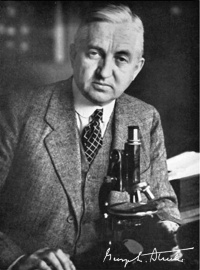
[[File:George_L._Streeter.jpg|thumb|200px|alt=Embryology History George Streeter|link=Embryology History - George Streeter|George Linius Streeter (1873-1948)]] | [[File:George_L._Streeter.jpg|thumb|200px|alt=Embryology History George Streeter|link=Embryology History - George Streeter|George Linius Streeter (1873-1948)]] | ||
By George L. Streeter. Wistar Institute of Anatomy, Philadelphia. | By [[Embryology History - George Streeter|George L. Streeter]]. | ||
Wistar Institute of Anatomy, Philadelphia. | |||
A morphological study of the corpus callosum and the commissure of the {{hippocampus}}, based on a series of wax-plate reconstructions of human embryos varying from 80 to 150 mm. in length. All three structures cross the median line in that portion of the brain wall developed from the lamina terminalis. | |||
In 80 mm. embryos the corpus callosum consists of a round bundle of fibers lying directly on the commissure of the hippocampus, representing the condition found in non-placental animals. The succeeding growth consists in the lengthening of the fornix and caudal migration of the hippocampal commissure, the latter remaining in close relation to the caudal end of the corpus callosum, which in the meantime, by increase in number of fibers, has extended anterior to the anterior commissure and posterior so as to deck over the region of the third ventricle. | |||
The formation of a cavity in the septum lucidum occurs in embryos of about 95 mm. The anterior or olfactory division of the anterior commissure does not enter the olfactory bulb, but is traced to the cortex dorsal to the bulb. | |||
{{Footer}} | |||
[[Category:Hippocampus]][[Category:Neural]][[Category:Historic Embryology]][[Category:1900's]] | |||
Latest revision as of 11:14, 17 January 2020
| Embryology - 19 Apr 2024 |
|---|
| Google Translate - select your language from the list shown below (this will open a new external page) |
|
العربية | català | 中文 | 中國傳統的 | français | Deutsche | עִברִית | हिंदी | bahasa Indonesia | italiano | 日本語 | 한국어 | မြန်မာ | Pilipino | Polskie | português | ਪੰਜਾਬੀ ਦੇ | Română | русский | Español | Swahili | Svensk | ไทย | Türkçe | اردو | ייִדיש | Tiếng Việt These external translations are automated and may not be accurate. (More? About Translations) |
Streeter GL. Development of the interfore-brain commissures in the human embryo. (1907) Amer. J Anat. :56.

| Historic Disclaimer - information about historic embryology pages |
|---|
 |
Development of the Interfore-brain Commissures in the Human Embryo
Wistar Institute of Anatomy, Philadelphia.
A morphological study of the corpus callosum and the commissure of the hippocampus, based on a series of wax-plate reconstructions of human embryos varying from 80 to 150 mm. in length. All three structures cross the median line in that portion of the brain wall developed from the lamina terminalis.
In 80 mm. embryos the corpus callosum consists of a round bundle of fibers lying directly on the commissure of the hippocampus, representing the condition found in non-placental animals. The succeeding growth consists in the lengthening of the fornix and caudal migration of the hippocampal commissure, the latter remaining in close relation to the caudal end of the corpus callosum, which in the meantime, by increase in number of fibers, has extended anterior to the anterior commissure and posterior so as to deck over the region of the third ventricle.
The formation of a cavity in the septum lucidum occurs in embryos of about 95 mm. The anterior or olfactory division of the anterior commissure does not enter the olfactory bulb, but is traced to the cortex dorsal to the bulb.
Cite this page: Hill, M.A. (2024, April 19) Embryology Paper - Development of the interfore-brain commissures in the human embryo. Retrieved from https://embryology.med.unsw.edu.au/embryology/index.php/Paper_-_Development_of_the_interfore-brain_commissures_in_the_human_embryo
- © Dr Mark Hill 2024, UNSW Embryology ISBN: 978 0 7334 2609 4 - UNSW CRICOS Provider Code No. 00098G