Paper - Congenital hernia into the umbilical cord - two cases, one associated with persistent cloaca: Difference between revisions
mNo edit summary |
mNo edit summary |
||
| (9 intermediate revisions by the same user not shown) | |||
| Line 4: | Line 4: | ||
! Online Editor | ! Online Editor | ||
|- | |- | ||
| [[File:Mark_Hill.jpg|50px|left]] This historic 1938 paper by Burns and Ogryzlo describes two cases of congenital hernia into the | | [[File:Mark_Hill.jpg|50px|left]] This historic 1938 paper by Burns and Ogryzlo describes two clinical cases of congenital abnormalities including hernia into the {{placental cord}}, and a persistent {{cloaca}}. | ||
<br> | <br> | ||
See also - | See also - {{Ref-Florian1933}} | ||
<br><br><br> | <br><br><br> | ||
'''Modern Notes:''' | '''Modern Notes:''' {{cloaca}} | {{placental cord}} | ||
<br> | <br> | ||
{{Gastrointestinal Links}} | {{Gastrointestinal Tract Links}} | ||
|} | |} | ||
{{Historic Disclaimer}} | {{Historic Disclaimer}} | ||
=Congenital Hernia into the Umbilical Cord - Two cases, one associated with Persistent Cloaca= | |||
By C. W. Burns, M.D. and M. A. Ogryzlo, M.D. Winnipeg | |||
This was a male baby, about fifteen hours old when admitted to the Winnipeg General IIospit-al. It was normally delivered, born of healthy parents. The mother was thirty—two years old, para II, gravida II, the previous child having been normal. The baby was born on January 13, 1938, at three o’clock in the morning on a farm in rural Manitoba. | |||
Transportation to Winnipeg required a ten mile ride by sleigh in below zero weather, one hundred miles by rai1, and taxi conveyance to the hospitaL The local physiciam who had skilfully conducted the accouchement, prepared the tiny patient for its journey by wrapping it in cottonwool and blankets, and placing it in an ordinary parcel This made a comfortable and essentially private conveyance. A male relative accepted the responsibility of the journey. At the expected time a basket was calm1y deposited on the admitting office counter, and from it was extracted the little patient, apparently none the worse for its unique experience. | |||
Physical examination revealed a robust, full-term | Physical examination revealed a robust, full-term infant, weighing eight pounds and thirteen ounces, whose only abnormality was a large hernia into the umbilical cord, about the size of its head. This hernia contained practically the whole of the small bowel, and was covered only by a thin transparent membrane, throu h which the loops of intestine -and peristalsis could easi y be seen. The colour of the sac was a pearly grey, while the intestinal walls were a healthy red. The neck of the sac was about three inches in diameter, and a small cuff of skin about one inch wide was carried on to it from the abdominal wall. The membrane, though intact, showed local evidence of abrasions and peeling of its outer layer. | ||
infant, weighing eight pounds and thirteen ounces, whose | |||
only abnormality was a large hernia into the umbilical | |||
cord, about the size of its head. This hernia contained | |||
practically the whole of the small bowel, and was | |||
Since the child presented no other congenital anomaly an immediate operation was decided upon. A | Since the child presented no other congenital anomaly an immediate operation was decided upon. A normal passage of urine just before operation relieved any amciety of the bladder being abnormal. | ||
normal passage of urine just before operation relieved | |||
any amciety of the bladder being abnormal. | |||
Bther was administered as a general sanæsthetic in the usual manner, the period of anæsthesia being fifteen minutes. Observing strict aseptic technique, the loops of bowel were with diiiiculty reduced into the abdominal cavity. The sac was then opened and the abdominal wall closed with one continuous chromic cat-gut suture, catching the deep fascia along with the peritoneum. No attempt was made to separate the various layers, as this would only prolong the operation. The cuff of skin was then trimmed so as to cover over the wound, and this was closed with deep interrupted silkworm gut sutures. A small binder was applied to support the wound, and the infant was given a hypodermoclysis of 50 c.c. of 5 per cent glucose solution. | |||
of | |||
the | |||
Recovery was uneventful except for a slight wound infection, and the child Evas discharged after 23 days of hospitalizatiom weighing 8 pounds and 9 ounces. It has progressed normally since its discharge from hospital on February 5, 1938. The accompanying photographs taken before and after operation are typical of the condition. | |||
of the | |||
===Discussion=== | |||
In the early months of fetal life the greater portion of the intestines lie in the proximal part of the umbilical cord, which is called the extra-coelomic cavity. Normally the intestines withdraw into the abdominal cavity at about the tenth to the twelfth week, the umbilical ring closes, and the extra-coelomic cavity is thereby obliterated In rare cases the umbilical ring does not close, and in these instances variable portions of the intestines remain in the extra-coelomic cavity, which persists. | |||
The size of the hernial mass varies, depending on the amount of intestines or other abdominal viscera it may contain. The cyst wall is composed of an outer layer of amnion lined by peritoneum, being very thin and transparent, so that its contents are easily seen. These are usually coils of small intestine, but one may find almost any of the movable intraperitoneal Organs, depending on the size of the defect in the abdominal wall. As a rule there is a cuff of skin from one-half to one inch wide, which extends from the abdominal wall on to the neck of the sac. | |||
The incidence of this condition is low, being given by Tow<sup>1</sup> as 1 in 5,000 births for the true congenital umbilical hernia, as differentiated from post-natal hernia, which is common. It is commoner in male infants and is associated with prematurity. Hempelqlorgensen2 reported two cases in one family, occurring in boys born a year apart. The parents were normal. | |||
===Treatment=== | |||
In the case of a small hernia careful reduction of the contents, with strict aseptic handling of the sac, may succeed in producing a cure. A tight strapping may be applied until the wound has completely cicatrizedg This however may take weeks and is unreliable. | |||
When the hernia is large and not reducible, as, is frequently the case, surgical operation should be undertaken as soon as possible after birth. If the peritoneum is opened, one can more readily detect other abnormalities, such as persistent vitello-intestinal duct, or patent urachus, etc. The cuff of skin facilitates closing of the wound. Provided there are no other contraindications, a general ether anæsthetic, expertly administered, is apparently quite safe. | |||
Without operation the dangers are incarceration, with strangulation or volvulus, or infection of the sac wall, which has no blood supp1y, with the development of a fatal peritonitis. | |||
0ne of the most interesting cases reported is that of Beedk Here the cord had ruptured at a point about two inches from the abdominal wall. The bed was filthy, and the intestines were covered with fæcal matter, straw, and feathers. Two hours after birth these were cleansed gently but not thoroughly, and an appendicectomy performed. The loops of bowel were then replaced and the abdominal wall closed. Recovery was uneventful. | |||
==Case 2== | |||
This baby was four hours old on admission to the Winnipeg General IIospitaL It was a first-born, of healthy parents, the delivery being normal in all respects. The duration of the pregnancy was eight months. | |||
Bxamination revealed a premature infant weighing five pounds and nine ounces, with a gross defect of its anterior abdominal wall extending from the umbilicus down through the symphysis to where the anus should be. In the region of the umbilicus there was a large hernia into the umbilical cord, about three inches in diameter. Below this the whole abdominal wall was de1icient, and was occupied by a large everted sac covered with mucous membrane, and consisting of three parts, a pale central portion and two darker lateral masses. No skin intervened between the hernia into the cord and the everted mass. In the central portion near the umbilical hernia was a conical projection with an aperture at its summit, from which meconium was expressed This was the only opening that could be found, and it was thought at first to be prolapsed sigmoid. Below this was a smaller protrusion which could be invaginated against the imperforate anus to form a small cul—de-sac about 2 cm. deep. 0n each side, lying against the thighs, was an everted mass covered with darker mucous membrane, which was interpreted as representing two halves of the bladder. An attempt was made to locate the openings of the ureters but without success. No external genitalia were present so that the sex was not determined. | |||
Postmortem — The head, chest and limbs were normal. The lower half of the anterior abdominal wall was deiicient of skin and muscles and was covered only with the scab of the cord. The pubic bone was absent. Projecting from the usual site of the pubes and perineum was a red, rounded, everted pouch, three and a half inches in diameter, covered with mucous membrane and consisting of the three parts mentioned above. At the upper end of the middle portion was a conical projection with a small opening. No other orilices were apparent, and no external genitalia. | |||
The chest was negative except for a patent foramen ovale. The Oesophagus and stomach were enormously dilated, while the duodenum and small bowel appeared normal, but the ileum ended by opening into the conical projection in the middle of the large everted pouch previously described. Thd appendix was seen arising from the left side of the pouch inside the abdominal cavity. The liver was enlarged and contained a few small congenital cysts. The kidneys were large and cystic, and the ureters dilated. They opened into the everted lateral masses on each side near the thighs. This ectopic pouch represented the latter half of the mid-gut, the hind-gut, and the bladder, the latter in two halves. Attached to the lateral masses on each side, near the openings of the ureters, was a short tortuous cord of tissue, about 3 cm. long. A section near its lower end showed this to be canalized and lined by stratified epithelium two to four cells thick These may have represented rudimentary vaginæ, but ovarian tissue could not be demonstrated anywhere. | |||
Discussion | {| | ||
| [[File:BurnsOgryzlo1938 fig01.jpg|400px]] | |||
| [[File:BurnsOgryzlo1938 fig02.jpg|400px]] | |||
|- | |||
| Fig. 1. case 1. Showing hernia into umbilical cord. | |||
| Fig. 2. Gase 1. Appearance following repair. | |||
|- | |||
| [[File:BurnsOgryzlo1938 fig03.jpg|400px]] | |||
| [[File:BurnsOgryzlo1938 fig04.jpg|400px]] | |||
|- | |||
| Fig. 3. Case 2. Lateral view. (a) Hernia into umbilical cord. (b) Opening of small intestine. (c) Lateral mass, bladder. | |||
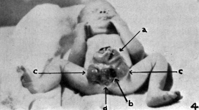
| Fig. 4. Case 2. Anterior view. Opening of ssmall intestine. (a) Hernia into umbilical cord. (b) Faeces emerging from opening of small intestine. (c) Lateral masses, bladder. (d) Imperforate anus. | |||
|} | |||
===Discussion=== | |||
The above anomaly can be exp1ained on the basis of an extroverted bladder, with persistence of the cloaca, and a hernia into the umbilical cord. | |||
In the early stages of development (4 weeks) the cloaca forms a triangle-shaped cavity at the tai1 end of the embryo, and receives the openings of the· allantois and rectum, as well as the Wolffian duct (Fig. 5a). 1ts ventral wall is largely formed by the cloacal membrane which extends upwards on the ventral wall of the allantois and on to the body stalk or umbilical cord. This membrane consists only of the two primitive layers, entoderm and ectoderm. | |||
The cloacal orifice of the rectum then migrates backwards, leaving the ventral part of the cloaca to form the bladder and urogenital sinus, and gives the appearance of these two being separated from the rectum by a septum, called the urorectal septum (Fig. 5b). When the recta1 orifice reaches the anal depressions it becomes separated from the urogenital sinus (Fig. 5c). | |||
the cloaca | |||
the | |||
the | |||
In the meantime the cloacal membrane is invaded from each side by mesodermal tissue | In the meantime the cloacal membrane is invaded from each side by mesodermal tissue growing between the two 1ayers, which then fuses along the midline, and gives rise to the infra-umbilical part of the abdominal wall as well as the anterior wall of the bladder, etc. This mesoderm arises from the primary mesenchyme and as processes of secondary mesoderm passing around the cloacal membrane from the hind end of the primitive streak. The urorectal septum in the male gives rise to the greater part of the floor of the penile urethra and perineum (Fig. 5d), while in the female the Uterus and vagina develop in it. | ||
growing between the two 1ayers, which then fuses along the midline, and gives rise to the | |||
infra-umbilical part of the abdominal wall as | |||
well as the anterior wall of the bladder, etc. | |||
This mesoderm arises from the primary mesenchyme and as processes of secondary mesoderm | |||
passing around the cloacal membrane from the | |||
hind end of the primitive | |||
septum in the male gives rise | |||
of the floor of the penile urethra and perineum | |||
( | |||
vagina develop in it. | |||
[[File:BurnsOgryzlo1938 fig05.jpg|600px]] | |||
Fig. | '''Fig. 5.''' Case 2. — Differentiation of cloaca. | ||
Al. - Allantois. B. — Bladder. cl.— cloaca. Cm.— Cloacal membrane. R. — Rectum. Ug. — Urogenital sinus. Ur. — Urorectal septum. P. — Penis. | |||
Many theories have been put forward to account for extroversions of the bladder. Roughly | Many theories have been put forward to account for extroversions of the bladder. Roughly these fall into three groups: | ||
these fall into three groups: | |||
(a) Mechanical — where the reason has been given as rupture of the cloacal membrane, or, persistence of the membrane so that it is not comp1etely replaced by mesoderm followed by rupture, or, short or absent umbilical cord. | |||
membrane by mesoderm | |||
(b) Pathological — where the cause has been given as due to ulceration of the abdominal wall and necrosis of the symphysis pubis with separation, or, fetal endometritis. | |||
(c) Arrested development — where it is due to arrest in development at an early stage of embryonal life, after the formation of the cloaca. | |||
It would seem in this instance that the development had become arrested at a very early stage, the urorectal septum having been incompletely developed. The invasion of the cloacal membrane by mesoderm has also not been completed, so that the whole infraumbilicalpart of the abdominal wall has failed to form. In practically all of these cases paired appendices and spina biiida are reported as being accompanying features. Ours had only one appendix and no spina bifida, the spinal canal being fused well along its entire length. | |||
Various degrees of ectopia vesicæ have been treated surgically with good results. In this case the malformation was so gross that no surgical procedure could be contemplated. The child was put on a formula and treated along the lines of premature infants, but it survived only fifteen days. | |||
==Summary== | |||
Two congenital anomalies have been briefly presented These are in no way alike, but both are exceedingly interesting, the first from the point of view of surgical treatment, the second from a scientiiic standpoint. No attempt has been made to discuss the theories of causation in the second case, and those interested in this aspect of the subject are referred to the bibliography cited. | |||
==Bibliography== | |||
1. Tow: Diseases of the Newborn, Oxford Medical Publioation, New York, 1937, D. 224.« . | 1. Tow: Diseases of the Newborn, Oxford Medical Publioation, New York, 1937, D. 224.« . | ||
Z. | Z. Hempel-Jorgensen, P.: Familial congenital umbilical hernia. TIERE. I. Laeger» 1929, M: 273. | ||
hernia. TIERE. I. Laeger» 1929, M: 273. | |||
Z. Rand, E. N.: lnfant disemboweled at birthz appendectomy suocessfuh J. Am. M. Ase» 1913. Cl: 199. | Z. Rand, E. N.: lnfant disemboweled at birthz appendectomy suocessfuh J. Am. M. Ase» 1913. Cl: 199. | ||
4. | 4. Freshman B.: congenital umbilical hernia, Wie Dei-need 1933. L: 701. | ||
Dei-need 1933. L: 701. | |||
5. {{Ref-Keith1902}}, P. 431. | |||
6. Keith, Sir A. Anomalies of the hind end of the body, Brit. M. J. 1908, L: 1736. 1804, 1857. | |||
7. Keith, Sir A. Malformations of the body from a new point of view, Brit. M. J» 1932, 1: 435, 489. | |||
8. WooD-Jones, R: Flxtroversion of the bladder and some problems in eonnection with it, J. Anat. es Physiol» 1912, 46: 193. | |||
9. JoHNsToN, T. B.: Extroversion of the bladder complicated by the presence of intestinal openings on the surface of the extroverted area, J. Phys. 1914, 48: 89. | |||
of | |||
10. VoN Geldern, C. B.: The etiology of extrophy of the bladder, Arcla Sarg» 1924, s: 61. | |||
II. Verm, J. R. AND MCFDTRIDGEY B. M.: Dxtrophy of the bladder, J. Pages» 1934, 4: 95. | |||
12. Potter, A. H. Ectopia vesicæ, imperforate rectum and anus. true hermaphroditism and other ano malies, Am. J. Sarg» 1936, s1: 172. | |||
13. Wyburg G. M. Development of the infra-umbilical portion of the abdominal wall, with remarlcs on etiolzoogy of ectopia vesicæ, J. Anat. ck Physiol» 1937, 71: 1. | |||
[[File:BurnsOgryzlo1938 fig01-4.jpg|600px]] | |||
{{Footer}} | {{Footer}} | ||
Latest revision as of 10:47, 14 November 2018
| Embryology - 24 Apr 2024 |
|---|
| Google Translate - select your language from the list shown below (this will open a new external page) |
|
العربية | català | 中文 | 中國傳統的 | français | Deutsche | עִברִית | हिंदी | bahasa Indonesia | italiano | 日本語 | 한국어 | မြန်မာ | Pilipino | Polskie | português | ਪੰਜਾਬੀ ਦੇ | Română | русский | Español | Swahili | Svensk | ไทย | Türkçe | اردو | ייִדיש | Tiếng Việt These external translations are automated and may not be accurate. (More? About Translations) |
Burns CW. and Ogryzlo MA. Congenital hernia into the umbilical cord; two cases, one associated with persistent cloaca. (1938) Can Med Assoc J. 39(5): 438-41. PMID 20321146
| Historic Disclaimer - information about historic embryology pages |
|---|
| Pages where the terms "Historic" (textbooks, papers, people, recommendations) appear on this site, and sections within pages where this disclaimer appears, indicate that the content and scientific understanding are specific to the time of publication. This means that while some scientific descriptions are still accurate, the terminology and interpretation of the developmental mechanisms reflect the understanding at the time of original publication and those of the preceding periods, these terms, interpretations and recommendations may not reflect our current scientific understanding. (More? Embryology History | Historic Embryology Papers) |
Congenital Hernia into the Umbilical Cord - Two cases, one associated with Persistent Cloaca
By C. W. Burns, M.D. and M. A. Ogryzlo, M.D. Winnipeg
This was a male baby, about fifteen hours old when admitted to the Winnipeg General IIospit-al. It was normally delivered, born of healthy parents. The mother was thirty—two years old, para II, gravida II, the previous child having been normal. The baby was born on January 13, 1938, at three o’clock in the morning on a farm in rural Manitoba.
Transportation to Winnipeg required a ten mile ride by sleigh in below zero weather, one hundred miles by rai1, and taxi conveyance to the hospitaL The local physiciam who had skilfully conducted the accouchement, prepared the tiny patient for its journey by wrapping it in cottonwool and blankets, and placing it in an ordinary parcel This made a comfortable and essentially private conveyance. A male relative accepted the responsibility of the journey. At the expected time a basket was calm1y deposited on the admitting office counter, and from it was extracted the little patient, apparently none the worse for its unique experience.
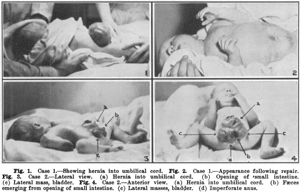
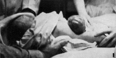
Physical examination revealed a robust, full-term infant, weighing eight pounds and thirteen ounces, whose only abnormality was a large hernia into the umbilical cord, about the size of its head. This hernia contained practically the whole of the small bowel, and was covered only by a thin transparent membrane, throu h which the loops of intestine -and peristalsis could easi y be seen. The colour of the sac was a pearly grey, while the intestinal walls were a healthy red. The neck of the sac was about three inches in diameter, and a small cuff of skin about one inch wide was carried on to it from the abdominal wall. The membrane, though intact, showed local evidence of abrasions and peeling of its outer layer.
Since the child presented no other congenital anomaly an immediate operation was decided upon. A normal passage of urine just before operation relieved any amciety of the bladder being abnormal.
Bther was administered as a general sanæsthetic in the usual manner, the period of anæsthesia being fifteen minutes. Observing strict aseptic technique, the loops of bowel were with diiiiculty reduced into the abdominal cavity. The sac was then opened and the abdominal wall closed with one continuous chromic cat-gut suture, catching the deep fascia along with the peritoneum. No attempt was made to separate the various layers, as this would only prolong the operation. The cuff of skin was then trimmed so as to cover over the wound, and this was closed with deep interrupted silkworm gut sutures. A small binder was applied to support the wound, and the infant was given a hypodermoclysis of 50 c.c. of 5 per cent glucose solution.
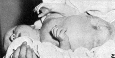
Recovery was uneventful except for a slight wound infection, and the child Evas discharged after 23 days of hospitalizatiom weighing 8 pounds and 9 ounces. It has progressed normally since its discharge from hospital on February 5, 1938. The accompanying photographs taken before and after operation are typical of the condition.
Discussion
In the early months of fetal life the greater portion of the intestines lie in the proximal part of the umbilical cord, which is called the extra-coelomic cavity. Normally the intestines withdraw into the abdominal cavity at about the tenth to the twelfth week, the umbilical ring closes, and the extra-coelomic cavity is thereby obliterated In rare cases the umbilical ring does not close, and in these instances variable portions of the intestines remain in the extra-coelomic cavity, which persists.
The size of the hernial mass varies, depending on the amount of intestines or other abdominal viscera it may contain. The cyst wall is composed of an outer layer of amnion lined by peritoneum, being very thin and transparent, so that its contents are easily seen. These are usually coils of small intestine, but one may find almost any of the movable intraperitoneal Organs, depending on the size of the defect in the abdominal wall. As a rule there is a cuff of skin from one-half to one inch wide, which extends from the abdominal wall on to the neck of the sac.
The incidence of this condition is low, being given by Tow1 as 1 in 5,000 births for the true congenital umbilical hernia, as differentiated from post-natal hernia, which is common. It is commoner in male infants and is associated with prematurity. Hempelqlorgensen2 reported two cases in one family, occurring in boys born a year apart. The parents were normal.
Treatment
In the case of a small hernia careful reduction of the contents, with strict aseptic handling of the sac, may succeed in producing a cure. A tight strapping may be applied until the wound has completely cicatrizedg This however may take weeks and is unreliable.
When the hernia is large and not reducible, as, is frequently the case, surgical operation should be undertaken as soon as possible after birth. If the peritoneum is opened, one can more readily detect other abnormalities, such as persistent vitello-intestinal duct, or patent urachus, etc. The cuff of skin facilitates closing of the wound. Provided there are no other contraindications, a general ether anæsthetic, expertly administered, is apparently quite safe.
Without operation the dangers are incarceration, with strangulation or volvulus, or infection of the sac wall, which has no blood supp1y, with the development of a fatal peritonitis.
0ne of the most interesting cases reported is that of Beedk Here the cord had ruptured at a point about two inches from the abdominal wall. The bed was filthy, and the intestines were covered with fæcal matter, straw, and feathers. Two hours after birth these were cleansed gently but not thoroughly, and an appendicectomy performed. The loops of bowel were then replaced and the abdominal wall closed. Recovery was uneventful.
Case 2
This baby was four hours old on admission to the Winnipeg General IIospitaL It was a first-born, of healthy parents, the delivery being normal in all respects. The duration of the pregnancy was eight months.
Bxamination revealed a premature infant weighing five pounds and nine ounces, with a gross defect of its anterior abdominal wall extending from the umbilicus down through the symphysis to where the anus should be. In the region of the umbilicus there was a large hernia into the umbilical cord, about three inches in diameter. Below this the whole abdominal wall was de1icient, and was occupied by a large everted sac covered with mucous membrane, and consisting of three parts, a pale central portion and two darker lateral masses. No skin intervened between the hernia into the cord and the everted mass. In the central portion near the umbilical hernia was a conical projection with an aperture at its summit, from which meconium was expressed This was the only opening that could be found, and it was thought at first to be prolapsed sigmoid. Below this was a smaller protrusion which could be invaginated against the imperforate anus to form a small cul—de-sac about 2 cm. deep. 0n each side, lying against the thighs, was an everted mass covered with darker mucous membrane, which was interpreted as representing two halves of the bladder. An attempt was made to locate the openings of the ureters but without success. No external genitalia were present so that the sex was not determined.
Postmortem — The head, chest and limbs were normal. The lower half of the anterior abdominal wall was deiicient of skin and muscles and was covered only with the scab of the cord. The pubic bone was absent. Projecting from the usual site of the pubes and perineum was a red, rounded, everted pouch, three and a half inches in diameter, covered with mucous membrane and consisting of the three parts mentioned above. At the upper end of the middle portion was a conical projection with a small opening. No other orilices were apparent, and no external genitalia.
The chest was negative except for a patent foramen ovale. The Oesophagus and stomach were enormously dilated, while the duodenum and small bowel appeared normal, but the ileum ended by opening into the conical projection in the middle of the large everted pouch previously described. Thd appendix was seen arising from the left side of the pouch inside the abdominal cavity. The liver was enlarged and contained a few small congenital cysts. The kidneys were large and cystic, and the ureters dilated. They opened into the everted lateral masses on each side near the thighs. This ectopic pouch represented the latter half of the mid-gut, the hind-gut, and the bladder, the latter in two halves. Attached to the lateral masses on each side, near the openings of the ureters, was a short tortuous cord of tissue, about 3 cm. long. A section near its lower end showed this to be canalized and lined by stratified epithelium two to four cells thick These may have represented rudimentary vaginæ, but ovarian tissue could not be demonstrated anywhere.
|
|
| Fig. 1. case 1. Showing hernia into umbilical cord. | Fig. 2. Gase 1. Appearance following repair. |

|
|
| Fig. 3. Case 2. Lateral view. (a) Hernia into umbilical cord. (b) Opening of small intestine. (c) Lateral mass, bladder. | Fig. 4. Case 2. Anterior view. Opening of ssmall intestine. (a) Hernia into umbilical cord. (b) Faeces emerging from opening of small intestine. (c) Lateral masses, bladder. (d) Imperforate anus. |
Discussion
The above anomaly can be exp1ained on the basis of an extroverted bladder, with persistence of the cloaca, and a hernia into the umbilical cord.
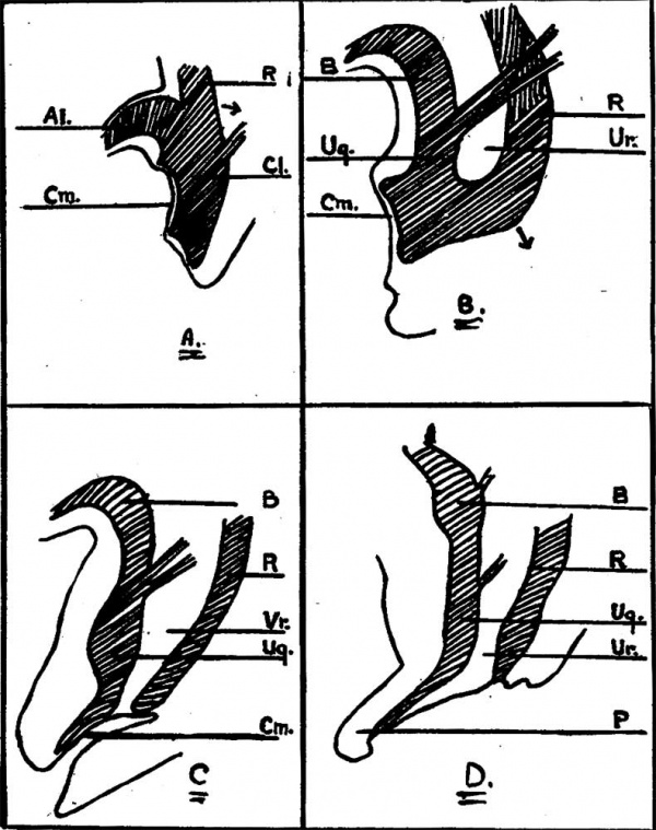
In the early stages of development (4 weeks) the cloaca forms a triangle-shaped cavity at the tai1 end of the embryo, and receives the openings of the· allantois and rectum, as well as the Wolffian duct (Fig. 5a). 1ts ventral wall is largely formed by the cloacal membrane which extends upwards on the ventral wall of the allantois and on to the body stalk or umbilical cord. This membrane consists only of the two primitive layers, entoderm and ectoderm.
The cloacal orifice of the rectum then migrates backwards, leaving the ventral part of the cloaca to form the bladder and urogenital sinus, and gives the appearance of these two being separated from the rectum by a septum, called the urorectal septum (Fig. 5b). When the recta1 orifice reaches the anal depressions it becomes separated from the urogenital sinus (Fig. 5c).
In the meantime the cloacal membrane is invaded from each side by mesodermal tissue growing between the two 1ayers, which then fuses along the midline, and gives rise to the infra-umbilical part of the abdominal wall as well as the anterior wall of the bladder, etc. This mesoderm arises from the primary mesenchyme and as processes of secondary mesoderm passing around the cloacal membrane from the hind end of the primitive streak. The urorectal septum in the male gives rise to the greater part of the floor of the penile urethra and perineum (Fig. 5d), while in the female the Uterus and vagina develop in it.
Fig. 5. Case 2. — Differentiation of cloaca.
Al. - Allantois. B. — Bladder. cl.— cloaca. Cm.— Cloacal membrane. R. — Rectum. Ug. — Urogenital sinus. Ur. — Urorectal septum. P. — Penis.
Many theories have been put forward to account for extroversions of the bladder. Roughly these fall into three groups:
(a) Mechanical — where the reason has been given as rupture of the cloacal membrane, or, persistence of the membrane so that it is not comp1etely replaced by mesoderm followed by rupture, or, short or absent umbilical cord.
(b) Pathological — where the cause has been given as due to ulceration of the abdominal wall and necrosis of the symphysis pubis with separation, or, fetal endometritis.
(c) Arrested development — where it is due to arrest in development at an early stage of embryonal life, after the formation of the cloaca.
It would seem in this instance that the development had become arrested at a very early stage, the urorectal septum having been incompletely developed. The invasion of the cloacal membrane by mesoderm has also not been completed, so that the whole infraumbilicalpart of the abdominal wall has failed to form. In practically all of these cases paired appendices and spina biiida are reported as being accompanying features. Ours had only one appendix and no spina bifida, the spinal canal being fused well along its entire length.
Various degrees of ectopia vesicæ have been treated surgically with good results. In this case the malformation was so gross that no surgical procedure could be contemplated. The child was put on a formula and treated along the lines of premature infants, but it survived only fifteen days.
Summary
Two congenital anomalies have been briefly presented These are in no way alike, but both are exceedingly interesting, the first from the point of view of surgical treatment, the second from a scientiiic standpoint. No attempt has been made to discuss the theories of causation in the second case, and those interested in this aspect of the subject are referred to the bibliography cited.
Bibliography
1. Tow: Diseases of the Newborn, Oxford Medical Publioation, New York, 1937, D. 224.« .
Z. Hempel-Jorgensen, P.: Familial congenital umbilical hernia. TIERE. I. Laeger» 1929, M: 273.
Z. Rand, E. N.: lnfant disemboweled at birthz appendectomy suocessfuh J. Am. M. Ase» 1913. Cl: 199.
4. Freshman B.: congenital umbilical hernia, Wie Dei-need 1933. L: 701.
5. Keith A. Human Embryology and Morphology. (1902) London: Edward Arnold., P. 431.
6. Keith, Sir A. Anomalies of the hind end of the body, Brit. M. J. 1908, L: 1736. 1804, 1857.
7. Keith, Sir A. Malformations of the body from a new point of view, Brit. M. J» 1932, 1: 435, 489.
8. WooD-Jones, R: Flxtroversion of the bladder and some problems in eonnection with it, J. Anat. es Physiol» 1912, 46: 193.
9. JoHNsToN, T. B.: Extroversion of the bladder complicated by the presence of intestinal openings on the surface of the extroverted area, J. Phys. 1914, 48: 89.
10. VoN Geldern, C. B.: The etiology of extrophy of the bladder, Arcla Sarg» 1924, s: 61.
II. Verm, J. R. AND MCFDTRIDGEY B. M.: Dxtrophy of the bladder, J. Pages» 1934, 4: 95.
12. Potter, A. H. Ectopia vesicæ, imperforate rectum and anus. true hermaphroditism and other ano malies, Am. J. Sarg» 1936, s1: 172.
13. Wyburg G. M. Development of the infra-umbilical portion of the abdominal wall, with remarlcs on etiolzoogy of ectopia vesicæ, J. Anat. ck Physiol» 1937, 71: 1.
Cite this page: Hill, M.A. (2024, April 24) Embryology Paper - Congenital hernia into the umbilical cord - two cases, one associated with persistent cloaca. Retrieved from https://embryology.med.unsw.edu.au/embryology/index.php/Paper_-_Congenital_hernia_into_the_umbilical_cord_-_two_cases,_one_associated_with_persistent_cloaca
- © Dr Mark Hill 2024, UNSW Embryology ISBN: 978 0 7334 2609 4 - UNSW CRICOS Provider Code No. 00098G

