Paper - A Note on the Development of the Septum Transversum and the Liver
| Embryology - 24 Apr 2024 |
|---|
| Google Translate - select your language from the list shown below (this will open a new external page) |
|
العربية | català | 中文 | 中國傳統的 | français | Deutsche | עִברִית | हिंदी | bahasa Indonesia | italiano | 日本語 | 한국어 | မြန်မာ | Pilipino | Polskie | português | ਪੰਜਾਬੀ ਦੇ | Română | русский | Español | Swahili | Svensk | ไทย | Türkçe | اردو | ייִדיש | Tiếng Việt These external translations are automated and may not be accurate. (More? About Translations) |
Thompson P. A note on the development of the septum transversum and the liver. (1908) J Anat Physiol. 42(2): 170-5. PMID 17232762
| Historic Disclaimer - information about historic embryology pages |
|---|
| Pages where the terms "Historic" (textbooks, papers, people, recommendations) appear on this site, and sections within pages where this disclaimer appears, indicate that the content and scientific understanding are specific to the time of publication. This means that while some scientific descriptions are still accurate, the terminology and interpretation of the developmental mechanisms reflect the understanding at the time of original publication and those of the preceding periods, these terms, interpretations and recommendations may not reflect our current scientific understanding. (More? Embryology History | Historic Embryology Papers) |
A Note on the Development of the Septum Transversum and the Liver
By Peter Thompson, M.D.,
Professor of Anatomy, King's College, London.
Introduction
In the description of a human embryo of twenty-three paired somites, which appeared in the April number of the Journal, it was only possible to mention briefly the arrangement of the septum transversum and the liver. In this note it is intended to amplify the account of those structures and to illustrate their form and relations to surrounding organs.
To W. His (1) we owe the first clear account of the septum transversum, and, since the publication of his work, several valuable papers have added to our knowledge of its comparative embryology. One may perhaps mention particularly the work of Ravn (2) on birds and rabbits, Wolfel (3) on ruminants, Lockwood (4«), Brachet (5), and Uskow (6) on rabbits, and Robinson (7) on rats and mice; whilst Mall (8), Bromann (9), and others have given us detailed accounts of the development of the diaphragm in man, and have dealt with the arrangement of the septum transversum in embryos of various stages of development.
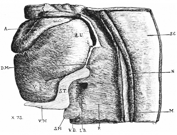
In the specimen now under consideration, which, it may be recalled, was 2.5 mm. in length, the septum transversum is seen before the cells of the liver bud have invaded the vessels which lie embedded within it. Fig. 1 is a drawing of a model reconstructed by the wax-plate method of a part of the embryo with the heart in situ. With the exception of the heart and spinal cord, the model is in sagittal section through the middle line, so that a view is obtained of the right half of the body from within. It shows the arrangement of a thick mesoblastic septum (S.T.) which is continuous cranialwards with the dorsal mesocardium and ventrally with the structure known as the ventral mesentery. The latter is continuous with the ventral body wall opposite the attachment of the root of the amnion. It will be clear, therefore, that the general disposition of the mesoderm here takes the form of an oblique septum, placed between the heart and the alimentary canal, and in front of the umbilical orifice. Leaving out of account the dorsal mesocardium, which subsequently disappears, the oblique septum consists of two parts: (1) a thick mass into which the liver bud is seen to be growing, and (2) the ventral mesentery. It is placed opposite the third and fourth cervical somites.
There is, however, a third part continuous with the septum just described, but not seen in the model, because it is situated on either side of the entodermal tube. To follow the disposition of this part of the septum clearly, it is to be noted that the thick mass into which the liver bud is growing is crescentic in shape, with the convexity ventralwards and the concavity dorsalwards, the arrangement being somewhat in the form of a collar round the floor and sides of the entodermal tube. The two cornua of the crescentic mass have embedded within them the vessels which are passing along the lateral aspect of the alimentary canal to the venous end of the heart, viz.—the umbilical and vitelline veins on each side, as well as a part of each horn of the sinus venosus. Both the crescentic mass and its cornua are continuous laterally with the body wall.
Fig 1. - Part of model of embryo, with heart in situ. x 75.
S.C., spinal cord; N., notocord ; M., mesoderm between the two dorsal aortas ; A., aortic stem ; A.U., auricle ; D.M., dorsal mesocardium; S.T., septum trausversum; V.M,, ventral mesentery; S.M., wall of vitelline duct; V.D., cavity of vitelline duct lined by entoderm ; P., duodenum ; L.B., liver bud.
It is interesting to notice here that this third constituent of the septum transversum is the first part to be formed. It is laid down before the reversal of the pericardial area, at the anterior end of the embryonic rim, as a thickening of mesoderm,, which soon unites with the extra-embryonic mesoderm, so that when the reversal of the pericardial area has been accomplished, it occupies a position in front of the umbilicus. It is continuous, on the one hand, with the somatic and splanchnic layers of the pericardial mesoderm, and on the other with the mesoderm of the amnion and the yolk sac. In it the vitelline veins run to reach the venous end of the heart, and subsequently the ducts of Cuvier and the umbilical veins, when the lateral mesocardia are formed and when the more caudal part of the septum has enlarged and united with the lateral body wall.
The obliquely placed mesodermal septum becomes greatly influenced by the outgrowth of the hepatic bud. There is a marked increase of the septum at this point, and, as the model shows, there is now a thick cushion hollowed for the reception of the liver. It is this thickening, together with the parts forming the bed for the vessels passing to the sinus venosus, which His (1) described under the name of septum transversum or primary diaphragm.
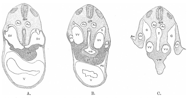
Fig. 2. - Schematic representation of three sections of embryo. A, at level of sinus venosus: B. at level of liver bud; C, at level of ventral mesentery.
N.,notochord ; Ao.,aoI-ta; S.V.,sinus venosus; S.T.,septum transversum ; C.,ca-rlom; D.C., ductus Cuv.; V., ventricle; P.C., pericanlial cavity; 8., stomach; H.B., liver bud; V.V., vltelline vein; U.V., umbilical vein; Am., amnion; V.M., ventral mesentery; I., small intestine.
From a consideration of its mode of formation it seems convenient, however, to describe the mesodermal septum, as seen in an embryo at the beginning of the third week, as consisting of the following parts: (1) the part forming the bed for the passage of vessels to the sinus venosus; (2) the thick hepatic part associated with the development of the liver; and (3) the ventral mesentery. The “pulmonary ridge” (F. P. Mall, 8), out of which the pleuro-pericardial and the pleuro-peritoneal membranes are formed, which Mall regards as an extension of the septum transversum, is not yet differentiated.
The relations of the oblique septum to the coelom are well known, but it may here be pointed out that whereas the septum is first laid down anterior to the cavity of the pericardium, after the reversal of the pericardial area it lies behind it, forming the floor of the pericardium, and later, as in the present specimen, dorso-posterior to it. The coelomic cavity on either side is indeed flexed round the dorsal and anterior free edge of the septum, so that the single pericardial cavity is below and in front and the trunk cavity above and behind. Uniting these are the two narrow passages of the coelom, known as the dorsal parietal recesses of His and Ravn. Each recess communicates with the pericardial cavity by the pleuro—pericardial opening, and with the abdominal coelom by the pleuroperitoneal opening. These recesses form the beginnings of the pleural cavities, towards Which, even at this early stage, the already paired lung buds are seen to be growing. It will be clear that the caudal boundary of each parietal recess is formed by the cornu of the transverse septum.
It is interesting to find that the arrangement of the mesoblastic septum in the human embryo is very similar to what has been described in some other mammals. Professor Robinson (7), in a paper on the development of the lungs of rats and mice, points out that “the opening of the fore-gut into the umbilical sac is placed immediately behind the heart, and is separated from the latter organ by a layer of mesoblast formed by the union of the splanchnic and somatic layers, which meet at this place. From this point of union a mesoblastic septum extends forward and dorsally to tl1e anterior end of the body cavity, which it divides into a ventro-anterior portion, the pericardium, and a dorso-posterior portion, the pleuro-peritoneal cavity. The septum contains the vitelline veins and the ducts of Cuvier, and gives origin eventually to the Walls of the pericardial sac and ventral half of the central portion of the diaphragm.” Professor Robinson’s description of the mesoblastic septum thus refers to the stage after complete separation of pericardium from pleura, later, therefore, than that described above in the human embryo; and he believed at the time that there were lateral infoldings in the pericardial area, which he has more recently shown us is not correct, as far as mammals are concerned.
Ravn (2) distinguishes in the chick four constituent parts of the whole septum transversum: (1) the middle mass into which the liver grows; (2) the two lateral mesocardia; (3) the unpaired median primary ventral ligament of the liver; and (4) the two lateral closing folds of the septum transversum. These latter folds extend inwards from the body wall to the septum transversum, between the lateral mesocardia and the primary ventral ligament of the liver, and are concerned in the closure of the “ventral parietal recesses,” which are present in birds in addition to the “ dorsal parietal recesses.”
The Liver Anlage
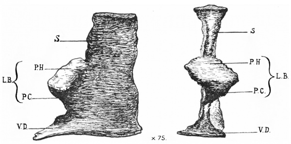
Fig. 3 represents drawings of a model of the liver bud and part of the entodermal tube, from the left side and from the front. The bud grows out from the alimentary tube opposite the third and fourth cervical somites as a median structure with thick walls enclosing a cavity in wide communication with the alimentary canal. Its relations with the septum transversum are seen in fig. 1. When seen from the front, it would appear that the greater part of the outgrowth is to the left of the middle line.
Fig. 3. — Model of entodermal tube showing the hepatic bud. x 75. Seen from the left side and the front.
S., stomach; L.B., liver bud ; P.H., pars hepatica; I’.C., pars cystica; V.D.. vitelline duct.
The gall-bladder has not yet formed, and there is no sign of the dorsal or ventral pancreas.
The liver bud exhibits an extremely early phase of development. It may be divided into two parts, cephalic and caudal, the “pars hepatica ” and the “pars cystica” of Maurer. In most mammals, according to F. Maurer (10), it is possible to distinguish the cranial division of the anlage from the caudal. The former gives rise to the group of liver cells, and the latter to the gall-bladder. An interesting model showing the further differentiation of the liver bud and its relation to the ventral pancreas in a human embryo of 4'9 mm. has been recently described and figured by Dr Ingalls (11). He finds that the two parts of the ventral pancreatic anlage arise, one on the right side, the larger of the two, and one on the left, as outgrowths between the pars hepatica and the gut, and that they unite caudally to the pars cystica. A little later there is only one ventral pancreas anlage, and it is uncertain, according to Dr Ingalls, how much the left side contributes to it.
Bibliography
(1) His, W., Anatomie Menschlichen Embryonen, Leipzig, 1885.
(2) RAVN, E., (a) “Ueber die Bildung der Scheiderwand zwischexi Brust- v. Bauchhtihle in Siiugetierembryonen,” Arclriv fiir Anat. u. Entwick, 1889. (b) “ Ueber die Entwickelung des Septum transversum ” Anat. Anz., Bd. xv., 1898, 1899.
(3) WOLFE., K., “ Beitrelge zur Entwickelung des Zwerchfells und des Magens bei Wiederkiiuern,” Anat. Anz., Bd. xxx., 1907.
(4) LOCKWOOD, C. B., “The Development of the Pericardium, Diaphragm, and Great Veins,” Proc. Roy. Soc., 1888.
(5) BRACKET, “Recherches sur le développement du diaphragme et du foie,” Joum. dc l’A'natomz'e et de la Phys. 1895.
(6) Usxow, M., “ Ueber die lflntwickelung des Zwerchfells des Pericardiums und des Ctiloms,” Archiv fair milcrosk. Anat., 1883.
(7) ROBINSON, A., “Observations on the Earlier Stages in the Development of the Lungs of Rats and Mice,” Joum. of Anat. and Phys., 1889.
(8) MALL, P. “ 0n The Development of the Human Diaphragm,” Bulletin of e o ns op ms 08p’t a, v0. x11., .
(9) BROMANN, “Ueber die Entwickelungsges. des Zwerchfells beim Menschen,” Verhan. d. anat. Gesell. auf d. 16 Verb. in Halle a. S., 1902.
(10) MAURER, F., “Article on the Development of the Alimentary Canal,” Hertun'g’s Handbuck, Sechste his achte Lieferung.
(11) INGALLS, N. W., “Beschreibung eines menschlichen Embryos von 4'9 mm.,” Arch. f. milcrosk. Anat. und Entiwiclm, Bd. lxx., 1907.
<pubmed>17232762</pubmed>
Cite this page: Hill, M.A. (2024, April 24) Embryology Paper - A Note on the Development of the Septum Transversum and the Liver. Retrieved from https://embryology.med.unsw.edu.au/embryology/index.php/Paper_-_A_Note_on_the_Development_of_the_Septum_Transversum_and_the_Liver
- © Dr Mark Hill 2024, UNSW Embryology ISBN: 978 0 7334 2609 4 - UNSW CRICOS Provider Code No. 00098G


