Palate Development: Difference between revisions
mNo edit summary |
mNo edit summary |
||
| Line 2: | Line 2: | ||
== Introduction == | == Introduction == | ||
[[File:Stage18 em11.jpg|thumb|300px|Human Embryo Face ([[Week 7]], [[Carnegie stage 18]], 44 - 48 days, CRL 13 - 17 mm)]] | [[File:Stage18 em11.jpg|thumb|300px|Human Embryo Face ([[Week 7]], [[Carnegie stage 18]], 44 - 48 days, CRL 13 - 17 mm)]] | ||
The palate has | The palate anatomically separates the nasal cavity from the oral cavity and structurally has a bony (hard) anterior component and a muscular (soft) posterior component. The oral side of the palate is covered with a squamous stratified (pluristratified) epithelium. The surface of the hard palate of most mammalian species is further thrown into a series of transversal palatal ridges or ''rugae palatinae''. Both the palatal ridge number and arrangement are also species specific. | ||
Neural crest has a major contribution to the palate development with two main and separate times of development, during embryonic (primary) and an early fetal (secondary). There are a number of molecular, mechanical and morphological steps in involving the fusion of contributing structures including a key epithelial to mesenchymal transition. | |||
The primary palate is formed by two parts: | The primary palate is formed by two parts: | ||
| Line 14: | Line 15: | ||
# posterior soft palate - muscular. | # posterior soft palate - muscular. | ||
This separation of events into embryonic and fetal period corresponds closely to the classification of abnormalities associated with the palate. | |||
| Line 71: | Line 71: | ||
* '''week 9''' - secondary palate shelves fuse, separating oral and nasal cavities. | * '''week 9''' - secondary palate shelves fuse, separating oral and nasal cavities. | ||
Embryonic Period | ===Embryonic Period=== | ||
* (week 4) - pharyngeal arch formation in rostrocaudal sequence (1, 2, 3, 4 and 6) | * (week 4) - pharyngeal arch formation in rostrocaudal sequence (1, 2, 3, 4 and 6) | ||
* First pharyngeal arch - upper maxillary (pair) and lower mandibular prominences | * First pharyngeal arch - upper maxillary (pair) and lower mandibular prominences | ||
| Line 83: | Line 83: | ||
</gallery> | </gallery> | ||
Fetal Period | ===Fetal Period=== | ||
{| | {| | ||
| | | | ||
| Line 120: | Line 120: | ||
===Frontonasal Process=== | ===Frontonasal Process=== | ||
The frontonasal process (FNP) forms the majority of the superior part of the early face primordia. It later fuses with the maxillary component of the first pharyngeal arch to form the upper jaw. Failure of this fusion event during the embryonic period leads to cleft lip. Under the surface ectoderm the process mesenchyme consists of two cell populations; neural crest cells, forming the connective tissues; and the mesoderm forming the endothelium of the vascular network. | The frontonasal process (FNP) forms the majority of the superior part of the early face primordia. It later fuses with the maxillary component of the first pharyngeal arch to form the upper jaw. Failure of this fusion event during the embryonic period leads to cleft lip. Under the surface ectoderm the process mesenchyme consists of two cell populations; neural crest cells, forming the connective tissues; and the mesoderm forming the endothelium of the vascular network. | ||
A chicken developmental model study has identified a specific surface region, the Frontonasal Ectodermal Zone (FEZ), initially induced by bone morphogenetic proteins that appears to regulate the future growth and patterning of the frontonasal process. The specific frontonasal ectodermal zone was located in the frontonasal process ectoderm flanking a boundary between Sonic hedgehog (Shh) and Fibroblast growth factor 8 (Fgf8) expression domains.<ref><pubmed>18028903</pubmed></ref> | A chicken developmental model study has identified a specific surface region, the Frontonasal Ectodermal Zone (FEZ), initially induced by bone morphogenetic proteins that appears to regulate the future growth and patterning of the frontonasal process. The specific frontonasal ectodermal zone was located in the frontonasal process ectoderm flanking a boundary between Sonic hedgehog (Shh) and Fibroblast growth factor 8 (Fgf8) expression domains.<ref><pubmed>18028903</pubmed></ref> | ||
== Embryonic Palate== | == Embryonic Palate== | ||
Revision as of 11:55, 16 May 2014
| Embryology - 25 Apr 2024 |
|---|
| Google Translate - select your language from the list shown below (this will open a new external page) |
|
العربية | català | 中文 | 中國傳統的 | français | Deutsche | עִברִית | हिंदी | bahasa Indonesia | italiano | 日本語 | 한국어 | မြန်မာ | Pilipino | Polskie | português | ਪੰਜਾਬੀ ਦੇ | Română | русский | Español | Swahili | Svensk | ไทย | Türkçe | اردو | ייִדיש | Tiếng Việt These external translations are automated and may not be accurate. (More? About Translations) |
Introduction

The palate anatomically separates the nasal cavity from the oral cavity and structurally has a bony (hard) anterior component and a muscular (soft) posterior component. The oral side of the palate is covered with a squamous stratified (pluristratified) epithelium. The surface of the hard palate of most mammalian species is further thrown into a series of transversal palatal ridges or rugae palatinae. Both the palatal ridge number and arrangement are also species specific.
Neural crest has a major contribution to the palate development with two main and separate times of development, during embryonic (primary) and an early fetal (secondary). There are a number of molecular, mechanical and morphological steps in involving the fusion of contributing structures including a key epithelial to mesenchymal transition.
The primary palate is formed by two parts:
- maxillary components of the first pharyngeal arch (lateral)
- frontonasal prominence (midline)
The secondary palate can also be divided in two anatomical parts:
- anterior hard palate - ossified (contributions from the maxilla and palatine bones)
- posterior soft palate - muscular.
This separation of events into embryonic and fetal period corresponds closely to the classification of abnormalities associated with the palate.
Some Recent Findings

|
| More recent papers |
|---|
 This table allows an automated computer search of the external PubMed database using the listed "Search term" text link.
More? References | Discussion Page | Journal Searches | 2019 References | 2020 References Search term: Cleft Palate <pubmed limit=5>Cleft Palate</pubmed> Search term: Palate Embryology <pubmed limit=5>Palate Embryology</pubmed> |
Textbooks

- The Developing Human: Clinically Oriented Embryology (8th Edition) by Keith L. Moore and T.V.N Persaud - Moore & Persaud Chapter Chapter 10 The Pharyngeal Apparatus pp201 - 240.
- Larsen’s Human Embryology by GC. Schoenwolf, SB. Bleyl, PR. Brauer and PH. Francis-West - Chapter 12 Development of the Head, the Neck, the Eyes, and the Ears pp349 - 418.
Movies
|
|
|
|
|
|
|

- Links: Movies | Ultrasound
Development Overview
- week 4 - pharyngeal arch formation, first pharngeal arch contributes mandible and maxilla.
- week 6 - 7 - primary palate formation maxillary processes and frontonasal prominence.
- week 9 - secondary palate shelves fuse, separating oral and nasal cavities.
Embryonic Period
- (week 4) - pharyngeal arch formation in rostrocaudal sequence (1, 2, 3, 4 and 6)
- First pharyngeal arch - upper maxillary (pair) and lower mandibular prominences
- Late embryonic period - maxillary prominences fuse with frontonasal prominence forming upper jaw (maxilla and upper lip)


Fetal Period
|
|
Pharyngeal Arch Development
Major features to identify for each: arch, pouch, groove and membrane. Contribute to the formation of head and neck and in the human appear at the 4th week. The first arch contributes the majority of upper and lower jaw structures.
- Pharynx - begins at the buccopharyngeal membrane (oral membrane), apposition of ectoderm with endoderm (no mesoderm between).


- branchial arch (Gk. branchia= gill)
- arch consists of all 3 trilaminar embryo layers
- ectoderm- outside
- mesoderm- core of mesenchyme
- endoderm- inside
Neural Crest
- Mesenchyme invaded by neural crest generating connective tissue components
- cartilage, bone, ligaments
- arises from midbrain and hindbrain region
Face Development

Begins week 4 centered around stomodeum, external depression at oral membrane
5 initial primordia from neural crest mesenchyme
- single frontonasal prominence (FNP) - forms forehead, nose dorsum and apex
- nasal placodes develop later bilateral, pushed medially
- paired maxillary prominences - form upper cheek and upper lip
- paired mandibular prominences - lower cheek, chin and lower lip
Frontonasal Process
The frontonasal process (FNP) forms the majority of the superior part of the early face primordia. It later fuses with the maxillary component of the first pharyngeal arch to form the upper jaw. Failure of this fusion event during the embryonic period leads to cleft lip. Under the surface ectoderm the process mesenchyme consists of two cell populations; neural crest cells, forming the connective tissues; and the mesoderm forming the endothelium of the vascular network.
A chicken developmental model study has identified a specific surface region, the Frontonasal Ectodermal Zone (FEZ), initially induced by bone morphogenetic proteins that appears to regulate the future growth and patterning of the frontonasal process. The specific frontonasal ectodermal zone was located in the frontonasal process ectoderm flanking a boundary between Sonic hedgehog (Shh) and Fibroblast growth factor 8 (Fgf8) expression domains.[6]
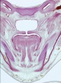
Embryonic Palate
|
Human primary palate
|

|
- EM Links: Image - stage 16 | Image - stage 17 | Image - stage 18 | Image - stage 19 | Palate Development
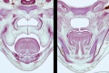
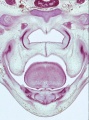
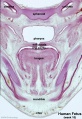
Fetal Palate
Secondary palate, fusion in the human embryo in week 9. This requires the early palatal shelves growth, elevation and fusion during the early embryonic period. The fusion event is to both each other and the primary palate. palatal shelf elevation | secondary palate
Hard and soft palate
hard palate
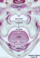
hard palate labeled
soft palate
soft palate labeled
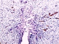
Detail - hard and soft palate junction
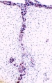
Detail - hard palate seam


Ventral aspect of hard palate of human embryo of 80 mm
Head Growth
- continues postnatally - fontanelle allow head distortion on birth and early growth
- bone plates remain unfused to allow growth, puberty growth of face
Animal Palate
Mouse Palate
- E11 - protrude from bilateral maxillary processes
- E12.5 - secondary palatal development begins
- E12.5-E14 - grow vertically along the developing tongue
- E14.5 - they elevate, meet, and fuse at the midline, to form an intact palate shelf, reflex opening and closing movements of the mouth
- E15.5 - palatal fusion is complete, mesenchymal condensation followed by osteogenic differentiation occurs.

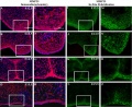
Mouse (E13.5) Palatal Shelf Wnt5a, Osr2 and Pax9 Expression.[8]
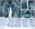
Image - Mouse E13.5 Bmp7 palate
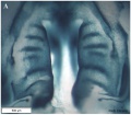
Image - palate
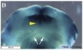
Image - palate detail


|

|
| Mouse ruga pattern (E16) | Mouse - Spry1 cleft palate |
- Links: Mouse Development | Bone Morphogenetic Protein | Wnt | Pax
Dog Palate

Newborn dog with cleft palate
Molecular
MMP25 PMID 20809987
Image - Mouse E13.5 Bmp7 palate PMID 23516636
Image - palate Bmp7 palate PMID 23516636
Image - palate detail Bmp7 palate PMID 23516636

- Links: Bone Morphogenetic Protein
Abnormalities

The way in which the upper jaw forms from fusion of the smaller upper prominence of the first pharyngeal arch leads to a common congenital defect in this region called "clefting", which may involve either the upper lip, the palate or both structures.
| Australian Palate Abnormalities (2002-2003) |
|---|
| Cleft lip with or without cleft palate (9.2 per 10,000 births) ICD-10 Q36.0, Q36.1, Q36.9, Q37.0–Q37.5, Q37.8, Q37.9 |
A congenital anomaly characterised by a partial or complete clefting of the upper lip, with or without clefting of the alveolar ridge or the hard palate. Excludes a midline cleft of the upper or lower lip and an oblique facial fissure (going towards the eye).
|
| Cleft palate without cleft lip (8.1 per 10,000 births) ICD-10 Q35.0–Q35.9 |
A congenital anomaly characterised by a closure defect of the hard and/or soft palate behind the foramen incisivum without a cleft lip. This anomaly includes sub-mucous cleft palate, but excludes cleft palate with a cleft lip, a functional short palate and high narrow palate.
|
|
International Classification of Diseases - Cleft Palate
Cleft lip and cleft palate (Q35-Q37)
Use additional code (Q30.2), if desired, to identify associated malformations of the nose. Excludes Robin's syndrome ( Q87.0 )
| Q37 | Cleft palate with cleft lip |
| Q37.0 | Cleft hard palate with bilateral cleft lip |
| Q37.1 | Cleft hard palate with unilateral cleft lip |
| Cleft hard palate with cleft lip NOS | |
| Q37.2 | Cleft soft palate with bilateral cleft lip |
| Q37.3 | Cleft soft palate with unilateral cleft lip |
| Cleft soft palate with cleft lip NOS | |
| Q37.4 | Cleft hard and soft palate with bilateral cleft lip |
| Q37.5 | Cleft hard and soft palate with unilateral cleft lip |
| Cleft hard and soft palate with cleft lip NOS | |
| Q37.8 | Unspecified cleft palate with bilateral cleft lip |
| Q37.9 | Unspecified cleft palate with unilateral cleft lip |
| Cleft palate with cleft lip NOS |
Embryonic Human Cleft Palate

|
| Stage16 (ventral view) |
Cleft Lip

|
|
| Cleft Lip Genes[10] | ||||||||||||||||||||||||||||||||||||||||||||||||||||||||||||||||||||||||||||||||||||||||||||||||||||||||||||||
|---|---|---|---|---|---|---|---|---|---|---|---|---|---|---|---|---|---|---|---|---|---|---|---|---|---|---|---|---|---|---|---|---|---|---|---|---|---|---|---|---|---|---|---|---|---|---|---|---|---|---|---|---|---|---|---|---|---|---|---|---|---|---|---|---|---|---|---|---|---|---|---|---|---|---|---|---|---|---|---|---|---|---|---|---|---|---|---|---|---|---|---|---|---|---|---|---|---|---|---|---|---|---|---|---|---|---|---|---|---|---|
Midline Cleft Lip Genes
| ||||||||||||||||||||||||||||||||||||||||||||||||||||||||||||||||||||||||||||||||||||||||||||||||||||||||||||||
|
Cleft Lip (+/− cleft palate) Genes
Cleft Palate An 8 month old infant with an extensive cleft palate associated with Bamforth- Lazarus syndrome.[11]
Cleft Risk VariantsTwo genes were identified from a recent genome-wide study.[4]
Ten most frequently reported Birth Anomalies
(From the Victorian Perinatal Data Collection Unit in the Australian state of Victoria between 2003-2004)
FolateA recent study of periconceptional folate supplementation using the Cochrane Pregnancy and Childbirth Group's Trials Register (July 2010) identified no statistically significant evidence of any effects on prevention of cleft palate and cleft lip at birth.[12] References
Journals
ReviewsIndian J Plast Surg. 2009 October; 42(Suppl):Cleft Lip and Palate Issue <pubmed>22186724</pubmed> <pubmed>19131313</pubmed> <pubmed>16962647</pubmed> <pubmed>3074914</pubmed> <pubmed>8714286</pubmed> Articles<pubmed></pubmed> <pubmed></pubmed> <pubmed></pubmed> <pubmed>20149609</pubmed> <pubmed>19341725</pubmed> Search PubMedSearch Pubmed: palate development | cleft palate development | Additional Images
Historic
Terms
External LinksExternal Links Notice - The dynamic nature of the internet may mean that some of these listed links may no longer function. If the link no longer works search the web with the link text or name. Links to any external commercial sites are provided for information purposes only and should never be considered an endorsement. UNSW Embryology is provided as an educational resource with no clinical information or commercial affiliation.
Glossary Links
Cite this page: Hill, M.A. (2024, April 25) Embryology Palate Development. Retrieved from https://embryology.med.unsw.edu.au/embryology/index.php/Palate_Development
|


