Neural Tube Movie: Difference between revisions
mNo edit summary |
mNo edit summary |
||
| Line 22: | Line 22: | ||
:'''Links:''' [[Media:Neuraltube_001.mp4|MP4 version]] | [[Media:Neuraltube_001.mov|Quicktime version]] | [[Neural_System_Development|Neural Development]] | [[Lecture_-_Ectoderm_Development|Lecture - Early Neural Development]] | :'''Links:''' [[Media:Neuraltube_001.mp4|MP4 version]] | [[Media:Neuraltube_001.mov|Quicktime version]] | [[Neural_System_Development|Neural Development]] | [[Lecture_-_Ectoderm_Development|Lecture - Early Neural Development]] | ||
{| | {| | ||
| Line 31: | Line 27: | ||
| valign="bottom"|{{Neural tube movie}} | | valign="bottom"|{{Neural tube movie}} | ||
|} | |} | ||
|- | |||
|} | |||
{{Movie footer}} | {{Movie footer}} | ||
[[Category:Week 3]] [[Category:Neural]] | [[Category:Week 3]] [[Category:Neural]] | ||
Revision as of 13:10, 8 May 2014
| Embryology - 19 Apr 2024 |
|---|
| Google Translate - select your language from the list shown below (this will open a new external page) |
|
العربية | català | 中文 | 中國傳統的 | français | Deutsche | עִברִית | हिंदी | bahasa Indonesia | italiano | 日本語 | 한국어 | မြန်မာ | Pilipino | Polskie | português | ਪੰਜਾਬੀ ਦੇ | Română | русский | Español | Swahili | Svensk | ไทย | Türkçe | اردو | ייִדיש | Tiếng Việt These external translations are automated and may not be accurate. (More? About Translations) |
| <mediaplayer width='480' height='480' image="http://php.med.unsw.edu.au/embryology/images/6/6a/Neuraltube_001_icon.jpg">File:Neuraltube_001.mp4</mediaplayer> |
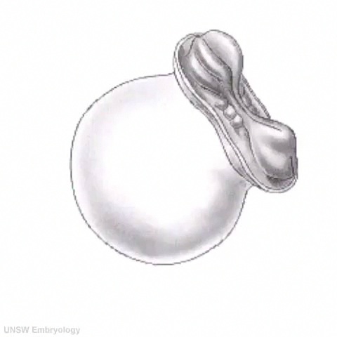
Early Neural DevelopmentThis animation of early neural development from week 3 onward shows the neural groove fusing to form the neural tube. View - Dorsolateral of the whole early embryo and yolk sac. Cranial (head) to top and caudal (tail) to bottom. Yolk sac is shown to the left.
|
Glossary Links: A | B | C | D | E | F | G | H | I | J | K | L | M | N | O | P | Q | R | S | T | U | V | W | X | Y | Z | Numbers | Symbols | Movies
Cite this page: Hill, M.A. (2024, April 19) Embryology Neural Tube Movie. Retrieved from https://embryology.med.unsw.edu.au/embryology/index.php/Neural_Tube_Movie
- © Dr Mark Hill 2024, UNSW Embryology ISBN: 978 0 7334 2609 4 - UNSW CRICOS Provider Code No. 00098G