The printable version is no longer supported and may have rendering errors. Please update your browser bookmarks and please use the default browser print function instead.
Introduction
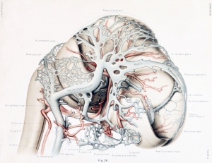
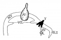
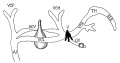
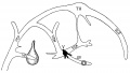
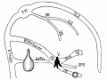
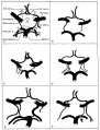
Human Embryo vascular development (week 8, stage 20 Carnegie Embryo No. 460)
This page relates specifically to vascular development associated with the central nervous system. There is an additional page that looks at vascular development and remodelling.
See the historic articles on human vascular development by:
Mall FP. On the development of the blood-vessels of the brain in the human embryo. (1905) Amer. J Anat. 4(1): 1–18.
Streeter GL. The developmental alterations in the vascular system of the brain of the human embryo. (1921) Contrib. Embryol., Carnegie Inst. Wash. 8:7-38.
Human embryo 50 mm long (Carnegie Collection, No. 96.
Some Recent Findings
- Formation of the circle of Willis during human embryonic development[1] "The circle of Willis (CW) is a circulatory anastomosis that supplies blood to the brain and adjacent structures. We examined the timing of formation of CW in 20 Japanese human embryo samples by using 3-dimensional reconstruction of serial histological sections. The CW was closed in 1 (n = 6), 2 (n = 8), 2 (n = 3) and 2 (n = 3) samples at Carnegie stages 20, 21, 22, and 23, respectively. The CW was unclosed in 13 samples (unclosed at ACOM alone, 6 samples; ACOM and bilateral P1, 4; left PCOM and right P1, 1; right PCOM and right P1, 1; ACOM and left PCOM, 1). It was difficult to predict whether the circle would close during further development, as such variations frequently exist in adults."
- Foxc1 is required for early stage telencephalic vascular development[2] "The brain vascular system arises from the perineural vascular plexus (PNVP) which sprouts radially into the neuroepithelium and subsequently branches off laterally to form a secondary plexus in the subventricular zone (SVZ), the subventricular vascular plexus (SVP). The process of SVP formation remains to be fully elucidated. We investigated the role of Foxc1 in early stage vascular formation in the ventral telencephalon. Results: The Foxc1 loss of function mutant mouse, Foxc1ch/ch , showed enlarged telencephalon and hemorrhaging in the ventral telencephalon by E11.0. The mutant demonstrated blood vessel dilation and aggregation of endothelial cells in the SVZ after the invasion of endothelial cells through the radial path, which lead to failure of SVP formation. During this early stage of vascular development, Foxc1 was expressed in endothelial cells and pericytes, as well as in cranial mesenchyme surrounding the neural tube."
- Review - The human brain intracerebral microvascular system: development and structure.[3] "The capillary from the meningeal inner pial lamella play a crucial role in the development and structural organization of the cerebral cortex extrinsic and intrinsic microvascular compartments. Only pial capillaries are capable of perforating through the cortex external glial limiting membrane (EGLM) to enter into the nervous tissue, although incapable of perforating the membrane to exit the brain. Circulatory dynamics and functional demands determine which capillaries become arterial and which capillaries become venous."
|
| More recent papers
|
|
This table allows an automated computer search of the external PubMed database using the listed "Search term" text link.
- This search now requires a manual link as the original PubMed extension has been disabled.
- The displayed list of references do not reflect any editorial selection of material based on content or relevance.
- References also appear on this list based upon the date of the actual page viewing.
References listed on the rest of the content page and the associated discussion page (listed under the publication year sub-headings) do include some editorial selection based upon both relevance and availability.
More? References | Discussion Page | Journal Searches | 2019 References | 2020 References
Search term: Neural Vascular System Development
<pubmed limit=5>Neural Vascular System Development</pubmed>
|
Cerebral Blood Supply Development
|
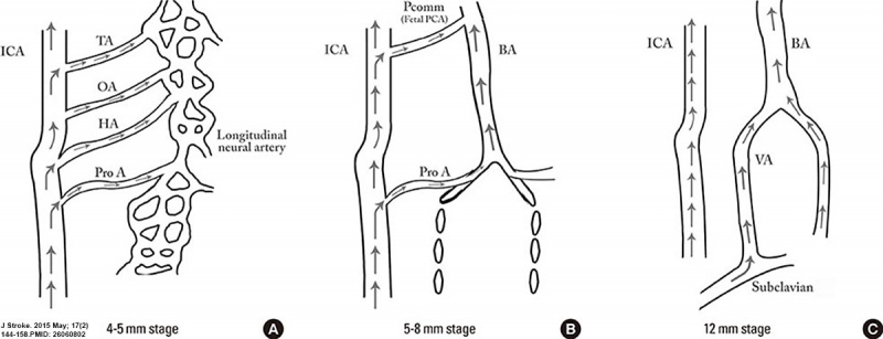
Embryonic stage
- 4-5 mm - hindbrain (i.e., future posterior fossa) is supplied by two parallel neural arteries (or channels). These arteries obtain their blood supply from carotid-vertebrobasilar anastomoses given by the trigeminal artery (TA), the otic artery (OA), hypoglossal artery (HA), and the proatlantal artery (ProA)
- 5-8 mm - basilar artery (BA) forms from the consolidation of the neural arteries.
- 7-12 mm - vertebral arteries (VA) forms from transverse anastomoses between cervical intersegmental arteries, beginning with the ProA and proceeding downward to the 6th intersegmental artery,
- 11-12 mm - (35 days) development of the middle cerebral artery (MCA) is first identified as small buds originating proximal to the anterior cerebral artery (ACA) on the anterior division of the primitive internal carotid artery (ICA).
- 16-18 mm - middle cerebral artery (MCA) becomes more prominent, the plexi fuse into a single artery and further branches pierce the cerebral hemisphere.
- 18 mm - stem of the ACA gives rise to the olfactory artery.
- 21-24 mm - formation of the anterior communicating artery (ACOMM).
|
- A - In early phases of development the posterior circulation relies almost entirely from blood supply coming from the anterior circulation through carotid-vertebrobasilar anastomoses.
- B and C - As the posterior fossa structures and the occipital lobe grow, the posterior circulation becomes progressively independent from the anterior circulation with obliteration of the anterior-posterior anastomoses from caudal to rostral maintaining in the majority of adult only one connection between the distal basilar arteries with the carotid artery via the posterior communicating artery.
|
(above text modified from reference[4])
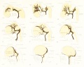
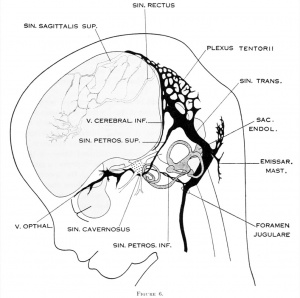
Cerebral Veins
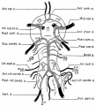
Figures from the 1905 study by Mall.[5]
Fig 14. Veins of the head of an embryo four weeks old.
Fig 15. Veins of the head during the fifth week.
Fig 16. Veins of the head beginning of third month.
Fig 17. Veins of the brain of an older foetus.
Molecular
Foxc1
- required for early stage telencephalic vascular development[2]
References
Online Textbooks
Reviews
<pubmed></pubmed>
<pubmed></pubmed>
<pubmed></pubmed>
<pubmed></pubmed>
<pubmed>22993505</pubmed>
<pubmed>20561492</pubmed>
Articles
<pubmed></pubmed>
<pubmed></pubmed>
<pubmed></pubmed>
<pubmed></pubmed>
<pubmed>468705</pubmed>
Search PubMed
Search Pubmed: neural vascular system development |
External Links
External Links Notice - The dynamic nature of the internet may mean that some of these listed links may no longer function. If the link no longer works search the web with the link text or name. Links to any external commercial sites are provided for information purposes only and should never be considered an endorsement. UNSW Embryology is provided as an educational resource with no clinical information or commercial affiliation.
Additional Images
Historic
| Historic Disclaimer - information about historic embryology pages
|
| Pages where the terms "Historic" (textbooks, papers, people, recommendations) appear on this site, and sections within pages where this disclaimer appears, indicate that the content and scientific understanding are specific to the time of publication. This means that while some scientific descriptions are still accurate, the terminology and interpretation of the developmental mechanisms reflect the understanding at the time of original publication and those of the preceding periods, these terms, interpretations and recommendations may not reflect our current scientific understanding. (More? Embryology History | Historic Embryology Papers)
|
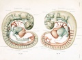
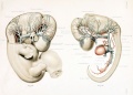
The Developmental Alterations in the Vascular System of the Brain of the Human Embryo (1921)
Gillilan, LA. Significant superficial anastomoses in the arterial blood supply to the human brain. J Comp Neurol. 1959 Jun;112:55-74. PMID 13850118
Fig. 1. Different patterns of the Circle of Willis
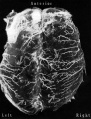
Fig. 2. Complex of arteries on the Ventral Surface of the Brain
Fig. 3. Fetal Brain (6 month) Large Superficial Arteries
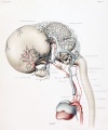
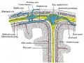
Gray, Henry. Anatomy of the Human Body Philadelphia: Lea & Febiger, 1918.
Fig. 769. Diagrammatic representation of a section across the top of the skull
Terms
| Cardiovascular Terms
|
Cardiovascular System Development See also Heart terms, Immune terms and Blood terms.
- angioblast - the stem cells in blood islands generating endothelial cells which will form the walls of both arteries and veins. (More? Blood Vessel)
- angiogenesis - the formation of new blood vessels from pre-existing vessels following from vasculogenesis in the embryo. (More? Blood Vessel)
- anlage (German, anlage = primordium) structure or cells which will form a future more developed or differentiated adult structure.
- blood islands - earliest sites of blood vessel and blood cell formation, seen mainly on yolk sac chorion.
- cardinal veins - paired main systemic veins of early embryo, anterior, common, posterior.
- cardiogenic region - region above prechordal plate in mesoderm where heart tube initially forms.
- ectoderm - the layer (of the 3 germ cell layers) which form the nervous system from the neural tube and neural crest and also generates the epithelia covering the embryo.
- endoderm - the layer (of the 3 germ cell layers) which form the epithelial lining of the gastrointestinal tract (GIT) and accessory organs of GIT in the embryo.
- endocardium - lines the heart. Epithelial tissue lining the inner surface of heart chambers and valves.
- endothelial cells - single layer of cells closest to lumen that line blood vessels.
- extraembryonic mesoderm - mesoderm lying outside the trilaminar embryonic disc covering the yolk sac, lining the chorionic sac and forming the connecting stalk. Contributes to placental villi development.
- haemocytoblasts - stem cells for embryonic blood cell formation.
- anastomose - to connect or join by a connection (anastomosis) between tubular structures.
- chorionic villi - the finger-like extensions which are the functional region of the placental barrier and maternal/fetal exchange. Develop from week 2 onward as: primary, secondary, tertiary villi.
- estrogens - support the maternal endometrium.
- growth factor - usually a protein or peptide that will bind a cell membrane receptor and then activates an intracellular signaling pathway. The function of the pathway will be to alter the cell directly or indirectly by changing gene expression. (eg VEGF, shh)
- intra-aortic hematopoietic cluster - (IAHC) blood stem cells associated with the endothelial layer of aorta and large arteries.
- maternal decidua - region of uterine endometrium where blastocyst implants. undergoes modification following implantation, decidual reaction.
- maternal sinusoids - placental spaces around chorionic villi that are filled with maternal blood. Closest maternal/fetal exchange site.
- Megakaryocytopoiesis - the process of bone marrow progenitor cells developMENT into mature megakaryocytes.
- mesoderm - the middle layer of the 3 germ cell layers of the embryo. Mesoderm outside the embryo and covering the amnion, yolk and chorion sacs is extraembryonic mesoderm.
- myocardium - muscular wall of the heart. Thickest layer formed by spirally arranged cardiac muscle cells.
- pericardium - covers the heart. Formed by 3 layers consisting of a fibrous pericardium and a double layered serous pericardium (parietal layer and visceral epicardium layer).
- pericytes - (Rouget cells) cells located at the abluminal surface of microvessels close to endothelial cells, mainly found associated with CNS vessels and involved in vessel formation, remodeling and stabilization.
- pharyngeal arches (=branchial arches, Gk. gill) series of cranial folds that form most structures of the head and neck. Six arches form but only 4 form any structures. Each arch has a pouch, membrane and groove.
- placenta - (Greek, plakuos = flat cake) refers to the discoid shape of the placenta, embryonic (villous chorion)/maternal organ (decidua basalis)
- placental veins - paired initially then only left at end of embryonic period, carry oxygenated blood to the embryo (sinus venosus).
- protein hormone - usually a protein distributed in the blood that binds to membrane receptors on target cells in different tissues. Do not easliy cross placental barrier.
- sinus venosus - cavity into which all major embryonic paired veins supply (vitelline, placental, cardinal).
- splanchnic mesoderm - portion of lateral plate mesoderm closest to the endoderm when coelom forms.
- steroid hormone - lipid soluble hormone that easily crosses membranes to bind receptors in cytoplasm or nucleus of target cells. Hormone+Receptor then binds DNA activating or suppressing gene transcription. Easliy cross placental barrier.
- syncitiotrophoblast extraembryonic cells of trophoblastic shell surrounding embryo, outside the cytotrophoblast layer, involved with implantation of the blastocyst by eroding extracellular matrix surrounding maternal endometrial cells at site of implantation, also contribute to villi. (dark staining, multinucleated).
- truncus arteriosus - an embryological heart outflow structure, that forms in early cardiac development and will later divides into the pulmonary artery and aorta. Term is also used clinically to describe the malformation where only one artery arises from the heart and forms the aorta and pulmonary artery.
- vascular endothelial growth factor - (VEGF) A secreted protein growth factor family, which stimulates the proliferation of vasular endotheial cells and therefore blood vessel growth. VEGF's have several roles in embryonic development. The VEGF family has 7 members (VEGF-A, VEGF-B, VEGF-C, VEGF-D, VEGF-E, VEGF-F, and PlGF) that have a common VEGF homology domain. PIGF is the placental growth factor. They act through 3 VEGF tyrosine kinase membrane receptors (VEGFR-1 to 3) with seven immunoglobulin-like domains in the extracellular domain, a single transmembrane region, and an intracellular tyrosine kinase sequence.
- vasculogenesis - the formation of new blood vessels from mesoderm forming the endothelium. Compared to angiogenesis that is the process of blood vessel formation from pre-existing vessels.
- vitelline blood vessels - blood vessels associated with the yolk sac.
- waste products - products of cellular metabolism and cellular debris, e.g.- urea, uric acid, bilirubin.
|
Glossary Links
- Glossary: A | B | C | D | E | F | G | H | I | J | K | L | M | N | O | P | Q | R | S | T | U | V | W | X | Y | Z | Numbers | Symbols | Term Link
Cite this page: Hill, M.A. (2024, April 19) Embryology Neural - Vascular Development. Retrieved from https://embryology.med.unsw.edu.au/embryology/index.php/Neural_-_Vascular_Development
- What Links Here?
- © Dr Mark Hill 2024, UNSW Embryology ISBN: 978 0 7334 2609 4 - UNSW CRICOS Provider Code No. 00098G