The printable version is no longer supported and may have rendering errors. Please update your browser bookmarks and please use the default browser print function instead.
Notice - Mark Hill
Currently this is only a template page and is being updated (this notice removed when completed).
Introduction
Neural development is one of the earliest systems to begin and the last to be completed after birth. This development generates the most complex structure within the embryo and the long time period of development means in utero insult during pregnancy may have consequences to development of the nervous system.
The early central nervous system begins as a simple neural plate that folds to form a groove then tube, open initially at each end. Failure of these opening to close contributes a major class of neural abnormalities (neural tube defects).
Within the neural tube stem cells generate the 2 major classes of cells that make the majority of the nervous system : neurons and glia. Both these classes of cells differentiate into many different types generated with highly specialized functions and shapes. This section covers the establishment of neural populations, the inductive influences of surrounding tissues and the sequential generation of neurons establishing the layered structure seen in the brain and spinal cord.
- Neural development beginnings quite early, therefore also look at notes covering Week 3- neural tube and Week 4-early nervous system.
- Development of the neural crest and sensory systems (hearing/vision/smell) are only introduced in these notes and are covered in other notes sections.
Some Recent Findings
- Isthmus organizer for mesencephalon and metencephalon[1] " Brain vesicles formation is the first sign of regionalization. Classical transplantation using quail and chick embryos revealed that the mesencephalon-metencephalon boundary (isthmus) functions as an organizer of the mesencephalon and metencephalon. Fgf8 is accepted as a main organizing molecule of the isthmus. Strong Fgf8 signal activates the Ras-ERK signaling pathway to differentiate the cerebellum. In this review, the historical background of the means of identifying the isthmus organizer and the molecular mechanisms of signal transduction for tectum and cerebellum differentiation is reviewed."
- Role of Lmx1b and Wnt1 in mesencephalon and metencephalon development[2] "The isthmus is the organizing center for the tectum and cerebellum. Fgf8 and Wnt1 are secreted molecules expressed around the isthmus. The function of Fgf8 has been well analyzed, and now accepted as the most important organizing signal. Involvement of Wnt1 in the isthmic organizing activity was suggested by analysis of Wnt1 knockout mice. But its role in isthmic organizing activity is still obscure. Recently, it has been shown that Lmx1b is expressed in the isthmic region and that it may occupy higher hierarchical position in the gene expression cascade in the isthmus. We have carried out misexpression experiment of Lmx1b and Wnt1, and considered their role in the isthmic organizing activity. Lmx1b or Wnt1 misexpression caused expansion of the tectum and cerebellum. Fgf8 was repressed in a cells that misexpress Lmx1b, but Fgf8 expression was induced around Lmx1b-misexpressing cells. As Lmx1b induced Wnt1 and Wnt1 induced Fgf8 expression in turn, Wnt1 may be involved in non cell-autonomous induction of Fgf8 expression by Lmx1b. Wnt1 could not induce Lmx1b expression so that Lmx1b may be put at the higher hierarchical position than Wnt1 in gene expression cascade in the isthmus. We have examined the relationship among isthmus related genes, and discuss the mechanism of the formation and maintenance of isthmic organizing activity."
- Early mesencephalon/metencephalon patterning and development of the cerebellum[3] "Fate mapping studies in chick have shown that at early stages the cerebellum derives from cells in the mesencephalon and metencephalon (mes-met). Transplantation studies in chick have implicated the mes-met junction (isthmus) as a source of secreted factors that organize development of the entire mes-met, perhaps by stimulating proliferation and specifying positional values across the region. Fgf-8 has been implicated as a major factor involved in the isthmus organizing activity. Gene expression studies indicate that the anterior and posterior expression domains of the homeobox genes Otx-2 and Gbx-2, respectively, are the earliest indication of a division of the brain."
|
Development Overview
Neuralation begins at the trilaminar embryo with formation of the notochord and somites, both of which underly the ectoderm and do not contribute to the nervous system, but are involved with patterning its initial formation. The central portion of the ectoderm then forms the neural plate that folds to form the neural tube, that will eventually form the entire central nervous system.
- Early developmental sequence: Epiblast - Ectoderm - Neural Plate - Neural groove and Neural Crest - Neural Tube and Neural Crest
Neural Tube Development
| Neural Tube
|
Primary Vesicles
|
Secondary Vesicles
|
Adult Structures
|
| week 3
|
week 4
|
week 5
|
adult
|
neural plate
neural groove
neural tube
Brain
|
prosencephalon (forebrain)
|
telencephalon
|
Rhinencephalon, Amygdala, hippocampus, cerebrum (cortex), hypothalamus, pituitary | Basal Ganglia, lateral ventricles
|
| diencephalon
|
epithalamus, thalamus, Subthalamus, pineal, posterior commissure, pretectum, third ventricle
|
| mesencephalon (midbrain)
|
mesencephalon
|
tectum, Cerebral peduncle, cerebral aqueduct, pons
|
| rhombencephalon (hindbrain)
|
metencephalon
|
cerebellum
|
| myelencephalon
|
medulla oblongata, isthmus
|
| spinal cord, pyramidal decussation, central canal
|
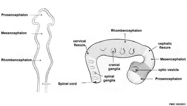
Early Brain Vesicles
Primary Vesicles
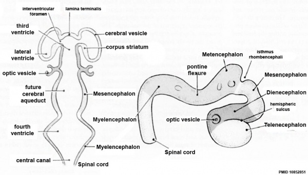
Secondary Vesicles
References
- ↑ <pubmed>18494704</pubmed>
- ↑ <pubmed>12399317</pubmed>
- ↑ <pubmed>9509514</pubmed>
Reviews
<pubmed>19206138</pubmed>
Articles
Search PubMed
Search Pubmed: Telencephalon Embryology | Telencephalon Development |
Glossary Links
- Glossary: A | B | C | D | E | F | G | H | I | J | K | L | M | N | O | P | Q | R | S | T | U | V | W | X | Y | Z | Numbers | Symbols | Term Link
Cite this page: Hill, M.A. (2024, April 19) Embryology Neural - Telencephalon Development. Retrieved from https://embryology.med.unsw.edu.au/embryology/index.php/Neural_-_Telencephalon_Development
- What Links Here?
- © Dr Mark Hill 2024, UNSW Embryology ISBN: 978 0 7334 2609 4 - UNSW CRICOS Provider Code No. 00098G


