Neural - Prosencephalon Development
| Embryology - 19 Apr 2024 |
|---|
| Google Translate - select your language from the list shown below (this will open a new external page) |
|
العربية | català | 中文 | 中國傳統的 | français | Deutsche | עִברִית | हिंदी | bahasa Indonesia | italiano | 日本語 | 한국어 | မြန်မာ | Pilipino | Polskie | português | ਪੰਜਾਬੀ ਦੇ | Română | русский | Español | Swahili | Svensk | ไทย | Türkçe | اردو | ייִדיש | Tiếng Việt These external translations are automated and may not be accurate. (More? About Translations) |
Introduction
Neural development is one of the earliest systems to begin and the last to be completed after birth. This development generates the most complex structure within the embryo and the long time period of development means in utero insult during pregnancy may have consequences to development of the nervous system.
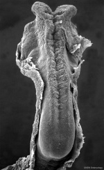
The early central nervous system begins as a simple neural plate that folds to form a groove then tube, open initially at each end. Failure of these opening to close contributes a major class of neural abnormalities (neural tube defects).
Within the neural tube stem cells generate the 2 major classes of cells that make the majority of the nervous system : neurons and glia. Both these classes of cells differentiate into many different types generated with highly specialized functions and shapes. This section covers the establishment of neural populations, the inductive influences of surrounding tissues and the sequential generation of neurons establishing the layered structure seen in the brain and spinal cord.
- Neural development beginnings quite early, therefore also look at notes covering Week 3- neural tube and Week 4-early nervous system.
- Development of the neural crest and sensory systems (hearing/vision/smell) are only introduced in these notes and are covered in other notes sections.
Some Recent Findings
|
| More recent papers |
|---|
|
This table allows an automated computer search of the external PubMed database using the listed "Search term" text link.
More? References | Discussion Page | Journal Searches | 2019 References | 2020 References Search term: Prosencephalon Development | Prosencephalon Embryology | Prosencephalon |
| Older papers |
|---|
| These papers originally appeared in the Some Recent Findings table, but as that list grew in length have now been shuffled down to this collapsible table.
See also the Discussion Page for other references listed by year and References on this current page. |
Development Overview
Neuralation begins at the trilaminar embryo with formation of the notochord and somites, both of which underly the ectoderm and do not contribute to the nervous system, but are involved with patterning its initial formation. The central portion of the ectoderm then forms the neural plate that folds to form the neural tube, that will eventually form the entire central nervous system.
- Early developmental sequence: Epiblast - Ectoderm - Neural Plate - Neural groove and Neural Crest - Neural Tube and Neural Crest
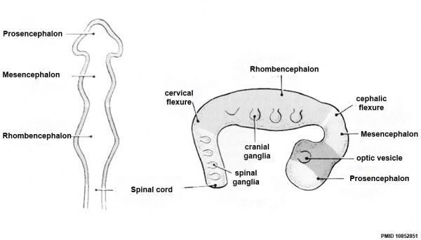
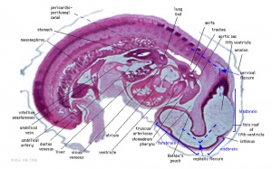
| Neural Tube | Primary Vesicles | Secondary Vesicles | Adult Structures |
|---|---|---|---|
| week 3 | week 4 | week 5 | adult |
| prosencephalon (forebrain) | telencephalon | Rhinencephalon, Amygdala, hippocampus, cerebrum (cortex), hypothalamus, pituitary | Basal Ganglia, lateral ventricles | |
| diencephalon | epithalamus, thalamus, Subthalamus, pineal, posterior commissure, pretectum, third ventricle | ||
| mesencephalon (midbrain) | mesencephalon | tectum, Cerebral peduncle, cerebral aqueduct, pons | |
| rhombencephalon (hindbrain) | metencephalon | cerebellum | |
| myelencephalon | medulla oblongata, isthmus | ||
| spinal cord, pyramidal decussation, central canal | |||
Primary Vesicles
Additional Images
Historic
| Human Embryology And Morphology (1921) |
|---|
| Keith, A. Human Embryology And Morphology (1921) Longmans, Green & Co.:New York.
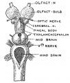
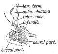
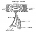
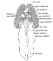
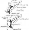
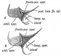
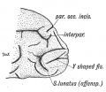
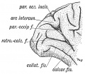
9 Fore-Brain and 10 Fore-Brain Cerebral Vesicles |
References
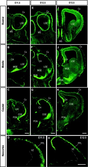
- ↑ Barber M, Di Meglio T, Andrews WD, Hernández-Miranda LR, Murakami F, Chédotal A & Parnavelas JG. (2009). The role of Robo3 in the development of cortical interneurons. Cereb. Cortex , 19 Suppl 1, i22-31. PMID: 19366869 DOI.
- ↑ Zhang Z, Liu S, Lin X, Teng G, Yu T, Fang F & Zang F. (2011). Development of laminar organization of the fetal cerebrum at 3.0T and 7.0T: a postmortem MRI study. Neuroradiology , 53, 177-84. PMID: 20981415 DOI.
Reviews
Hoch RV, Rubenstein JL & Pleasure S. (2009). Genes and signaling events that establish regional patterning of the mammalian forebrain. Semin. Cell Dev. Biol. , 20, 378-86. PMID: 19560042 DOI.
Lindwall C, Fothergill T & Richards LJ. (2007). Commissure formation in the mammalian forebrain. Curr. Opin. Neurobiol. , 17, 3-14. PMID: 17275286 DOI.
Bertrand N & Dahmane N. (2006). Sonic hedgehog signaling in forebrain development and its interactions with pathways that modify its effects. Trends Cell Biol. , 16, 597-605. PMID: 17030124 DOI.
Rhinn M, Picker A & Brand M. (2006). Global and local mechanisms of forebrain and midbrain patterning. Curr. Opin. Neurobiol. , 16, 5-12. PMID: 16418000 DOI.
Marín O & Rubenstein JL. (2003). Cell migration in the forebrain. Annu. Rev. Neurosci. , 26, 441-83. PMID: 12626695 DOI.
Rallu M, Corbin JG & Fishell G. (2002). Parsing the prosencephalon. Nat. Rev. Neurosci. , 3, 943-51. PMID: 12461551 DOI.
Articles
Ohkubo Y, Chiang C & Rubenstein JL. (2002). Coordinate regulation and synergistic actions of BMP4, SHH and FGF8 in the rostral prosencephalon regulate morphogenesis of the telencephalic and optic vesicles. Neuroscience , 111, 1-17. PMID: 11955708
Hidalgo-Sánchez M & Alvarado-Mallart RM. (2002). Temporal sequence of gene expression leading caudal prosencephalon to develop a midbrain/hindbrain phenotype. Dev. Dyn. , 223, 141-7. PMID: 11803577 DOI.
Crossley PH, Martinez S, Ohkubo Y & Rubenstein JL. (2001). Coordinate expression of Fgf8, Otx2, Bmp4, and Shh in the rostral prosencephalon during development of the telencephalic and optic vesicles. Neuroscience , 108, 183-206. PMID: 11734354
Ulfig N, Neudörfer F & Bohl J. (1999). Distribution patterns of vimentin-immunoreactive structures in the human prosencephalon during the second half of gestation. J. Anat. , 195 ( Pt 1), 87-100. PMID: 10473296
Hidalgo-Sánchez M, Simeone A & Alvarado-Mallart RM. (1999). Fgf8 and Gbx2 induction concomitant with Otx2 repression is correlated with midbrain-hindbrain fate of caudal prosencephalon. Development , 126, 3191-203. PMID: 10375509
Sztriha L, Várady E, Hertecant J & Nork M. (1998). Mediobasal and mantle defect of the prosencephalon: lobar holoprosencephaly, schizencephaly and diabetes insipidus. Neuropediatrics , 29, 272-5. PMID: 9810564 DOI.
Nakamura H, Matsui KA, Takagi S & Fujisawa H. (1991). Projection of the retinal ganglion cells to the tectum differentiated from the prosencephalon. Neurosci. Res. , 11, 189-97. PMID: 1661870
Couly GF & Le Douarin NM. (1985). Mapping of the early neural primordium in quail-chick chimeras. I. Developmental relationships between placodes, facial ectoderm, and prosencephalon. Dev. Biol. , 110, 422-39. PMID: 4018406
Search PubMed
Search Pubmed: Prosencephalon Embryology | Prosencephalon Development |
Glossary Links
- Glossary: A | B | C | D | E | F | G | H | I | J | K | L | M | N | O | P | Q | R | S | T | U | V | W | X | Y | Z | Numbers | Symbols | Term Link
Cite this page: Hill, M.A. (2024, April 19) Embryology Neural - Prosencephalon Development. Retrieved from https://embryology.med.unsw.edu.au/embryology/index.php/Neural_-_Prosencephalon_Development
- © Dr Mark Hill 2024, UNSW Embryology ISBN: 978 0 7334 2609 4 - UNSW CRICOS Provider Code No. 00098G
