|
|
| Line 48: |
Line 48: |
|
| |
|
| ===Search PubMed=== | | ===Search PubMed=== |
| '''Search Pubmed:''' [http://www.ncbi.nlm.nih.gov/sites/entrez?db=pubmed&cmd=search&term=] | | '''Search Pubmed:''' [http://www.ncbi.nlm.nih.gov/sites/entrez?db=pubmed&cmd=search&term=Cerebrum%20Development Cerebrum Development] |
|
| |
|
| ==External Links== | | ==External Links== |
Revision as of 18:25, 14 December 2010
Notice - Mark Hill
Currently this page is only a template and will be updated (this notice removed when completed).
Introduction
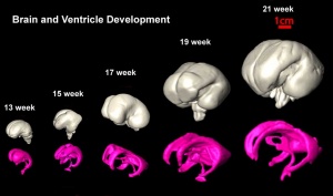
Human cerebrum and underlying ventricular development imaged by
MRI[1]Some Recent Findings
- Correlation of diffusion tensor imaging with histology in the developing human frontal cerebrum[2] "Transient early cerebral laminar organization resulting from normal developmental events has been revealed in human beings through histology and imaging studies. DTI studies have postulated that the fractional anisotropy (FA)-based differentiation of different laminar structures reflects both differing cellular density over the glial fibers and fiber alignment in respective regions. The aim of this study was to correlate FA values in these transient zones with histology. Brain DTI was performed on 50 freshly aborted human fetuses with gestational ages (GA) ranging from 12 to 42 weeks. Regions of interest were placed on the cortical plate, subplate, intermediate and germinal matrix (GMx) zones of the frontal lobe to quantify FA values. Glial fibrillary acidic protein (GFAP), neurofilament (NF) and neuron-specific enolase (NSE) immunohistochemical analyses were performed for the cortical plate, intermediate zone and GMx. In the cortical plate, a significant positive correlation was observed between FA values and percentage area of GFAP expression in fetuses <or=28 weeks of GA (r = 0.56, p = 0.01). FA values showed a significant positive correlation with the percentage area of NF expression in the intermediate zone (r = 0.54, p = 0.05). A significant positive correlation was also observed between FA and the number of NSE-positive cells per mm(2) in the GMx (r = 0.76, p < 0.01) and subplate (r = 0.59, p = 0.03) zones. The results of our study suggest that the FA can be used as noninvasive marker of neurodevelopmental events in the frontal lobe of human fetal brain."
- Development of laminar organization of the fetal cerebrum[3] "Heads of 131 fetal specimens of 14-40 weeks gestational age (GA) were scanned by 3.0T MRI. Eleven fetal specimens of 14-27 weeks GA were scanned by 7.0T MRI. On T(1)-weighted 3.0T MRI, layers could be visualized at 14 weeks GA and appeared clearer after 18 weeks GA. On 7.0T MRI, four zones could be recognized at 14 weeks GA. During 15-22 weeks GA, when laminar organization appeared typical, seven layers including the periventricular zone and external capsule fibers could be differentiated, which corresponded to seven zones in histological stained sections. At 23-28 weeks GA, laminar organization appeared less typical, and borderlines among them appeared obscured. After 30 weeks GA, it disappeared and turned into mature-like structures. The developing lamination appeared the most distinguishable at the parieto-occipital part of brain and peripheral regions of the hippocampus. The migrating thalamocortical afferents were probably delineated as a high signal layer located at the lower, middle, and upper part of the subplate zone at 16-28 weeks GA on T(1)-weighted 3.0T MRI."
|
Development Overview
Neuralation begins at the trilaminar embryo with formation of the notochord and somites, both of which underly the ectoderm and do not contribute to the nervous system, but are involved with patterning its initial formation. The central portion of the ectoderm then forms the neural plate that folds to form the neural tube, that will eventually form the entire central nervous system.
- Early developmental sequence: Epiblast - Ectoderm - Neural Plate - Neural groove and Neural Crest - Neural Tube and Neural Crest
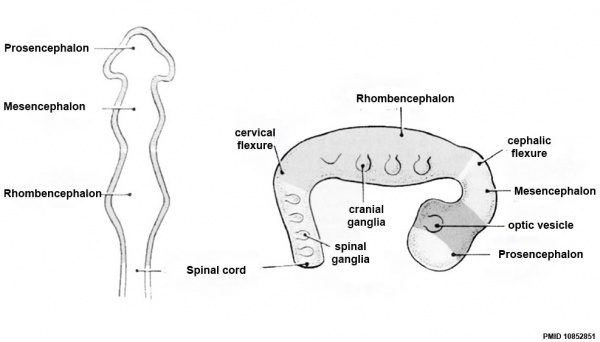
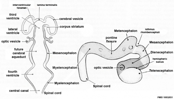
Neural Tube Development
| Neural Tube
|
Primary Vesicles
|
Secondary Vesicles
|
Adult Structures
|
| week 3
|
week 4
|
week 5
|
adult
|
neural plate
neural groove
neural tube
Brain
|
prosencephalon (forebrain)
|
telencephalon
|
Rhinencephalon, Amygdala, hippocampus, cerebrum (cortex), hypothalamus, pituitary | Basal Ganglia, lateral ventricles
|
| diencephalon
|
epithalamus, thalamus, Subthalamus, pineal, posterior commissure, pretectum, third ventricle
|
| mesencephalon (midbrain)
|
mesencephalon
|
tectum, Cerebral peduncle, cerebral aqueduct, pons
|
| rhombencephalon (hindbrain)
|
metencephalon
|
cerebellum
|
| myelencephalon
|
medulla oblongata, isthmus
|
| spinal cord, pyramidal decussation, central canal
|
Early Brain Vesicles
Primary Vesicles
Secondary Vesicles
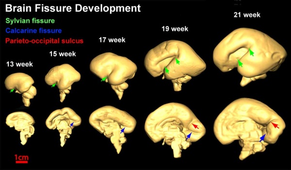
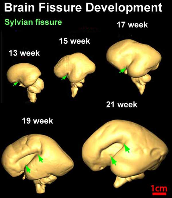
Brain Fissures
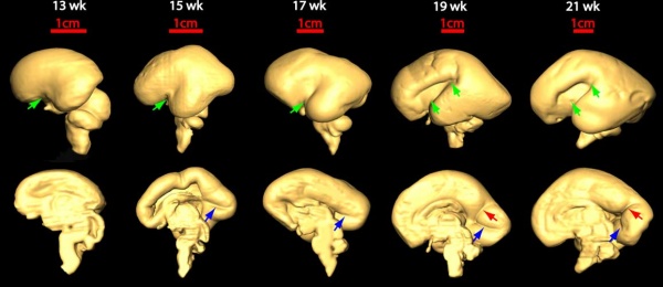
Fissures are the major indentations, sulci (singular sulcus), that divide the brain surface into lobes and appear during fetal development as the brain grows. The images below show MRI analysis of the developing human fetal brain.
- Links: Magnetic Resonance Imaging
References
- ↑ <pubmed>20108226</pubmed>
- ↑ <pubmed>19622880</pubmed>
- ↑ <pubmed>20981415</pubmed>
Reviews
<pubmed></pubmed>
Articles
<pubmed></pubmed>
Search PubMed
Search Pubmed: Cerebrum Development
External Links
External Links Notice - The dynamic nature of the internet may mean that some of these listed links may no longer function. If the link no longer works search the web with the link text or name. Links to any external commercial sites are provided for information purposes only and should never be considered an endorsement. UNSW Embryology is provided as an educational resource with no clinical information or commercial affiliation.
Glossary Links
- Glossary: A | B | C | D | E | F | G | H | I | J | K | L | M | N | O | P | Q | R | S | T | U | V | W | X | Y | Z | Numbers | Symbols | Term Link
Cite this page: Hill, M.A. (2024, April 23) Embryology Neural - Cerebrum Development. Retrieved from https://embryology.med.unsw.edu.au/embryology/index.php/Neural_-_Cerebrum_Development
- What Links Here?
- © Dr Mark Hill 2024, UNSW Embryology ISBN: 978 0 7334 2609 4 - UNSW CRICOS Provider Code No. 00098G


