Musculoskeletal System - Shoulder Development: Difference between revisions
mNo edit summary |
mNo edit summary |
||
| (One intermediate revision by the same user not shown) | |||
| Line 62: | Line 62: | ||
{{Shoulder Timeline table}} | {{Shoulder Timeline table}} | ||
{{Shoulder Timeline table2}} | |||
Acromial secondary ossification centers began appear at age 10 and clavicular ones, while uncommon, began forming at age 11. Fusion of acromial primary and secondary ossification centers began at age 14 and was generally complete after age 16.{{#pmid:25319561|PMID25319561}} A similar time course has been shown in Japanese subjects.{{#pmid:26321557|PMID26321557}} | |||
:'''Links:''' {{Upper limb ossification timeline}} | |||
==Embryonic Shoulder== | ==Embryonic Shoulder== | ||
{| | {| | ||
Latest revision as of 14:04, 10 April 2019
| Embryology - 19 Apr 2024 |
|---|
| Google Translate - select your language from the list shown below (this will open a new external page) |
|
العربية | català | 中文 | 中國傳統的 | français | Deutsche | עִברִית | हिंदी | bahasa Indonesia | italiano | 日本語 | 한국어 | မြန်မာ | Pilipino | Polskie | português | ਪੰਜਾਬੀ ਦੇ | Română | русский | Español | Swahili | Svensk | ไทย | Türkçe | اردو | ייִדיש | Tiếng Việt These external translations are automated and may not be accurate. (More? About Translations) |

Introduction


The skeletal shoulder consists of: the clavicle (collarbone), the scapula (shoulder blade), and the humerus. Development of his region occurs through both forms of ossification processes.
The mesoderm forms nearly all the connective tissues of the musculoskeletal system. Each tissue (cartilage, bone, and muscle) goes through many different mechanisms of differentiation.
The musculoskeletal system consists of skeletal muscle, bone, and cartilage and is mainly mesoderm in origin with some neural crest contribution.
The intraembryonic mesoderm can be broken into paraxial, intermediate and lateral mesoderm relative to its midline position. During the 3rd week the paraxial mesoderm forms into "balls" of mesoderm paired either side of the neural groove, called somites.
Somites appear bilaterally as pairs at the same time and form earliest at the cranial (rostral,brain) end of the neural groove and add sequentially at the caudal end. This addition occurs so regularly that embryos are staged according to the number of somites that are present. Different regions of the somite differentiate into dermomyotome (dermal and muscle component) and sclerotome (forms vertebral column). An example of a specialized musculoskeletal structure can be seen in the development of the limbs.
Skeletal muscle forms by fusion of mononucleated myoblasts to form mutinucleated myotubes. Bone is formed through a lengthy process involving ossification of a cartilage formed from mesenchyme. Two main forms of ossification occur in different bones, intramembranous (eg skull) and endochondrial (eg limb long bones) ossification. Ossification continues postnatally, through puberty until mid 20s. Early ossification occurs at the ends of long bones.
Musculoskeletal and limb abnormalities are one of the largest groups of congenital abnormalities.
- Historic Embryology: 1902 Shoulder Girdle
Some Recent Findings
|
| More recent papers |
|---|
 This table allows an automated computer search of the external PubMed database using the listed "Search term" text link.
More? References | Discussion Page | Journal Searches | 2019 References | 2020 References Search term: Shoulder Development | Scapula Development | Clavicle Development |
| Older papers |
|---|
| These papers originally appeared in the Some Recent Findings table, but as that list grew in length have now been shuffled down to this collapsible table.
See also the Discussion Page for other references listed by year and References on this current page.
|
Textbooks
- The Developing Human: Clinically Oriented Embryology (8th Edition) by Keith L. Moore and T.V.N Persaud - Moore & Persaud Chapter 15 the skeletal system
- Larsen’s Human Embryology by GC. Schoenwolf, SB. Bleyl, PR. Brauer and PH. Francis-West - Chapter 11 Limb Dev (bone not well covered in this textbook)
- Before we Are Born (5th ed.) Moore and Persaud Chapter 16,17: p379-397, 399-405
- Essentials of Human Embryology Larson Chapter 11 p207-228
Timeline
| Carnegie Stage | Event |
|---|---|
| 17 | chondrogenic progenitor of the humerus and the medial border of the scapula can be observed. |
| 18 | chondrogenic progenitor for rest of the scapula appears. |
| 19 | glenohumeral joint will begin delaminating and showing a looser central band (interzone). Denser lateral bands will join the humeral head (caput humeri) and the margins of the articular surface of the scapula, thus forming the glenoid labrum (glenoid ligament). |
| 21 | long head of the biceps tendon present |
| 22 | glenoid labrum (glenoid ligament) present |
| 23 | coracohumeral ligament present |
| Week | |
| Fetal Week 10 | osteogenic process begins in the humeral head. Primitive glenohumeral ligament present |
| Fetal Week 11 | osteogenic process begins in the scapula |
| Links: shoulder | joint | limb | timeline Data from human histological study.[7] | |
| Bone | Centres | Time of appearance of centre | Union of primary and secondary centres; remarks. |
|---|---|---|---|
| Clavicle | Diaphysis | 6th week | There are two centres in the shaft, a medial and a lateral. These blend on the 45th day (Mall). Shaft and epiphysis unite between the 20th and 25th years. |
| Sternal epiphysis | 18th to 20th year | ||
| Scapula | Primary centres: | The chief centre appears near the lateral angle. The subcoracoid centre appears at the base of the coracoid process and also gives rise to a part of the superior margin of the glenoid fossa. The coracoid process joins the body about the age of puberty. The acromial epiphysis centres (two or three in number) fuse with one another soon after their appearance and with the spine between the 22nd and 25th years (Quain); 20th year (Wilms). The subcoracoid and the epiphysis of the coracoid process, the glenoid fossa, the inferior angle, and the vertebral margin join between the 18th and 24th years in the order mentioned (Sappey). | |
| 1. That of the body, the spine, and the base of the glenoid cavity. | 8th week (Mall) 1 | ||
| 2. Goraooid process | 1st year | ||
| 3. Subcoracoid | 10th to 12th year | ||
| Epiphyses: | |||
| Acromial epiphyses | 15th to 18th year | ||
| Epiphysis of the inferior angle. | 16 to 18th year | ||
| Epiphyses of the vertebral border. | 18th to 20th year | ||
| Epiphyses of upper surface of coracoid. | 16th to 18th year. | ||
| Epiphysis of surface of glenoid fossa. | 16th to 18th year. | ||
| Links: limb | bone | upper limb ossification timeline | lower limb ossification timeline | Historic - Chapter 11 Development of the Skeleton | timeline | Category:Timeline Table Data Reference[8] | |||
Acromial secondary ossification centers began appear at age 10 and clavicular ones, while uncommon, began forming at age 11. Fusion of acromial primary and secondary ossification centers began at age 14 and was generally complete after age 16.[9] A similar time course has been shown in Japanese subjects.[10]
Embryonic Shoulder

|
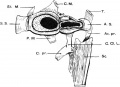
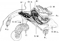
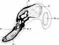
Drawing of a model of the right shoulder girdle of the 17mm. (Robinson) embryo, viewed from behind.Human embryonic shoulder girdle (17mm CRL)[11]
The sternal segment (S.S.) has been cut coronally to expose the interior, and the acromial segment has been cut horizontally for the same purpose. The black area in each is bone, the dotted area surrounding the bone is precartilage, and the area surrounding this is perichondrium. Above the junction of the two segments, two circles and a black dot are seen; the circles represent supraclavicular nerves, the black dot represents the cephalic vein. The scapula has been purposely shortened. Legend
|
| Human embryonic shoulder girdle (17mm CRL)[11] | 17mm CRL (Robinson) embryo |
Myogenesis
- Early myogenic progenitor cells in the dermomyotome can be initially identified by the transcription factor Pax3.
- Subsequent myogenic program development then depends on the myogenic determination factors (Myf5, MyoD, and MRF4), both Myf5 and MyoD are expressed in the limbs.
- Final differentiation of these cells into post-mitotic muscle fibers in the limb bud is regulated by another myogenic determination factor, Myogenin.
Some text modified from[12]
Links: Muscle Development
Limb Bone


Bone formation within the limb occurs by endochondral ossification of a pre-existing cartilage template. Ossification then replaces the existing cartilage except in the regions of articulation, where cartilage remains on the surface of the bone within the joint. Therefore bone development in the limb is initially about cartilage development or chondrogenesis.
In addition, there are two quite separate aspects to this development.
- Pattern - where the specific regions will commence to form cartilage, which will be different for each cartilage element.
- Chondrogenesis - the differentiation of mesoderm to form cartilage, which will be essentially the same program for all cartilage templates.
A recent study has identified that the overlying limb surface ectoderm potentially inhibits limb early chondrogenesis through Wnt6 signaling.[13]
Links: Cartilage Development | Bone Development
Clavicle Development
(collarbone)
Postnatal Growth
A paper has characterised the postnatal growth of male and female clavicles (data shown below).[14]
- 18 years of age the mean clavicle length +/-SD for females was 149+/-12 mm and for males it was 161+/-11 mm.
- statistically significant difference (P=0.049) was noted between the length of right and left clavicles it was not clinically significant (0.036 mm).
- A steady growth rate was noted for both genders from birth to the age of 12 years (8.4 mm/y).
- Above the age of 12 years there were significant differences in the growth of the clavicles of girls (2.6 mm/y) versus boys (5.4 mm/y) (P<0.001).
- Females achieve 80% of their clavicle length by 9 years of age and boys by 12 years of age.
Scapula Development
(shoulder blade)
Humerus Development
Pelvis

The skeletal pelvis consists of: the sacrum and coccyx (axial skeleton), and pelvic girdle formed by a pair of hip bones (appendicular skeleton). Before puberty, he pelvic girdle also consists of three unfused bones: the ilium, ischium, and pubis. In chicken, the entire pelvic girdle originates from the somatopleure mesoderm (somite levels 26 to 35) and the ilium, but not of the pubis and ischium, depends on somitic and ectodermal signals.[15]
- Links: Pelvis Development
Molecular
Fibroblast Growth Factors
- Fgf8 - morphogen gradient forms by a source-sink mechanism with freely diffusing molecules.[16]
T-box Transcription Factors
References
- ↑ Kicheva A & Briscoe J. (2010). Limbs made to measure. PLoS Biol. , 8, e1000421. PMID: 20644713 DOI.
- ↑ Young M, Selleri L & Capellini TD. (2019). Genetics of scapula and pelvis development: An evolutionary perspective. Curr. Top. Dev. Biol. , 132, 311-349. PMID: 30797513 DOI.
- ↑ Viciano J, Urbani V & D'Anastasio R. (2017). Congenital Anatomical Variant of the Clavicle. Anat Rec (Hoboken) , 300, 1401-1408. PMID: 28296289 DOI.
- ↑ Nagashima H, Sugahara F, Watanabe K, Shibata M, Chiba A & Sato N. (2016). Developmental origin of the clavicle, and its implications for the evolution of the neck and the paired appendages in vertebrates. J. Anat. , 229, 536-48. PMID: 27279028 DOI.
- ↑ Pu Q, Huang R & Brand-Saberi B. (2016). Development of the shoulder girdle musculature. Dev. Dyn. , 245, 342-50. PMID: 26676088 DOI.
- ↑ Capellini TD, Vaccari G, Ferretti E, Fantini S, He M, Pellegrini M, Quintana L, Di Giacomo G, Sharpe J, Selleri L & Zappavigna V. (2010). Scapula development is governed by genetic interactions of Pbx1 with its family members and with Emx2 via their cooperative control of Alx1. Development , 137, 2559-69. PMID: 20627960 DOI.
- ↑ Hita-Contreras F, Sánchez-Montesinos I, Martínez-Amat A, Cruz-Díaz D, Barranco RJ & Roda O. (2018). Development of the human shoulder joint during the embryonic and early fetal stages: anatomical considerations for clinical practice. J. Anat. , 232, 422-430. PMID: 29193070 DOI.
- ↑ Keibel F. and Mall FP. Manual of Human Embryology I. (1910) J. B. Lippincott Company, Philadelphia.
- ↑ Kothary P & Rosenberg ZS. (2015). Skeletal developmental patterns in the acromial process and distal clavicle as observed by MRI. Skeletal Radiol. , 44, 207-15. PMID: 25319561 DOI.
- ↑ Fujii K, Takeda Y & Miyatake K. (2015). Development of secondary ossification centres of the acromion in Japanese youth: a computed tomographic study. J Orthop Surg (Hong Kong) , 23, 229-32. PMID: 26321557 DOI.
- ↑ 11.0 11.1 Fawcett. (1913). The Development and Ossification of the Human Clavicle. J Anat Physiol , 47, 225-34. PMID: 17232952
- ↑ Giordani J, Bajard L, Demignon J, Daubas P, Buckingham M & Maire P. (2007). Six proteins regulate the activation of Myf5 expression in embryonic mouse limbs. Proc. Natl. Acad. Sci. U.S.A. , 104, 11310-5. PMID: 17592144 DOI.
- ↑ 13.0 13.1 Geetha-Loganathan P, Nimmagadda S, Christ B, Huang R & Scaal M. (2010). Ectodermal Wnt6 is an early negative regulator of limb chondrogenesis in the chicken embryo. BMC Dev. Biol. , 10, 32. PMID: 20334703 DOI.
- ↑ McGraw MA, Mehlman CT, Lindsell CJ & Kirby CL. (2009). Postnatal growth of the clavicle: birth to 18 years of age. J Pediatr Orthop , 29, 937-43. PMID: 19934713 DOI.
- ↑ Malashichev Y, Christ B & Pröls F. (2008). Avian pelvis originates from lateral plate mesoderm and its development requires signals from both ectoderm and paraxial mesoderm. Cell Tissue Res. , 331, 595-604. PMID: 18087724 DOI.
- ↑ Yu SR, Burkhardt M, Nowak M, Ries J, Petrásek Z, Scholpp S, Schwille P & Brand M. (2009). Fgf8 morphogen gradient forms by a source-sink mechanism with freely diffusing molecules. Nature , 461, 533-6. PMID: 19741606 DOI.
Reviews
Huang R, Christ B & Patel K. (2006). Regulation of scapula development. Anat. Embryol. , 211 Suppl 1, 65-71. PMID: 17006658 DOI.
Hall BK. (2001). Development of the clavicles in birds and mammals. J. Exp. Zool. , 289, 153-61. PMID: 11170011
Klima M. (1987). Early development of the shoulder girdle and sternum in marsupials (Mammalia: Metatheria). Adv Anat Embryol Cell Biol , 109, 1-91. PMID: 3324657
Articles
Currarino G & Herring JA. (2009). Congenital pseudarthrosis of the clavicle. Pediatr Radiol , 39, 1343-9. PMID: 19763557 DOI.
Kreitner KF, Schweden F, Schild HH, Riepert T & Nafe B. (1997). [Computerized tomography of the epiphyseal union of the medial clavicle: an auxiliary method of age determination during adolescence and the 3d decade of life?]. Rofo , 166, 481-6. PMID: 9272998 DOI.
Ogden JA, Conlogue GJ & Bronson ML. (1979). Radiology of postnatal skeletal development. III. The clavicle. Skeletal Radiol. , 4, 196-203. PMID: 531584
Search PubMed
Search Pubmed: Shoulder Development | Clavicle Development | Scapula Development
Additional Images
Historic Images
| Historic Disclaimer - information about historic embryology pages |
|---|
|

Adult appendicular skeleton
Human embryonic shoulder girdle (CRL 17mm)
Human embryonic shoulder girdle (CRL 18mm)
Human embryonic shoulder girdle (CRL 27mm)


Glossary Links
- Glossary: A | B | C | D | E | F | G | H | I | J | K | L | M | N | O | P | Q | R | S | T | U | V | W | X | Y | Z | Numbers | Symbols | Term Link
Cite this page: Hill, M.A. (2024, April 19) Embryology Musculoskeletal System - Shoulder Development. Retrieved from https://embryology.med.unsw.edu.au/embryology/index.php/Musculoskeletal_System_-_Shoulder_Development
- © Dr Mark Hill 2024, UNSW Embryology ISBN: 978 0 7334 2609 4 - UNSW CRICOS Provider Code No. 00098G