Musculoskeletal System - Bone Development Timeline
Introduction
The adult human skeleton has about 206 different bones which are formed from fusion of significantly more bones in the postnatal developing neonate and child (about 275). Two main forms of ossification occur in different bones, intramembranous (eg skull) and endochondral (eg vertebra) ossification. Ossification in general continues postnatally, through puberty until mid 20s.

Axial skeleton (80 bones in skull, vertebra, ribs, sternum) |

Appendicular skeleton (126 bones in limbs, shoulders, pelvis) |
Endochondral ossification within the limb begins at Carnegie stage 18 and also occurs throughout embryo skeleton. This process is the replacement of a cartilage "template" with bone (week 5-12) that continues through postnatal development, with a second surge of growth at puberty.
These notes summarise the timecourse of development of some of these bones in humans.
Some Recent Findings

|
Ossification Stages

The process of ossification as determined postnatally clinically has been divided into a series of stages.[2]
- Stage 1 - non-ossified epiphysis
- Stage 2 - discernible ossification centre
- Stage 3 - partial fusion
- Stage 4 - total fusion
- Stage 5 - an additional stage recently added is the disappearance of the epiphyseal scar after total fusion.
Clavicle Ossification
The following identifies the process of ossification of the medial clavicular epiphyseal cartilage.[3]
- Stage 3 - 16 years
- Stage 4 - 20 years (women), 21 years (men)
- Stage 5 - 26 years
Long Bone Ossification

Humerus
Appearance and fusion of bone secondary ossification centres, proximal is closer to body and distal is further away from the body.
Appearance
- Proximal epiphysis gestation week 36 - 4 years
- Distal epiphysis 6 months - 10 years
Fusion
- Proximal epiphysis 12 - 20 years
- Distal epiphysis 11 - 19 years
Femur
Appearance
- Proximal epiphysis 1 - 12 years
- Distal epiphysis Gestation week 36 - 40
Fusion
- Proximal epiphysis 11 - 19 years
- Distal epiphysis 14 - 19 years
Data from reference Table 1.[4]
Historic Data

Upper Limb Ossification Table - Part 1 |

Upper Limb Ossification Table - Part 2 |
Manual of Human Embryology by Franz Keibel and Franklin P. Mall (1910) Chapter XI. Development of the Skeleton and of the Connective Tissues
Hip

- Triradiate cartilage 14 - 16 years.
- Y-shaped growth plate region within the developing hip seen in childhood x-rays.
Mandible Ossification

Prenatal
Week 6 - Intramembranous ossification center develops lateral to Meckel's cartilage.
Week 7 - Coronoid process begins differentiating.
Week 8 - Coronoid process fuses with main mandibular mass.
Week 10 (approx) - Both condylar and coronoid processes are recognizable and anterior portion of Meckel's cartilage begins to ossify.
Weeks 12-14 - Secondary cartilages for the condyle, coronoid, and symphysis appear.
Weeks 14-16 - Deciduous tooth germs start to form.
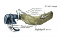
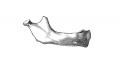
Embryo CRL 24 mm (outer aspect, about Carnegie stage 22)
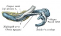
Embryo CRL 24 mm (inner aspect, about Carnegie stage 22)
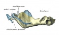
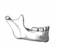
Embryo CRL 95 mm (outer aspect, about Fetal week 12, GA week 14)
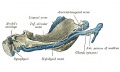
Embryo CRL 95 mm (inner aspectt, about Fetal week 12, GA week 14)


Birth

At birth mandible still has separate right and left halves.
Birth
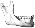
Childhood
Adult
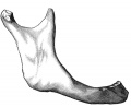
Old Age


Postnatal
Year 1 - Fusion of right and left halves of mandible at the symphysis.
Infancy and childhood - Increase in both size and shape of the mandible; eruption and replacement of teeth.
Year 12-14 - All permanent teeth emerged except third molars.
Data from reference[4] Table 4.
References
Online Textbooks
- Developmental Biology by Gilbert, Scott F. Sunderland (MA): Sinauer Associates, Inc.; c2000 Paraxial and intermediate mesoderm | Osteogenesis: The Development of Bones
- Molecular Biology of the Cell Alberts, Bruce; Johnson, Alexander; Lewis, Julian; Raff, Martin; Roberts, Keith; Walter, Peter New York and London: Garland Science; c2002 Search Molecular Biology of the CellBone Is Continually Remodeled by the Cells Within ItImage: Figure 22-52. Deposition of bone matrix by osteoblasts.Image: Figure 22-56. The development of a long bone.
Reviews
<pubmed>19092089</pubmed> <pubmed>18279783</pubmed>
Articles
<pubmed>14152896</pubmed>
Search PubMed
- Bone Development
Search Pubmed: Human Bone Development Timeline | Human Bone Development
External Links
External Links Notice - The dynamic nature of the internet may mean that some of these listed links may no longer function. If the link no longer works search the web with the link text or name. Links to any external commercial sites are provided for information purposes only and should never be considered an endorsement. UNSW Embryology is provided as an educational resource with no clinical information or commercial affiliation.
Terms
- growth recovery lines (growth arrest lines, Harris lines, Parks lines) - lines visible on x-ray images of increased bone density that represent the position of the growth plate at the time of insult to the organism and formed on long bones due to growth arrest. Insults can include juvenile malnutrition, disease or trauma.
Glossary Links
- Glossary: A | B | C | D | E | F | G | H | I | J | K | L | M | N | O | P | Q | R | S | T | U | V | W | X | Y | Z | Numbers | Symbols | Term Link
Cite this page: Hill, M.A. (2024, April 25) Embryology Musculoskeletal System - Bone Development Timeline. Retrieved from https://embryology.med.unsw.edu.au/embryology/index.php/Musculoskeletal_System_-_Bone_Development_Timeline
- © Dr Mark Hill 2024, UNSW Embryology ISBN: 978 0 7334 2609 4 - UNSW CRICOS Provider Code No. 00098G