Mouse Zygote 2 Movie: Difference between revisions
No edit summary |
No edit summary |
||
| Line 2: | Line 2: | ||
{| | {| | ||
| width=400px|<mediaplayer width='380' height='400' image="http://embryology.med.unsw.edu.au/embryology/images/d/d1/Mouse_zygote_division_02_icon.jpg">File:Mouse_zygote_division_02.mp4</mediaplayer> | | width=400px|<mediaplayer width='380' height='400' image="http://embryology.med.unsw.edu.au/embryology/images/d/d1/Mouse_zygote_division_02_icon.jpg">File:Mouse_zygote_division_02.mp4</mediaplayer> | ||
| [[File:Mouse_zygote_division_02_icon.jpg|100px|right]] | | valign=top| [[File:Mouse_zygote_division_02_icon.jpg|100px|right]] | ||
Movie shows mitotic division of the early mouse embryo starting at the zygote stage. | Movie shows mitotic division of the early mouse embryo starting at the zygote stage. | ||
| Line 10: | Line 10: | ||
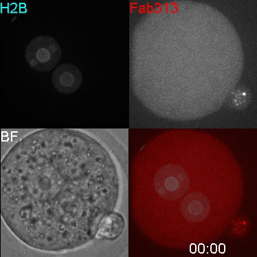
Maximum projections of labeled histone protein (H2B-mRFP; top left) and phosphorylated H3 (H3S10ph, Fab313-488; top right) are shown with brightfield (BF; bottom left) and in merged images (bottom right; H2B and Fab313 are shown in cyan and red, respectively). | Maximum projections of labeled histone protein (H2B-mRFP; top left) and phosphorylated H3 (H3S10ph, Fab313-488; top right) are shown with brightfield (BF; bottom left) and in merged images (bottom right; H2B and Fab313 are shown in cyan and red, respectively). | ||
Fab313-488 is preferentially concentrated on maternal condensed chromosomes at the first division (03:19–05:19) | Fab313-488 is preferentially concentrated on maternal condensed chromosomes at the first division (03:19–05:19) | ||
| Line 15: | Line 16: | ||
DNA in eukaryotes is wrapped around a histone octamer containing H2A, H2B, H3, and H4, forming a nucleosome, which is the fundamental unit of chromatin. | DNA in eukaryotes is wrapped around a histone octamer containing H2A, H2B, H3, and H4, forming a nucleosome, which is the fundamental unit of chromatin. | ||
'''Links:''' [[ | |||
:'''Links:''' [[Media:Mouse_zygote_division_02.mp4|MP4version]] | [[Media:Mouse_zygote_division_02.mov|Quicktime version]] | [[Mouse Zygote 1 Movie]] | [[Cell Division - Mitosis]] | [[Zygote]] | [[Morula]] | [[Week 1]] | |||
|} | |} | ||
{| | {| | ||
Revision as of 08:44, 7 March 2013
| Embryology - 16 Apr 2024 |
|---|
| Google Translate - select your language from the list shown below (this will open a new external page) |
|
العربية | català | 中文 | 中國傳統的 | français | Deutsche | עִברִית | हिंदी | bahasa Indonesia | italiano | 日本語 | 한국어 | မြန်မာ | Pilipino | Polskie | português | ਪੰਜਾਬੀ ਦੇ | Română | русский | Español | Swahili | Svensk | ไทย | Türkçe | اردو | ייִדיש | Tiếng Việt These external translations are automated and may not be accurate. (More? About Translations) |
| <mediaplayer width='380' height='400' image="http://embryology.med.unsw.edu.au/embryology/images/d/d1/Mouse_zygote_division_02_icon.jpg">File:Mouse_zygote_division_02.mp4</mediaplayer> | Movie shows mitotic division of the early mouse embryo starting at the zygote stage.
Maximum projections of labeled histone protein (H2B-mRFP; top left) and phosphorylated H3 (H3S10ph, Fab313-488; top right) are shown with brightfield (BF; bottom left) and in merged images (bottom right; H2B and Fab313 are shown in cyan and red, respectively).
DNA in eukaryotes is wrapped around a histone octamer containing H2A, H2B, H3, and H4, forming a nucleosome, which is the fundamental unit of chromatin.
|
| Mouse embryo was injected with H2B-mRFP mRNA and Fab313-488. Images of 51 focal planes (2-µm intervals) were captured at 10-min intervals using an inverted microscope (IX-71) with an UPlan-Apochromat 40× NA 1.0 oil immersion objective lens. Maximum projections of H2B-mRFP (top left) and Fab313-488 (top right) are shown with brightfield (BF; bottom left) and merged images (bottom right; H2B and Fab311 are shown in cyan and red, respectively).
The elapsed time from the start of acquisition (days:hours:minutes) is indicated. Fab313-488 is preferentially concentrated on maternal condensed chromosomes at the first division (03:19–05:19), as it recognizes histone H3S10ph next to di- or trimethylated H3K9. |
Reference
<pubmed>19995936</pubmed>| JCB
Published December 14, 2009 // JCB vol. 187 no. 6 781-790 The Rockefeller University Press, doi: 10.1083/jcb.200904137 Visualizing histone modifications in living cells: spatiotemporal dynamics of H3 phosphorylation during interphase
Copyright
Rockefeller University Press - Copyright Policy This article is distributed under the terms of an Attribution–Noncommercial–Share Alike–No Mirror Sites license for the first six months after the publication date (see http://www.jcb.org/misc/terms.shtml). After six months it is available under a Creative Commons License (Attribution–Noncommercial–Share Alike 4.0 Unported license, as described at https://creativecommons.org/licenses/by-nc-sa/4.0/ ). (More? Help:Copyright Tutorial)
Glossary Links
- Glossary: A | B | C | D | E | F | G | H | I | J | K | L | M | N | O | P | Q | R | S | T | U | V | W | X | Y | Z | Numbers | Symbols | Term Link
Cite this page: Hill, M.A. (2024, April 16) Embryology Mouse Zygote 2 Movie. Retrieved from https://embryology.med.unsw.edu.au/embryology/index.php/Mouse_Zygote_2_Movie
- © Dr Mark Hill 2024, UNSW Embryology ISBN: 978 0 7334 2609 4 - UNSW CRICOS Provider Code No. 00098G