Molecular Development - microRNA: Difference between revisions
mNo edit summary |
mNo edit summary |
||
| Line 1: | Line 1: | ||
{{Header}} | |||
==Introduction== | ==Introduction== | ||
Micro RNA (miRNA) are 22 nucleotide non-coding RNAs. These small pieces of RNA have been identified as important negative regulators in both development and adult cell processes involving gene expression. | Micro RNA (miRNA) are 22 nucleotide non-coding RNAs. These small pieces of RNA have been identified as important negative regulators in both development and adult cell processes involving gene expression. | ||
| Line 10: | Line 11: | ||
|-bgcolor="F5FAFF" | |-bgcolor="F5FAFF" | ||
| | | | ||
* '''Maternal peripheral blood natural killer cells incorporate placenta-associated microRNAs during pregnancy'''<ref name=PMID24682221><pubmed>24682221</pubmed>| [http://www.ncbi.nlm.nih.gov/pmc/articles/PMC4432927 PMC4432927] | [http://www.spandidos-publications.com/ijmm/35/6/1511 Int J Mol Med.}</ref> "Although recent studies have demonstrated that microRNAs (miRNAs or miRs) regulate fundamental natural killer (NK) cellular processes, including cytotoxicity and cytokine production, little is known about the miRNA‑gene regulatory relationships in maternal peripheral blood NK (pNK) cells during pregnancy. ...Twenty‑five miRNAs, including six C19MC miRNAs, were significantly upregulated in the third‑ compared to first‑trimester pNK cells. The rapid clearance of C19MC miRNAs also occurred in the pNK cells following delivery. Nine miRNAs, including eight C19MC miRNAs, were significantly downregulated in the post‑delivery pNK cells compared to those of the third‑trimester. DNA microarray analysis identified 69 NK cell function‑related genes that were differentially expressed between the first‑ and third‑trimester pNK cells. On pathway and network analysis, the observed gene expression changes of pNK cells likely contribute to the increase in the cytotoxicity, as well as the cell cycle progression of third‑ compared to first‑trimester pNK cells. Thirteen of the 69 NK cell function‑related genes were significantly downregulated between the first‑ and third‑trimester pNK cells. Nine of the 13 downregulated NK‑function‑associated genes were in silico target candidates of 12 upregulated miRNAs, including C19MC miRNA miR‑512‑3p. The results of this study suggest that the transfer of placental C19MC miRNAs into maternal pNK cells occurs during pregnancy." | |||
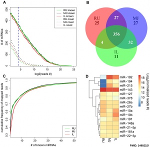
* '''Potential Regulatory Role of MicroRNAs in the Development of Bovine Gastrointestinal Tract during Early Life'''<ref name=PMID24682221><pubmed>24682221</pubmed>| [http://www.plosone.org/article/info%3Adoi%2F10.1371%2Fjournal.pone.0092592 PLoS One.]</ref> "This study aimed to investigate the potential regulatory role of miRNAs in the development of gastrointestinal tract (GIT) during the early life of dairy calves. Rumen and small intestinal (mid-jejunum and ileum) tissue samples were collected from newborn (30 min after birth; n = 3), 7-day-old (n = 6), 21-day-old (n = 6), and 42-day-old (n = 6) dairy calves. The miRNA profiling was performed using Illumina RNA-sequencing and the temporal and regional differentially expressed miRNAs were further validated using qRT-PCR. ...The present study revealed temporal and regional changes in miRNA expression and a correlation between miRNA expression and microbial population in the GIT during the early life, which provides further evidence for another mechanism by which host-microbial interactions play a role in regulating gut development." | * '''Potential Regulatory Role of MicroRNAs in the Development of Bovine Gastrointestinal Tract during Early Life'''<ref name=PMID24682221><pubmed>24682221</pubmed>| [http://www.plosone.org/article/info%3Adoi%2F10.1371%2Fjournal.pone.0092592 PLoS One.]</ref> "This study aimed to investigate the potential regulatory role of miRNAs in the development of gastrointestinal tract (GIT) during the early life of dairy calves. Rumen and small intestinal (mid-jejunum and ileum) tissue samples were collected from newborn (30 min after birth; n = 3), 7-day-old (n = 6), 21-day-old (n = 6), and 42-day-old (n = 6) dairy calves. The miRNA profiling was performed using Illumina RNA-sequencing and the temporal and regional differentially expressed miRNAs were further validated using qRT-PCR. ...The present study revealed temporal and regional changes in miRNA expression and a correlation between miRNA expression and microbial population in the GIT during the early life, which provides further evidence for another mechanism by which host-microbial interactions play a role in regulating gut development." | ||
* '''A survey of small RNAs in human sperm'''<ref><pubmed>21989093</pubmed></ref> "Bioinformatic analysis revealed the presence of multiple classes of small RNAs in human spermatozoa. The primary classes resolved included microRNA (miRNAs) (≈7%), Piwi-interacting piRNAs (≈17%), repeat-associated small RNAs (≈65%). A minor subset of short RNAs within the transcription start site/promoter fraction (≈11%) frames the histone promoter-associated regions enriched in genes of early embryonic development. These have been termed quiescent RNAs. CONCLUSIONS A complex population of male derived sncRNAs that are available for delivery upon fertilization was revealed. Sperm miRNA-targeted enrichment in the human oocyte is consistent with their role as modifiers of early post-fertilization. The relative abundance of piRNAs and repeat-associated RNAs suggests that they may assume a role in confrontation and consolidation. This may ensure the compatibility of the genomes at fertilisation." | * '''A survey of small RNAs in human sperm'''<ref><pubmed>21989093</pubmed></ref> "Bioinformatic analysis revealed the presence of multiple classes of small RNAs in human spermatozoa. The primary classes resolved included microRNA (miRNAs) (≈7%), Piwi-interacting piRNAs (≈17%), repeat-associated small RNAs (≈65%). A minor subset of short RNAs within the transcription start site/promoter fraction (≈11%) frames the histone promoter-associated regions enriched in genes of early embryonic development. These have been termed quiescent RNAs. CONCLUSIONS A complex population of male derived sncRNAs that are available for delivery upon fertilization was revealed. Sperm miRNA-targeted enrichment in the human oocyte is consistent with their role as modifiers of early post-fertilization. The relative abundance of piRNAs and repeat-associated RNAs suggests that they may assume a role in confrontation and consolidation. This may ensure the compatibility of the genomes at fertilisation." | ||
Revision as of 09:50, 12 June 2015
| Embryology - 16 Apr 2024 |
|---|
| Google Translate - select your language from the list shown below (this will open a new external page) |
|
العربية | català | 中文 | 中國傳統的 | français | Deutsche | עִברִית | हिंदी | bahasa Indonesia | italiano | 日本語 | 한국어 | မြန်မာ | Pilipino | Polskie | português | ਪੰਜਾਬੀ ਦੇ | Română | русский | Español | Swahili | Svensk | ไทย | Türkçe | اردو | ייִדיש | Tiếng Việt These external translations are automated and may not be accurate. (More? About Translations) |
Introduction
Micro RNA (miRNA) are 22 nucleotide non-coding RNAs. These small pieces of RNA have been identified as important negative regulators in both development and adult cell processes involving gene expression.
Some Recent Findings

|
| More recent papers |
|---|
|
This table allows an automated computer search of the external PubMed database using the listed "Search term" text link.
More? References | Discussion Page | Journal Searches | 2019 References | 2020 References Search term: microRNA Embryology <pubmed limit=5>microRNA Embryology</pubmed> |
Functional miRNA Grouping
The following identifies microRNAs that have been identified with specific developmental processes, based upon a commercial collation of basic research data.
Pluripotency
- let-7a, let-7b, let-7c, let-7d, let-7e, let-7g, miR-101, miR-106b, miR-125b, miR-130a, miR-133b, miR-141, miR-15a, miR-17, miR-182, miR-183, miR-18a, miR-18b, miR-205, miR-20a, miR-20b, miR-21, miR-214, miR-22, miR-222, miR-23b, miR-24, miR-302a, miR-302c, miR-345, miR-424, miR-498, miR-518b, miR-520g.
Early Development
- Embryoid Bodies: miR-132, miR-181a, miR-9.
- Definitive Endoderm: miR-205, miR-375.
Ectoderm
- Neurological Development: let-7b, miR-103a, miR-106b, miR-10b, miR-124, miR-125b, miR-130a, miR-132, miR-134, miR-137, miR-16, miR-181a, miR-182, miR-183, miR-20a, miR-210, miR-219-5p, miR-22, miR-23b, miR-24, miR-26a, miR-302a, miR-302c, miR-7, miR-9, miR-96.
- Eye Development: miR-130a, miR-196a, miR-219-5p, miR-23b, miR-96.
- Epidermal Differentiation: let-7b, miR-205, miR-210, miR-23b, miR-26a.
- Inner Ear Development: miR-182, miR-183, miR-96.
Mesoderm
- Hematopoiesis: let-7e, miR-125a-5p, miR-142-3p, miR-223.
- T Cell Development: let-7a, let-7f, miR-106b, miR-142-5p, miR-146b-5p, miR-150, miR-15a, miR-15b, miR-16, miR-181a, miR-20a, miR-222, miR-26a.
- Erythropoiesis: let-7a, let-7b, let-7c, let-7d, let-7f, let-7g, let-7i, miR-126, miR-128a, miR-137, miR-155, miR-15b, miR-16, miR-17, miR-181a, miR-182, miR-185, miR-206, miR-21, miR-22, miR-222, miR-24, miR-26a, miR-96.
- Lymphopoiesis: let-7b, miR-125b, miR-128a, miR-16, miR-181a, miR-21, miR-24.
- Megakaryopoiesis: miR-106b, miR-10a, miR-10b, miR-122, miR-126, miR-127-5p, miR-129-5p, miR-134, miR-146a, miR-150, miR-155, miR-17, miR-18a, miR-192, miR-20a, miR-20b, miR-21, miR-22, miR-301a, miR-33a, miR-378, miR-92a, miR-93.
- Monocyte Differentiation: miR-155, miR-222, miR-424.
- Myelopoiesis: miR-103a, miR-128a, miR-17, miR-181a, miR-24.
- Angiogenesis: miR-126, miR-130a, miR-218, miR-222, miR-92a.
- Myogenesis: miR-1, miR-125b, miR-206, miR-26a.
- Osteogenesis: miR-141, miR-15b, miR-424.
- Adipogenesis: let-7b, let-7c, let-7e, miR-100, miR-101, miR-103a, miR-10b, miR-146b-5p, miR-155, miR-182, miR-192, miR-194, miR-196a, miR-21, miR-210, miR-214, miR-22, miR-24, miR-498, miR-96, miR-99a.
- Chondrogenesis: let-7f, miR-1, miR-132, miR-181a, miR-196a, miR-96, miR-99a.
- Heart Development: miR-1, miR-208, miR-488.
Endoderm
- Liver Development: let-7a, let-7b, let-7c, miR-10a, miR-122, miR-125b, miR-192, miR-21, miR-22, miR-23b, miR-92a, miR-99a.
- Pancreatic Development: miR-15a, miR-15b, miR-16, miR-195, miR-214, miR-375, miR-7, miR-9.
- Intestinal Development: let-7d, let-7e, miR-103a, miR-106b, miR-125b, miR-126, miR-130a, miR-141, miR-146b-5p, miR-17, miR-192, miR-194, miR-21, miR-215, miR-301a, miR-424.
Data: SABiosciences Cell Differentiation & Development miRNA PCR Array
References
Reviews
<pubmed>21850044</pubmed> <pubmed>21742789</pubmed> <pubmed>21576351</pubmed> <pubmed>21504869</pubmed> <pubmed>21486922</pubmed> <pubmed>19148191</pubmed>
Articles
<pubmed></pubmed> <pubmed></pubmed> <pubmed>23617334</pubmed> <pubmed>20520778</pubmed> <pubmed>18029362</pubmed>| Nucleic Acids Res. <pubmed></pubmed>
Search Pubmed
Search Pubmed Now: microRNA
External Links
External Links Notice - The dynamic nature of the internet may mean that some of these listed links may no longer function. If the link no longer works search the web with the link text or name. Links to any external commercial sites are provided for information purposes only and should never be considered an endorsement. UNSW Embryology is provided as an educational resource with no clinical information or commercial affiliation.
- Memorial Sloan-Kettering Cancer Center - microRNA.org - Targets and Expression
- SABiosciences - Cell Differentiation & Development miRNA PCR Array
- Sigma-Aldrich - miRNA Introduction
- miRNAMap - http://mirnamap.mbc.nctu.edu.tw/
Glossary Links
- Glossary: A | B | C | D | E | F | G | H | I | J | K | L | M | N | O | P | Q | R | S | T | U | V | W | X | Y | Z | Numbers | Symbols | Term Link
Cite this page: Hill, M.A. (2024, April 16) Embryology Molecular Development - microRNA. Retrieved from https://embryology.med.unsw.edu.au/embryology/index.php/Molecular_Development_-_microRNA
- © Dr Mark Hill 2024, UNSW Embryology ISBN: 978 0 7334 2609 4 - UNSW CRICOS Provider Code No. 00098G
