Mesoderm: Difference between revisions
| Line 63: | Line 63: | ||
==External Links== | ==External Links== | ||
==Take the Quiz== | |||
<quiz display=simple> | |||
{Mesenchyme refers to the middle layer of the trilaminar embryo | |||
|type="()"} | |||
- true | |||
+ false | |||
|| The the middle layer of the trilaminar embryo is the '''mesoderm''' (meaning middle layer), while most of these cells are mesemchymal in appearance, this term is used to describe the cell histological appearance/organization. | |||
{The intraembryonic coelom forms within : | |||
|type="()"} | |||
- somites | |||
+ lateral plate | |||
- neural tube | |||
- intermediate mesoderm | |||
||The intraembryonic coelom forms initially small spaces in the mesoderm layer and coalesce to form a single large "horseshoe-shaped" space within the '''lateral plate mesoderm''' around the embryonic disc. Both young somites (somitocoels) and the neural tube (neural tube lumen) do have cavities, but neither is called the intraembryonic coelom. | |||
{All paraxial mesoderm segments into somites. | |||
|type="()"} | |||
- true | |||
+ false | |||
|| While somites do form within paraxial mesoderm, this region remains unsegmented at the level of the head and therefore does not incorporate into somites. | |||
{Somites are developmental structures that contribute the following adult structures : | |||
|type="()"} | |||
- vertebra, notochord, dermis, skeletal muscle | |||
+ vertebra, intervertebral discs, dermis, skeletal muscle | |||
- kidney, body wall connective tissue, sensory ganglia | |||
- kidney, gastrointestinal tract smooth muscle, mesentry | |||
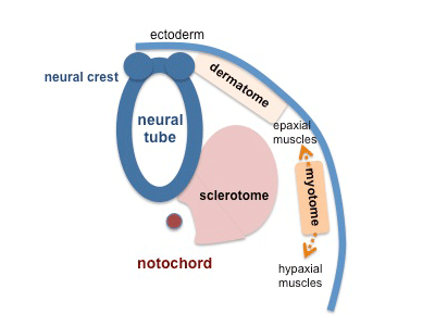
||Each somite has specific regions that contribute different components of the embryo. Sclerotome contributes the vertebral column ('''vertebra, intervertebral discs'''). Dermotome contributes the connective tissue layers of the skin ('''dermis''', hypodermis). Myotome ontributes the '''skeletal muscle''' of the body and limbs. | |||
</quiz> | |||
Revision as of 08:57, 10 August 2010
Introduction
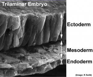
The middle layer of the early trilaminar embryo germ layers (ectoderm, mesoderm and endoderm) formed by gastrulation.
- Mesoderm Links: Lecture - Mesoderm Development | Notochord | Development Animation - Notochord | Somitogenesis | Musculoskeletal | Neural | Sonic hedgehog | Category:Mesoderm
Some Recent Findings
- Transcriptional profiling of the nucleus pulposus: say yes to notochord[1]"This editorial addresses the debate concerning the origin of adult nucleus pulposus cells in the light of profiling studies by Minogue and colleagues. In their report of several marker genes that distinguish nucleus pulposus cells from other related cell types, the authors provide novel insights into the notochordal nature of the former. Together with recently published work, their work lends support to the view that all cells present within the nucleus pulposus are derived from the notochord. Hence, the choice of an animal model for disc research should be based on considerations other than the cell loss and replacement by non-notochordal cells."
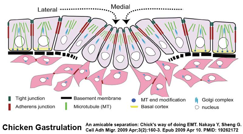
Mesoderm Formation during Gastrulation
| <Flowplayer width="390" height="510" autoplay="true">Mesoderm_001.flv</Flowplayer> | This animation shows the migration of mesoderm throughout the embryonic disc during gastrulation.
The pink arrow show how mesodermal cells spread out between the ectoderm and endoderm layers, forming the third layer of the trilaminar embryo. Axial process - the arrow running from the primitive node upward is the axial process which will later form the notochord. There are only 2 regions where no mesoderm is found: buccopharyngeal membrane and cloacal membrane.
Prechordal plate - lies above the buccopharyngeal membrane and is the cardiogenic mesoderm, that will form the heart. |
- Links: Gastrulation
Patterning
|
|
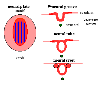
| Neural tube patterning | Somite patterning |
Molecular Factors
References
- ↑ <pubmed>20497604</pubmed>
Reviews
<pubmed>20568241</pubmed>
Articles
<pubmed>20565707</pubmed>
Search PubMed
Search NLM Online Textbooks: "Mesoderm" : Developmental Biology | The Cell- A molecular Approach | Molecular Biology of the Cell | Endocrinology
Search Pubmed: Mesoderm | Notochord
External Links
Take the Quiz
Glossary Links
- Glossary: A | B | C | D | E | F | G | H | I | J | K | L | M | N | O | P | Q | R | S | T | U | V | W | X | Y | Z | Numbers | Symbols | Term Link
Cite this page: Hill, M.A. (2024, April 23) Embryology Mesoderm. Retrieved from https://embryology.med.unsw.edu.au/embryology/index.php/Mesoderm
- © Dr Mark Hill 2024, UNSW Embryology ISBN: 978 0 7334 2609 4 - UNSW CRICOS Provider Code No. 00098G
