Menstrual Cycle - Histology: Difference between revisions
From Embryology
No edit summary |
|||
| Line 15: | Line 15: | ||
| 1 - 4 | | 1 - 4 | ||
| Click on image to see full size. | | Click on image to see full size. | ||
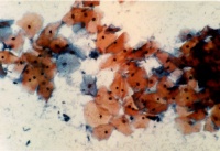
| Both '''stratum corneum''' (red) and '''stratum spinosum''' (blue) epithelial cells will mostly blood. Leukocytes and bacteria may also be present. | | Both '''stratum corneum''' (red) and '''stratum spinosum''' (blue) epithelial cells will mostly blood. | ||
Leukocytes and bacteria may also be present. | |||
| [[File:Human- menstrual uterine endometrium.jpg|200px]] | | [[File:Human- menstrual uterine endometrium.jpg|200px]] | ||
| Line 22: | Line 24: | ||
| 5 - 9 | | 5 - 9 | ||
| [[File:Smear-_early_proliferative.jpg|200px]] | | [[File:Smear-_early_proliferative.jpg|200px]] | ||
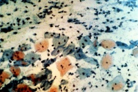
| | | Mainly large and small basophilic (blue) '''stratum spinosum''' cells. | ||
| | | | ||
| Line 29: | Line 31: | ||
| 9 - 13 | | 9 - 13 | ||
| [[File:Smear-_mid-proliferative.jpg|200px]] | | [[File:Smear-_mid-proliferative.jpg|200px]] | ||
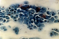
| '''Stratum spinosum''' (blue) increase in size. Dark precipate outside cells are bacteria. | | '''Stratum spinosum''' (blue) increase in size. | ||
Dark precipate outside cells are bacteria. | |||
| [[File:Human- mid-proliferative uterine endometrium.jpg|200px]] | | [[File:Human- mid-proliferative uterine endometrium.jpg|200px]] | ||
| Line 36: | Line 40: | ||
| 13-14 | | 13-14 | ||
| [[File:Smear-_late-proliferative.jpg|200px]] | | [[File:Smear-_late-proliferative.jpg|200px]] | ||
| mainly '''stratum corneum''' (red) which are large and flat | | mainly '''stratum corneum''' (red) which are large and flat. | ||
Appear due to high estrogen levels. | |||
| [[File:Human-_late_proliferative_uterine_endometrium.jpg|200px]] | | [[File:Human-_late_proliferative_uterine_endometrium.jpg|200px]] | ||
| Line 43: | Line 49: | ||
| 15 - 22 | | 15 - 22 | ||
| [[File:Smear-_secretory.jpg|200px]] | | [[File:Smear-_secretory.jpg|200px]] | ||
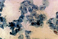
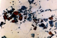
| '''stratum spinosum''' cells (blue) which are folded or with curled edges | | '''stratum spinosum''' cells (blue) which are folded or with curled edges. | ||
Appear immediately after ovulation due to increase in progesterone. | |||
'''Leukocytes''' (small black cells) becoming more numerous. | |||
| [[File:Human- secretory uterine endometrium.jpg|200px]] | | [[File:Human- secretory uterine endometrium.jpg|200px]] | ||
| Line 50: | Line 60: | ||
| 23 - 28 | | 23 - 28 | ||
| [[File:Smear-_late_secretory.jpg|200px]] | | [[File:Smear-_late_secretory.jpg|200px]] | ||
| '''stratum spinosum''' cells (blue) mainly with a few '''stratum corneum '''cells (red). Clustering of cells occurs at this stage | | '''stratum spinosum''' cells (blue) mainly with a few '''stratum corneum '''cells (red). | ||
Clustering of cells occurs at this stage. | |||
Both leukocytes and bacteria are prevelant. | |||
| [[File:Human- late secretory uterine endometrium.jpg|200px]] | | [[File:Human- late secretory uterine endometrium.jpg|200px]] | ||
Revision as of 18:14, 20 April 2010
Introduction
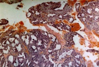
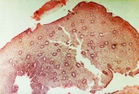
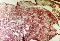
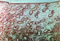
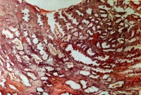
This page presents images from vaginal smears and uterine endometrium samples during different phases of the human menstrual cycle.
Links: Menstrual Cycle
Human vaginal smear histology images in sequence: early proliferative | mid-proliferative | late proliferative | secretory | late secretory
Human Uterus (D and C histology images) in sequence: menstrual | mid-proliferative | late proliferative | secretory | late secretory
Glossary Links
- Glossary: A | B | C | D | E | F | G | H | I | J | K | L | M | N | O | P | Q | R | S | T | U | V | W | X | Y | Z | Numbers | Symbols | Term Link
Cite this page: Hill, M.A. (2024, April 24) Embryology Menstrual Cycle - Histology. Retrieved from https://embryology.med.unsw.edu.au/embryology/index.php/Menstrual_Cycle_-_Histology
- © Dr Mark Hill 2024, UNSW Embryology ISBN: 978 0 7334 2609 4 - UNSW CRICOS Provider Code No. 00098G
