Manual of Human Embryology II - Figures: Difference between revisions
mNo edit summary |
mNo edit summary |
||
| (10 intermediate revisions by the same user not shown) | |||
| Line 184: | Line 184: | ||
<gallery> | <gallery> | ||
File:Keibel_Mall_2_279.jpg|Fig 279 | File:Keibel_Mall_2_279.jpg|Fig 279 | ||
</gallery> | |||
===The Development of the Liver=== | |||
[[Book_-_Manual_of_Human_Embryology_17-7#The_Development_of_the_Liver|The Development of the Liver]] | |||
<gallery> | |||
File:Keibel_Mall_2_288.jpg|Fig 288 | |||
File:Keibel_Mall_2_289.jpg|Fig 289 | |||
File:Keibel_Mall_2_290.jpg|Fig 290 | |||
File:Keibel_Mall_2_291.jpg|Fig 291 | |||
File:Keibel_Mall_2_292.jpg|Fig 292 | |||
File:Keibel_Mall_2_293.jpg|Fig 293 | |||
File:Keibel_Mall_2_294.jpg|Fig 294 | |||
File:Keibel_Mall_2_295.jpg|Fig 295 | |||
File:Keibel_Mall_2_296.jpg|Fig 296 | |||
File:Keibel_Mall_2_297.jpg|Fig 297 | |||
File:Keibel_Mall_2_298.jpg|Fig 298 | |||
File:Keibel_Mall_2_299.jpg|Fig 299 | |||
File:Keibel_Mall_2_300.jpg|Fig 300 | |||
File:Keibel_Mall_2_301.jpg|Fig 301 | |||
File:Keibel_Mall_2_302.jpg|Fig 302 | |||
File:Keibel_Mall_2_303.jpg|Fig 303 | |||
File:Keibel_Mall_2_304.jpg|Fig 304 | |||
File:Keibel_Mall_2_305.jpg|Fig 305 | |||
File:Keibel_Mall_2_306.jpg|Fig 306 | |||
</gallery> | |||
===Development of the Pancreas=== | |||
[[Book_-_Manual_of_Human_Embryology_17-8#Development_of_the_Pancreas|Development of the Pancreas]] | |||
<gallery> | |||
File:Keibel_Mall_2_307.jpg|Fig 307 | |||
File:Keibel_Mall_2_308.jpg|Fig 308 | |||
File:Keibel_Mall_2_309.jpg|Fig 309 | |||
File:Keibel_Mall_2_310.jpg|Fig 310 | |||
File:Keibel_Mall_2_311.jpg|Fig 311 | |||
File:Keibel_Mall_2_312.jpg|Fig 312 | |||
File:Keibel_Mall_2_313.jpg|Fig 313 | |||
</gallery> | </gallery> | ||
| Line 300: | Line 338: | ||
[[Book_-_Manual_of_Human_Embryology_18-2|II. The Development of the Heart]] | [[Book_-_Manual_of_Human_Embryology_18-2|II. The Development of the Heart]] | ||
===A. Origin of the Vascular System=== | |||
===B. Vascular System in Early Human Embryos=== | |||
<gallery> | <gallery> | ||
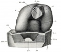
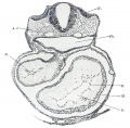
Keibel_Mall_2_372.jpg|Fig. 372. Section through the heart anlage of the Pfannenstiel-Kromer embryo Klb (Normentafel, Xo. 3, 5 to 6 primitive somites). | Keibel_Mall_2_372.jpg|Fig. 372. Section through the heart anlage of the Pfannenstiel-Kromer embryo Klb (Normentafel, Xo. 3, 5 to 6 primitive somites). | ||
Keibel_Mall_2_373.jpg|Fig. 373. | Keibel_Mall_2_373.jpg|Fig. 373. human embryo 2.5 mm GL heart | ||
Keibel_Mall_2_374.jpg|Fig. 374. Model of the heart of the embryo Halj of 3 mm. greatest length, 15 primitive somites. | Keibel_Mall_2_374.jpg|Fig. 374. Model of the heart of the embryo Halj of 3 mm. greatest length, 15 primitive somites. | ||
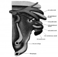
Keibel_Mall_2_375.jpg|Fig. 375. The model shown in Fig. 374 after removal of the pericardium, seen from behind. | Keibel_Mall_2_375.jpg|Fig. 375. The model shown in Fig. 374 after removal of the pericardium, seen from behind. | ||
Keibel_Mall_2_376.jpg|Fig. 376. Transverse section | Keibel_Mall_2_376.jpg|Fig. 376. Transverse section embryo Hah. | ||
Keibel_Mall_2_377.jpg|Fig. 377. | Keibel_Mall_2_377.jpg|Fig. 377. ventricular wall of the heart of embryo Hah, 3.5 mm. greatest length. | ||
Keibel_Mall_2_378.jpg|Fig. 378. Model of the heart of the embryo Hah of 5.2 mm. greatest length. | Keibel_Mall_2_378.jpg|Fig. 378. Model of the heart of the embryo Hah of 5.2 mm. greatest length. | ||
Keibel_Mall_2_379.jpg|Fig. 379. Model of the heart of embryo £U of 6.5 mm. greatest length. | Keibel_Mall_2_379.jpg|Fig. 379. Model of the heart of embryo £U of 6.5 mm. greatest length. | ||
| Line 341: | Line 383: | ||
<gallery> | <gallery> | ||
Keibel_Mall_2_465.jpg|Fig. 465 | Keibel_Mall_2_465.jpg|Fig. 465 | ||
</gallery> | |||
===Lymphatic=== | |||
<gallery> | |||
File:Keibel Mall 2 488.jpg|Fig. 488 | |||
File:Keibel Mall 2 493.jpg|Fig. 493 lymphatic system human embryo 30 mm | |||
File:Keibel Mall 2 494.jpg|Fig. 494 Frontal section jugular lymph-sacs human embryo 30 mm | |||
File:Keibel Mall 2 511.jpg|Fig. 511 | |||
File:Keibel Mall 2 512.jpg|Fig. 512 | |||
File:Keibel Mall 2 513.jpg|Fig. 513 | |||
File:Keibel Mall 2 514.jpg|Fig. 514 blood-vessels and lymphatics tadpole's tail 10 mm | |||
</gallery> | |||
===Spleen=== | |||
<gallery> | |||
File:Keibel Mall 2 515.jpg|Fig. 515 | |||
File:Keibel Mall 2 516.jpg|Fig. 516 | |||
File:Keibel Mall 2 517.jpg|Fig. 517 | |||
File:Keibel Mall 2 518.jpg|Fig. 518 | |||
File:Keibel Mall 2 519.jpg|Fig. 519 injected spleen pig embryo 12 cm | |||
</gallery> | </gallery> | ||
| Line 467: | Line 531: | ||
<gallery> | <gallery> | ||
Keibel_Mall_2_631.jpg|Fig. 631 posterior abdominal wall human embryo 19.4 mm | Keibel_Mall_2_631.jpg|Fig. 631 posterior abdominal wall human embryo 19.4 mm | ||
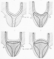
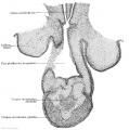
Keibel_Mall_2_632.jpg|Fig. 632 | Keibel_Mall_2_632.jpg|Fig. 632 Transverse section urogenital fold human embryo 50 mm | ||
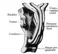
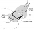
Keibel_Mall_2_633.jpg|Fig. 633 | Keibel_Mall_2_633.jpg|Fig. 633 Sagittal section human embryo 40 mm | ||
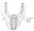
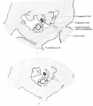
Keibel_Mall_2_634.jpg|Fig. 634 | Keibel_Mall_2_634.jpg|Fig. 634 Two transverse sections urogenital fold human embryo 22.5 mm | ||
Keibel_Mall_2_635.jpg|Fig. 635 | Keibel_Mall_2_635.jpg|Fig. 635 Transverse section human embryo 26 mm | ||
Keibel_Mall_2_636.jpg|Fig. 636 | Keibel_Mall_2_636.jpg|Fig. 636 Transverse section urogenital fold male embryo 26 mm | ||
</gallery> | </gallery> | ||
===IV. Development of the External Genitalia=== | ===IV. Development of the External Genitalia=== | ||
<gallery> | <gallery> | ||
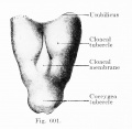
Keibel_Mall_2_637.jpg|Fig. 637 | Keibel_Mall_2_637.jpg|Fig. 637 Caudal end embryo 13 mm | ||
Keibel_Mall_2_638.jpg|Fig. 638 | Keibel_Mall_2_638.jpg|Fig. 638 Sagittal section embryo 24 mm | ||
Keibel_Mall_2_639.jpg|Fig. 639 | Keibel_Mall_2_639.jpg|Fig. 639 division of cloacal tubercle into phallus and genital tubercle | ||
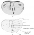
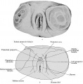
Keibel_Mall_2_640.jpg|Fig. 640 | Keibel_Mall_2_640.jpg|Fig. 640 indifferent external genitalia embryo 28 mm | ||
Keibel_Mall_2_641.jpg|Fig. 641 | Keibel_Mall_2_641.jpg|Fig. 641 female external genitalia embryo 32.5 mm | ||
Keibel_Mall_2_642.jpg|Fig. 642 | Keibel_Mall_2_642.jpg|Fig. 642 | ||
Keibel_Mall_2_643.jpg|Fig. 643 | Keibel_Mall_2_643.jpg|Fig. 643 | ||
Latest revision as of 09:37, 22 December 2018
Figures
XIV. The Development of the Nervous System
I. Histogenesis of Nervous Tissue
I. Histogenesis of Nervous Tissue
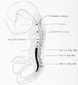
- Keibel Mall 2 008.jpg
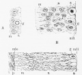
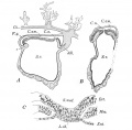
Fig. 8. Diagram showing distribution of neuroblasts in human embryo of four weeks.
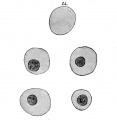
- Keibel Mall 2 009.jpg
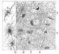
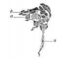
Fig. 9. Cluster of neuroblasts from nucleus of origin of n. oculomotorius, showing characteristic shape and grouping of cells.
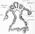
- Keibel Mall 2 010.jpg
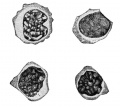
Fig. 10. Section through floor of mid-brain of human embryo one month old.
- Keibel Mall 2 011.jpg
Fig. 11. Isolated ganglion-cells, from embryonic spinal cord of frog, and growing in clotted lymph.
- Keibel Mall 2 012.jpg
Fig. 12. Three views, taken at intervals of Ik and 8i hours, of the same living nerve-fibres growing from a mass of spinal-cord tissue (frog embryo) out into clotted lymph.
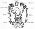
- Keibel Mall 2 013.jpg
Fig. 13. Transverse .sections through dorsal region of human embryos showing three stages in the development of the ganglion crest and the anlage of the spinal ganglia.
- Keibel Mall 2 014.jpg
Fig. 14. Section through spinal ganglion of human embryo 18 mm. long, about 6 weeks old .
- Keibel Mall 2 015.jpg
Fig. 15. Section through cervical spinal ganglion of human fetus 8.5 cm. long, about 3 months old, showing large ganglion-cells with eccentric nuclei.
- Keibel Mall 2 016.jpg
Fig. 16. Section through sixth cervical ganglion of human fetus 10.5 cm. long, about 4 months old.
- Keibel Mall 2 017.jpg
Fig. 17. Isolated cells teased from spinal ganglia of embryo pigs 20-40 mm. long, showing the variation in the form of the early ganglion-cells
- Keibel Mall 2 018.jpg
Fig. 18. Teased preparations from spinal ganglia of pig, showing development of sheath and capsule cells.
- Keibel Mall 2 019.jpg
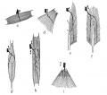
Fig. 19. Isolated fibres showing development of medullary sheath.
- Keibel Mall 2 020.jpg
Fig. 20. Isolated fibres of the sciatic nerve of sheep fetus 15 cm. long, treated with osmic acid and showing development of the nerve-sheath.
- Keibel Mall 2 021.jpg
Fig. 21. Section through hind-brain of new-born child, showing myelinization of fifth, sixth, seventh, and eighth cranial nerves and associated fibre tracts
II. Development of the Central Nervous System
II. Development of the Central Nervous System
- Keibel Mall 2 025.jpg
- Keibel Mall 2 026.jpg
- Keibel Mall 2 027.jpg
- Keibel Mall 2 028.jpg
- Keibel Mall 2 029.jpg
- Keibel Mall 2 031.jpg
- Keibel Mall 2 033.jpg
- Keibel Mall 2 034.jpg
- Keibel Mall 2 035.jpg
- Keibel Mall 2 036.jpg
- Keibel Mall 2 037.jpg
- Keibel Mall 2 038.jpg
- Keibel Mall 2 039.jpg
- Keibel Mall 2 040.jpg
- Keibel Mall 2 041.jpg
- Keibel Mall 2 042.jpg
- Keibel Mall 2 043.jpg
III. Peripheral Nervous System
III. Peripheral Nervous System
- Keibel Mall 2 090.jpg
Fig 90
- Keibel Mall 2 091.jpg
Fig 91
- Keibel Mall 2 092.jpg
Fig 92
- Keibel Mall 2 093.jpg
Fig 93
- Keibel Mall 2 094.jpg
Fig 94
- Keibel Mall 2 095.jpg
Fig 95
- Keibel Mall 2 096.jpg
Fig 96
- Keibel Mall 2 097.jpg
Fig 97
- Keibel Mall 2 098.jpg
Fig 98
XVII. The Development of the Digestive Tract and of the Organs of Respiration
XVII. The Development of the Digestive Tract and of the Organs of Respiration
The Early Development of the Entodermal Tract and the Formation of its Subdivisions
The Early Development of the Entodermal Tract and the Formation of its Subdivisions
The Development of the Sense-Organs
The Development of the Sense-Organs
The Mouth and Its Organs
The Development Of The Oesophagus
The Development Of The Oesophagus
The Development of the Stomach
The Development of the Stomach
- Keibel Mall 2 275.jpg
Fig 275
- Keibel Mall 2 276.jpg
Fig 276
- Keibel Mall 2 277.jpg
Fig 277
- Keibel Mall 2 278.jpg
Fig 278
The Development of the Small Intestine
The Development of the Small Intestine
- Keibel Mall 2 279.jpg
Fig 279
The Development of the Liver
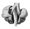
- Keibel Mall 2 288.jpg
Fig 288
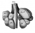
- Keibel Mall 2 289.jpg
Fig 289
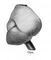
- Keibel Mall 2 290.jpg
Fig 290
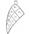
- Keibel Mall 2 291.jpg
Fig 291
Development of the Pancreas
- Keibel Mall 2 307.jpg
Fig 307
- Keibel Mall 2 308.jpg
Fig 308
- Keibel Mall 2 309.jpg
Fig 309
- Keibel Mall 2 310.jpg
Fig 310
- Keibel Mall 2 311.jpg
Fig 311
- Keibel Mall 2 312.jpg
Fig 312
- Keibel Mall 2 313.jpg
Fig 313
The Development of the Pharynx and of the Organs of Respiration
The Development of the Pharynx and of the Organs of Respiration
I. General Morphology of the Pharyngeal Pouches
I. General Morphology of the Pharyngeal Pouches
II. The Differentiation of the Pharyngeal Pouches - the Second Pharyngeal Pouch and the Tonsils
II. The Differentiation of the Pharyngeal Pouches - the Second Pharyngeal Pouch and the Tonsils
B. The Development of the Respiratory Apparatus
III. The Third to the Fifth Pharyngeal Pouches - the Branchiogenic Organs
III. The Third to the Fifth Pharyngeal Pouches - the Branchiogenic Organs
XVIII. The Development of the Blood, the Vascular System, and the Spleen
XVIII. The Development of the Blood, the Vascular System, and the Spleen
I. The Origin of the Angioblast and the Development of the Blood
I. The Origin of the Angioblast and the Development of the Blood
II. The Development of the Heart
II. The Development of the Heart
A. Origin of the Vascular System
B. Vascular System in Early Human Embryos
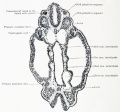
- Keibel Mall 2 377.jpg
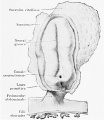
Fig. 377. ventricular wall of the heart of embryo Hah, 3.5 mm. greatest length.
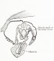
- Keibel Mall 2 378.jpg
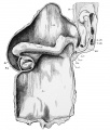
Fig. 378. Model of the heart of the embryo Hah of 5.2 mm. greatest length.
- Keibel Mall 2 379.jpg
Fig. 379. Model of the heart of embryo £U of 6.5 mm. greatest length.
- Keibel Mall 2 380.jpg
Fig. 380. Model of [thejjheart of embryo La of 9 mm. greatest length.
- Keibel Mall 2 381.jpg
Fig. 381. Sagittal section through the model shown in Fig. 380, the section passing to the left of the septum I.
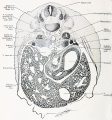
- Keibel Mall 2 382.jpg
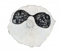
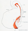
Fig. 382. Transverse section through the heart region of the embryo Wal of 8 mm. greatest length.
- Keibel Mall 2 383.jpg
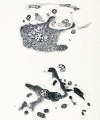
Fiag. 383. Section through the bulbus cordis of the embryo H6.
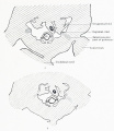
- Keibel Mall 2 384.jpg
Fig. 384. Model of the bulbus cordis of the embryo H6, divided longitudinally.
- Keibel Mall 2 385.jpg
Fig. 385. Section through the wall of the ventricle of embryo EU
- Keibel Mall 2 386.jpg
Fig. 386. Model of the heart of embryo S2 of 14.5 mm. greatest length.
- Keibel Mall 2 387.jpg
Fig. 387. Section through the heart of embryo Mi of 16.75 mm greatest length.
- Keibel Mall 2 388.jpg
Fig. 388. Model of the heart of an embryo of 310 mm greatest length.
- Keibel Mall 2 390.jpg
Fig. 389. Section through the heart of an embryo of 165 mm. greatest length.
C. Arteries
- Keibel Mall 2 462.jpg
Fig. 462
- Keibel Mall 2 463.jpg
Fig. 463
- Keibel Mall 2 464.jpg
Fig. 464
D. Veins
Lymphatic
- Keibel Mall 2 511.jpg
Fig. 511
Spleen
XIX Development of the Urinogenital Organs
I. The Development of the Excretory Glands and their Ducts
II. The Development of the Reproductive Glands and their Ducts
III. The Urogenital Union
IV. Development of the External Genitalia
| Historic Disclaimer - information about historic embryology pages |
|---|
| Pages where the terms "Historic" (textbooks, papers, people, recommendations) appear on this site, and sections within pages where this disclaimer appears, indicate that the content and scientific understanding are specific to the time of publication. This means that while some scientific descriptions are still accurate, the terminology and interpretation of the developmental mechanisms reflect the understanding at the time of original publication and those of the preceding periods, these terms, interpretations and recommendations may not reflect our current scientific understanding. (More? Embryology History | Historic Embryology Papers) |
Glossary Links
- Glossary: A | B | C | D | E | F | G | H | I | J | K | L | M | N | O | P | Q | R | S | T | U | V | W | X | Y | Z | Numbers | Symbols | Term Link
Cite this page: Hill, M.A. (2024, April 19) Embryology Manual of Human Embryology II - Figures. Retrieved from https://embryology.med.unsw.edu.au/embryology/index.php/Manual_of_Human_Embryology_II_-_Figures
- © Dr Mark Hill 2024, UNSW Embryology ISBN: 978 0 7334 2609 4 - UNSW CRICOS Provider Code No. 00098G
