Manual of Human Embryology II - Figures: Difference between revisions
mNo edit summary |
mNo edit summary |
||
| Line 180: | Line 180: | ||
Keibel_Mall_2_371.jpg|Fig. 371. Hepatic cylinders of a human embryo of 11 mm. Coll. F. P. Mali, No. 353. The blood-cells with small nuclei lie apparently in the substance of the liver-cells, -which have large nuclei. | Keibel_Mall_2_371.jpg|Fig. 371. Hepatic cylinders of a human embryo of 11 mm. Coll. F. P. Mali, No. 353. The blood-cells with small nuclei lie apparently in the substance of the liver-cells, -which have large nuclei. | ||
</gallery> | </gallery> | ||
===II. The Development of the Heart=== | |||
[[Book_-_Manual_of_Human_Embryology_18-2|II. The Development of the Heart]] | |||
<gallery> | |||
Keibel_Mall_2_372.jpg|Fig. 372. Section through the heart anlage of the Pfannenstiel-Kromer embryo Klb (Normentafel, Xo. 3, 5 to 6 primitive somites). | |||
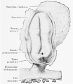
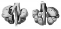
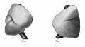
Keibel_Mall_2_373.jpg|Fig. 373. Model of the heart of a human embryo 2.5 mm. greatest length. | |||
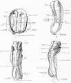
Keibel_Mall_2_374.jpg|Fig. 374. Model of the heart of the embryo Halj of 3 mm. greatest length, 15 primitive somites. | |||
Keibel_Mall_2_375.jpg|Fig. 375. The model shown in Fig. 374 after removal of the pericardium, seen from behind. | |||
Keibel_Mall_2_376.jpg|Fig. 376. Transverse section through the embryo Hah. | |||
Keibel_Mall_2_377.jpg|Fig. 377. Section through the ventricular wall of the heart of embryo Hah, 3.5 mm. greatest length. | |||
Keibel_Mall_2_378.jpg|Fig. 378. Model of the heart of the embryo Hah of 5.2 mm. greatest length. | |||
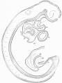
Keibel_Mall_2_379.jpg|Fig. 379. Model of the heart of embryo £U of 6.5 mm. greatest length. | |||
Keibel_Mall_2_380.jpg|Fig. 380. Model of [thejjheart of embryo La of 9 mm. greatest length. | |||
Keibel_Mall_2_381.jpg|Fig. 381. Sagittal section through the model shown in Fig. 380, the section passing to the left of the septum I. | |||
Keibel_Mall_2_382.jpg|Fig. 382. Transverse section through the heart region of the embryo Wal of 8 mm. greatest length. | |||
Keibel_Mall_2_383.jpg|Fiag. 383. Section through the bulbus cordis of the embryo H6. | |||
Keibel_Mall_2_384.jpg|Fig. 384. Model of the bulbus cordis of the embryo H6, divided longitudinally. | |||
Keibel_Mall_2_385.jpg|Fig. 385. Section through the wall of the ventricle of embryo EU | |||
Keibel_Mall_2_386.jpg|Fig. 386. Model of the heart of embryo S2 of 14.5 mm. greatest length. | |||
Keibel_Mall_2_387.jpg|Fig. 387. Section through the heart of embryo Mi of 16.75 mm greatest length. | |||
Keibel_Mall_2_388.jpg|Fig. 388. Model of the heart of an embryo of 310 mm greatest length. | |||
Keibel_Mall_2_390.jpg|Fig. 389. Section through the heart of an embryo of 165 mm. greatest length. | |||
</gallery> | |||
{{Human Embryology Manual 1 TOC}} | {{Human Embryology Manual 1 TOC}} | ||
Revision as of 11:51, 29 March 2014
Figures
XIV. The Development of the Nervous System
I. Histogenesis of Nervous Tissue
I. Histogenesis of Nervous Tissue
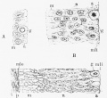
Fig. 1. Three early stages in the development of the wall of the neural tube,
- Keibel Mall 2 009.jpg
Fig. 2. Wall of the neurfl tube in a human embryo about two weeks old, showing its syncytial character.
- Keibel Mall 2 009.jpg
Fig. 3. Diagram showing the differentiation of the cells of the wall of the neural tube
- Keibel Mall 2 009.jpg
Fig. 4. Development of neuroglia framework.
- Keibel Mall 2 009.jpg
Fig. 5. Combined drawings, after Golgi and Benda methods, of the spinal cord of fetal pig, 20 cm. long
- Keibel Mall 2 009.jpg
Fig. 6. Section of spinal cord of suckling pig of two weeks
- Keibel Mall 2 009.jpg
Fig. 7. Neuroglia fibres in adult human spinal cord, showing their relation to 'the protoplasm of the neuroglia cell and its processes.
- Keibel Mall 2 009.jpg
Fig. 8. Diagram showing distribution of neuroblasts in human embryo of four weeks.
- Keibel Mall 2 009.jpg
Fig. 9. Cluster of neuroblasts from nucleus of origin of n. oculomotorius, showing characteristic shape and grouping of cells.
- Keibel Mall 2 010.jpg
Fig. 10. Section through floor of mid-brain of human embryo one month old.
- Keibel Mall 2 019.jpg
Fig. 11. Isolated ganglion-cells, from embryonic spinal cord of frog, and growing in clotted lymph.
- Keibel Mall 2 019.jpg
Fig. 12. Three views, taken at intervals of Ik and 8i hours, of the same living nerve-fibres growing from a mass of spinal-cord tissue (frog embryo) out into clotted lymph.
- Keibel Mall 2 019.jpg
Fig. 13. Transverse .sections through dorsal region of human embryos showing three stages in the development of the ganglion crest and the anlage of the spinal ganglia.
- Keibel Mall 2 019.jpg
Fig. 14. Section through spinal ganglion of human embryo 18 mm. long, about 6 weeks old .
- Keibel Mall 2 019.jpg
Fig. 15. Section through cervical spinal ganglion of human fetus 8.5 cm. long, about 3 months old, showing large ganglion-cells with eccentric nuclei.
- Keibel Mall 2 019.jpg
Fig. 16. Section through sixth cervical ganglion of human fetus 10.5 cm. long, about 4 months old.
- Keibel Mall 2 019.jpg
Fig. 17. Isolated cells teased from spinal ganglia of embryo pigs 20-40 mm. long, showing the variation in the form of the early ganglion-cells
- Keibel Mall 2 019.jpg
Fig. 18. Teased preparations from spinal ganglia of pig, showing development of sheath and capsule cells.
- Keibel Mall 2 019.jpg
Fig. 19. Isolated fibres showing development of medullary sheath.
- Keibel Mall 2 021.jpg
Fig. 20. Isolated fibres of the sciatic nerve of sheep fetus 15 cm. long, treated with osmic acid and showing development of the nerve-sheath.
- Keibel Mall 2 021.jpg
Fig. 21. Section through hind-brain of new-born child, showing myelinization of fifth, sixth, seventh, and eighth cranial nerves and associated fibre tracts

II. Development of the Central Nervous System
II. Development of the Central Nervous System
- Keibel Mall 2 025.jpg
- Keibel Mall 2 026.jpg
- Keibel Mall 2 027.jpg
- Keibel Mall 2 028.jpg
- Keibel Mall 2 029.jpg
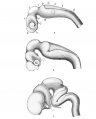
Fig. 30. Profile views of the brains of human embryos as seen during the third (A), fourth (B), and eighth (C) weeks
- Keibel Mall 2 031.jpg
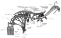
Fig. 32. Reconstruction showing the cranial nerves in a 10 mm human embryo
- Keibel Mall 2 033.jpg
- Keibel Mall 2 034.jpg
- Keibel Mall 2 035.jpg
- Keibel Mall 2 036.jpg
- Keibel Mall 2 037.jpg
- Keibel Mall 2 038.jpg
- Keibel Mall 2 039.jpg
- Keibel Mall 2 040.jpg
- Keibel Mall 2 041.jpg
- Keibel Mall 2 042.jpg
- Keibel Mall 2 043.jpg
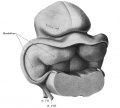
Fig. 44. Reconstruction showing cephalic portion of the rhombencephalon and adjoining midbrain at end of second month (human embryo 30 mm long (Mall collection. No. 86).


XVII. The Development of the Digestive Tract and of the Organs of Respiration
XVII. The Development of the Digestive Tract and of the Organs of Respiration
The Early Development of the Entodermal Tract and the Formation of its Subdivisions
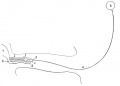
Fig 223
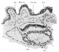
Fig 224
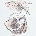
Fig 225
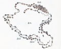
Fig 226
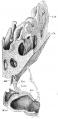
Fig 227
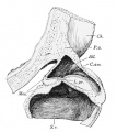
Fig 228
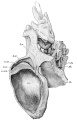
Fig 229
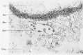
Fig 230
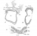
Fig 231
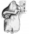
Fig 232
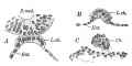
Fig 233
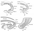
Fig 234
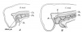
Fig 235
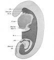
Fig 236
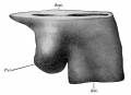
Fig 237
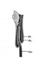
Fig 238
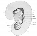
Fig 239
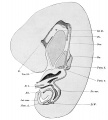
Fig 240
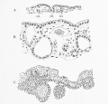
Fig 241
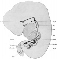
Fig 242
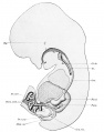
Fig 243
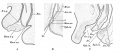
Fig 244
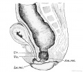
Fig 245
Fig 246
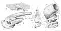
Fig 247
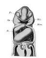
Fig 248


The Development of the Pharynx and of the Organs of Respiration
The Development of the Pharynx and of the Organs of Respiration
I. General Morphology of the Pharyngeal Pouches
I. General Morphology of the Pharyngeal Pouches
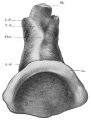
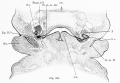
Fig. 314. Pharynx of the embryo Klb (Kromer-Pfannenstiel ; Normentafel, No. 3; 5-6 primitive segments, length, determined from the number of sections, 1.38 mm.)
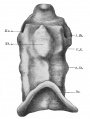
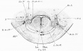
Fig. 315. Pharynx of the embryo Rob. Meyer No. 335 (9-10 pairs of primitive segments, length, determined from the number of sections, 1.70 mm.)
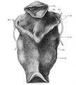
Fig. 316. Pharynx of the embryo Hah in the collection of the First Anatomical Institute, Vienna (about 15 pairs of primitive segments, length about 3 mm.)
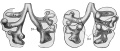
Fig. 317. Pharynx of the embryo Rob. Meyer No. 300 (Normentafel Xo. 7, 23 pairs of primitive segments, 2.5 mm. vertex-breech length) seen from the ventral surface.
Fig. 318.The model shown in Fig. 317 from the lateral surface.
Fig. 319. Pharyngeal pouches of the embryo Hah of the First Anatomical Institute, Vienna (5.2 mm., about the same stage as No. 16 of the Normentafel).
Fig. 320. Pharyngeal region of the embryo BR (Normentafel No. 42, 9.75 mm. vertex-breech measurement), from the ventral surface.


II. The Differentiation of the Pharyngeal Pouches - the Second Pharyngeal Pouch and the Tonsils
II. The Differentiation of the Pharyngeal Pouches - the Second Pharyngeal Pouch and the Tonsils
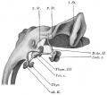
Fig. 321. Branchial derivatives of an embryo of 11.7 mm. (nape-length), somewhat simplified.
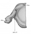
Fig. 322. Dorsal view of the left half of the pharynx of an embryo of 21 mm. (nape-length).
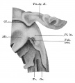
Fig. 323. Pharynx of an embryo of 24.4 mm. (nape-length), from the left side, somewhat simplified.
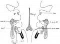
Fig. 324. Schema of the branchiogenic derivatives in man, adapted from the schemata of Groschuff and Kohn.
Figs. 325 and 326.Two sections through the embryo BR (compare Fig. 320).
Figs. 327 and 328. Two sections through the embryo BR (compare Fig. 320).
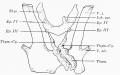
Fig. 329. The branchiogenic organs of an embryo of 26 mm., somewhat simplified. (After Verdun, 1898.)
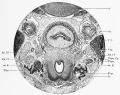
Fig. 330. Section through the laryngeal region of the embryo Nat2 of the First Anatomical Institute, Vienna (19.75 mm. vertex-breech length).


B. The Development of the Respiratory Apparatus
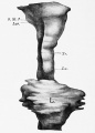
Fig. 331. Anlage of the respiratory tract of an embryo of 23 primitive segments
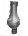
Fig. 332. Lung anlage of an embryo of 4.25 mm. vertex-breech measurement, from the ventral side.
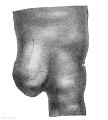
Fig. 333. The same model seen from the left side.
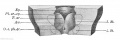
Fig. 334. Entrance to the larynx of an embryo of 8 mm
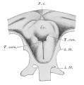
Fig. 335.Laryngeal entrance of an embryo of 28 to 29 days 8-9 mm
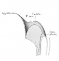
Fig. 336. Median section of the larynx shown in Fig. 335.
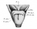
Fig. 337. — The entrance of the larynx in an embryo of 40-42 days 15-16 mm
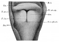
Fig. 338. Entrance of the larynx of an embryo of 30 mm
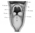
Fig. 339. Entrance of the larynx of an embryo of 16/23 cm male.
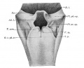
Fig. 340. Entrance of the larynx of an embryo of 29/43 cm male.
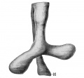
Fig. 341. Epithelial lung anlage of the embryo 18. 5 mm
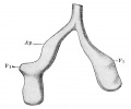
Fig. 342. Epithelial lung anlage of the embryo 7 mm
Figs. 343 and 344. lungs of an embryo at the beginning of the fifth week, ventral and dorsal views.
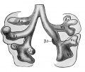
Fig. 343. lungs of an embryo at the beginning of the fifth week, ventral views.
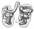
Fig. 344. lungs of an embryo at the beginning of the fifth week, dorsal views.
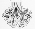
Fig. 345 Anlage of the lung of embryo 10.5 mm seen from in front with arteries and veins
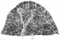
Fig. 346. Section through the lower lobe of the right lung of a fetus of 100 mm vertex-breech length

Fig. 347. The mesodermal anlage of the lungs of an embryo of 5 mm
Figs. 349 and 350. Mesodermal anlage of an embryo of about 13 mm seen from the ventral and the dorsal surface
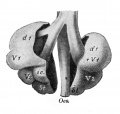
Fig. 349. Mesodermal anlage of an embryo of about 13 mm seen from the ventral surface
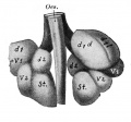
Fig. 350. Mesodermal anlage of an embryo of about 13 mm seen from the dorsal surface
Figs. 351 and 352. Lungs of an embryo of about 17.5 mm. seen from the right and from the left.
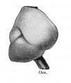
Fig. 351. Lungs of an embryo of about 17.5 mm. seen from the right.

Fig. 352. Lungs of an embryo of about 17.5 mm. seen from the left.
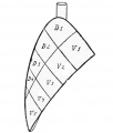
Fig. 353. Schema of the lobation of the lung.


III. The Third to the Fifth Pharyngeal Pouches - the Branchiogenic Organs
III. The Third to the Fifth Pharyngeal Pouches - the Branchiogenic Organs
XVIII. The Development of the Blood, the Vascular System, and the Spleen
XVIII. The Development of the Blood, the Vascular System, and the Spleen
I. The Origin of the Angioblast and the Development of the Blood
I. The Origin of the Angioblast and the Development of the Blood
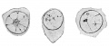
Fig. 354. Three primitive mesamoeboids from the yolk-sac of a human embryo of about 1 mm.
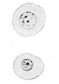
Fig. 355. Two blood-corpuscles of a human embryo of 4 mm.
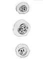
Fig. 356. Three blood-corpuscles of a human embryo of 7.5 mm.
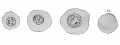
Fig. 357. Three blood-corpuscles from a human embryo of 9.4 mm.
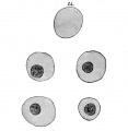
Fig. 359. Blood-corpuscles from a blood-vessel of a human embryo of eight months.
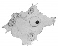
Fig. 360. An erythrocyte lying free in the mesenchyma.
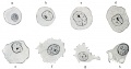
Fig. 361. Red blood-cells from the placental chorion of a human embryo of 15 mm.
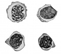
Fig. 363. Finely granular (neutrophile) leucocyte with compact nucleus, a so-called myelocyte.
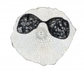
Fig. 364. A coarsely granular leucocyte (eosinophile) with a bilobate nucleus.
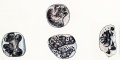
Fig. 365. Degenerating human leucocytes (Mastleucocyten of Maximow)
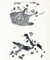
Fig. 366. Giant cells with processes from which blood-plates arise.
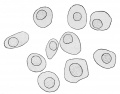
Fig. 367. Outlines of erythrocytes of a human embryo of 8 mm
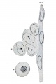
Fig. 368. Endothelium and blood-cells from the lower part of the aorta of a human embryo of 9.4 mm
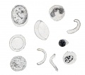
Fig. 369. Blood-corpuscles from the vessels of a human fetus of eight months.
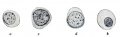
Fig. 370. Blood-cells from a hepatic vessel of a human embryo of 11 mm
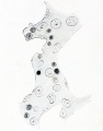
Fig. 371. Hepatic cylinders of a human embryo of 11 mm. Coll. F. P. Mali, No. 353. The blood-cells with small nuclei lie apparently in the substance of the liver-cells, -which have large nuclei.


II. The Development of the Heart
II. The Development of the Heart
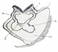
Fig. 372. Section through the heart anlage of the Pfannenstiel-Kromer embryo Klb (Normentafel, Xo. 3, 5 to 6 primitive somites).
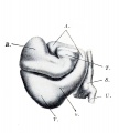
Fig. 373. Model of the heart of a human embryo 2.5 mm. greatest length.
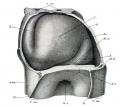
Fig. 374. Model of the heart of the embryo Halj of 3 mm. greatest length, 15 primitive somites.
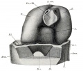
Fig. 375. The model shown in Fig. 374 after removal of the pericardium, seen from behind.
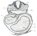
Fig. 376. Transverse section through the embryo Hah.
- Keibel Mall 2 377.jpg
Fig. 377. Section through the ventricular wall of the heart of embryo Hah, 3.5 mm. greatest length.
- Keibel Mall 2 378.jpg
Fig. 378. Model of the heart of the embryo Hah of 5.2 mm. greatest length.
- Keibel Mall 2 379.jpg
Fig. 379. Model of the heart of embryo £U of 6.5 mm. greatest length.
- Keibel Mall 2 380.jpg
Fig. 380. Model of [thejjheart of embryo La of 9 mm. greatest length.
- Keibel Mall 2 381.jpg
Fig. 381. Sagittal section through the model shown in Fig. 380, the section passing to the left of the septum I.
- Keibel Mall 2 382.jpg
Fig. 382. Transverse section through the heart region of the embryo Wal of 8 mm. greatest length.
- Keibel Mall 2 383.jpg
Fiag. 383. Section through the bulbus cordis of the embryo H6.
- Keibel Mall 2 384.jpg
Fig. 384. Model of the bulbus cordis of the embryo H6, divided longitudinally.
- Keibel Mall 2 385.jpg
Fig. 385. Section through the wall of the ventricle of embryo EU
- Keibel Mall 2 386.jpg
Fig. 386. Model of the heart of embryo S2 of 14.5 mm. greatest length.
- Keibel Mall 2 387.jpg
Fig. 387. Section through the heart of embryo Mi of 16.75 mm greatest length.
- Keibel Mall 2 388.jpg
Fig. 388. Model of the heart of an embryo of 310 mm greatest length.
- Keibel Mall 2 390.jpg
Fig. 389. Section through the heart of an embryo of 165 mm. greatest length.


| Historic Disclaimer - information about historic embryology pages |
|---|
 |
Glossary Links
- Glossary: A | B | C | D | E | F | G | H | I | J | K | L | M | N | O | P | Q | R | S | T | U | V | W | X | Y | Z | Numbers | Symbols | Term Link
Cite this page: Hill, M.A. (2024, April 25) Embryology Manual of Human Embryology II - Figures. Retrieved from https://embryology.med.unsw.edu.au/embryology/index.php/Manual_of_Human_Embryology_II_-_Figures
- © Dr Mark Hill 2024, UNSW Embryology ISBN: 978 0 7334 2609 4 - UNSW CRICOS Provider Code No. 00098G