MRI Stage 17 Movie 1: Difference between revisions
mNo edit summary |
mNo edit summary |
||
| Line 6: | Line 6: | ||
[[Media:Stage_17_MRI01.mp4|'''Click Here''' to play on mobile device]] | [[Media:Stage_17_MRI01.mp4|'''Click Here''' to play on mobile device]] | ||
|- | |- | ||
| This is a MRI off-axis (not in at the anatomical plane) sagittal section through the week 6 embryo. | |||
# spinal cord in longitudinal section (later cross-section). | # spinal cord in longitudinal section (later cross-section). | ||
# limb buds then appear | # limb buds then appear | ||
| Line 19: | Line 19: | ||
# central nervous system the secondary brain vesicles and flexures can be seen. | # central nervous system the secondary brain vesicles and flexures can be seen. | ||
# ventricular cavity of the CNS containing the choroid plexus. | # ventricular cavity of the CNS containing the choroid plexus. | ||
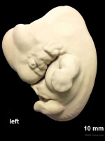
| | | valign=top| [[File:Stage17 model 03.jpg|150px]] | ||
[[Carnegie stage 17|Stage 17]]: Week 6, 42 - 44 days, 11 - 14 mm Gestational Age {{GA}} week 8 | [[Carnegie stage 17|Stage 17]]: Week 6, 42 - 44 days, 11 - 14 mm Gestational Age {{GA}} week 8 | ||
|- | |- | ||
Revision as of 10:32, 18 July 2016
| Embryology - 16 Apr 2024 |
|---|
| Google Translate - select your language from the list shown below (this will open a new external page) |
|
العربية | català | 中文 | 中國傳統的 | français | Deutsche | עִברִית | हिंदी | bahasa Indonesia | italiano | 日本語 | 한국어 | မြန်မာ | Pilipino | Polskie | português | ਪੰਜਾਬੀ ਦੇ | Română | русский | Español | Swahili | Svensk | ไทย | Türkçe | اردو | ייִדיש | Tiếng Việt These external translations are automated and may not be accurate. (More? About Translations) |
| <html5media height="560" width="700">File:Stage_17_MRI01.mp4</html5media> | |
This is a MRI off-axis (not in at the anatomical plane) sagittal section through the week 6 embryo.
|

Stage 17: Week 6, 42 - 44 days, 11 - 14 mm Gestational Age GA week 8 |
| |
| Week: | 1 | 2 | 3 | 4 | 5 | 6 | 7 | 8 |
| Carnegie stage: | 1 2 3 4 | 5 6 | 7 8 9 | 10 11 12 13 | 14 15 | 16 17 | 18 19 | 20 21 22 23 |
Reference
<pubmed>19635979</pubmed>
Copyright
Image source: The Kyoto Collection images are reproduced with the permission of Prof. Kohei Shiota and Prof. Shigehito Yamada, Anatomy and Developmental Biology, Kyoto University Graduate School of Medicine, Kyoto, Japan for educational purposes only and cannot be reproduced electronically or in writing without permission.
Cite this page: Hill, M.A. (2024, April 16) Embryology MRI Stage 17 Movie 1. Retrieved from https://embryology.med.unsw.edu.au/embryology/index.php/MRI_Stage_17_Movie_1
- © Dr Mark Hill 2024, UNSW Embryology ISBN: 978 0 7334 2609 4 - UNSW CRICOS Provider Code No. 00098G