Leonardo da Vinci - the anatomist (1930) Illustrations: Difference between revisions
mNo edit summary |
|||
| (4 intermediate revisions by the same user not shown) | |||
| Line 26: | Line 26: | ||
===Chapter VII Leonardo’s Anatomical Methods=== | ===Chapter VII Leonardo’s Anatomical Methods=== | ||
<gallery> | |||
File:McMurrich1930 fig10.jpg|Fig. 10. Transverse sections of the leg. (QV, 20.) | |||
File:McMurrich1930 fig11.jpg|Fig. 11. Figures in which the muscles of the leg are represented by cords or wires. (QV, 4.) | |||
File:McMurrich1930 fig12.jpg|Fig. 12. Figures of the surface anatomy of the leg with a comparison of the hip muscles of a man and a horse, the muscles being represented by cords or wires. (QV, 22.) | |||
</gallery> | |||
===Chapter IX Leonardo’s Canon of Proportions=== | |||
<gallery> | |||
File:McMurrich1930 fig13.jpg|Fig. 13. The figure of a man inscribed in a circle and in a square. A drawing in the Royal Academy, Venice (Anderson) | |||
File:McMurrich1930 fig14.jpg|Fig. 14. Figure illustrating the proportions of the head. (QVI, 1.) | |||
File:McMurrich1930 fig15.jpg|Fig. 15. Figure showing the lines of measurement used in determining the proportions of the leg. (QVI, llv.) | |||
File:McMurrich1930 fig16.jpg|Fig. 16. Figures illustrating the proportions of the face and eye. A drawing in the Royal Palace, Turin (Anderson) | |||
Fig. 13. The figure of a man inscribed in a circle and in a square. A drawing in the Royal Academy, Venice (Anderson) | File:McMurrich1930 fig17.jpg|Fig. 17. Proportions of the human body in the standing, kneeling and sitting postures. (QVI, 8.) | ||
File:McMurrich1930 fig18.jpg|Fiq. 18. The Bone Man from the Priifling five-figure series (1158). From Sudhoff, Studien, Heft 1, pi. 13, 1907 | |||
Fig. 14. Figure illustrating the proportions of the head. (QVI, 1.) | </gallery> | ||
===Chapter X The Skeleton=== | |||
Fig. 15. Figure showing the lines of measurement used in determining the proportions of the leg. (QVI, llv.) | <gallery> | ||
File:McMurrich1930 fig19.jpg|Fig. 19. Skeleton from a Provengal manuscript in the University Library, Basel, Codex D II, 11 (End of thirteenth century). From Sudhoff, Studien, Heft 4, pi. 1, 190S | |||
Fig. 16. Figures illustrating the proportions of the face and eye. A drawing in the Royal Palace, Turin (Anderson) | File:McMurrich1930 fig20.jpg|Fig. 20. Skeleton from the Dresden Codex No. 301 (1323). From Sudhoff, Studien, Heft 4, pi. 6, 1908 | ||
File:McMurrich1930 fig21.jpg|Fig. 21. Skeleton from the De arte phisicali of John Arderne (circa 1412). From Sudhoff, Studien, Heft 8, pi. 3, 1915 | |||
Fig. 17. Proportions of the human body in the standing, kneeling and sitting postures. (QVI, 8.) | File:McMurrich1930 fig22.jpg|Fig. 22. Skeleton by Richard Helain (1493). From Sudhoff, Archiv, vol. 1, p. 57, 1907 | ||
File:McMurrich1930 fig23.jpg|Fig. 23. Skeleton from the British Museum Additional Ms. No. 21618. From Sudhoff, Archiv, vol. 8, p. 140, 1915 | |||
Fiq. 18. The Bone Man from the Priifling five-figure series (1158). From Sudhoff, Studien, Heft 1, pi. 13, 1907 | File:McMurrich1930 fig24.jpg|Fig. 24. The vertebral column by Leonardo. (AnA, 8v.) | ||
File:McMurrich1930 fig25.jpg|Fig. 25. The cervical vertebra. (AnA, 8v.) | |||
Fig. 19. Skeleton from a Provengal manuscript in the University Library, Basel, Codex D II, 11 (End of thirteenth century). From Sudhoff, Studien, Heft 4, pi. 1, 190S | File:McMurrich1930 fig26.jpg|Fig. 26. The skull cut to show the frontal and maxillary sinuses. (AnB, 41v.) | ||
File:McMurrich1930 fig27.jpg|Fig. 27. The bones of the arm in supination and pronation, together with the scapula and biceps muscle. (AnA, lv.) | |||
Fig. 20. Skeleton from the Dresden Codex No. 301 (1323). From Sudhoff, Studien, Heft 4, pi. 6, 1908 | File:McMurrich1930 fig28.jpg|Fig. 28. The bones of the hand, with a dissection of the tendons and ligaments of the fingers. (AnA, lOv.) 124 | ||
File:McMurrich1930 fig29.jpg|Fig. 29. Various figures of the bones of the foot with a sketch of the bones of the shoulder. (AnA, 12.) 125 | |||
Fig. 21. Skeleton from the De arte phisicali of John Arderne (circa 1412). From Sudhoff, Studien, Heft 8, pi. 3, 1915 | </gallery> | ||
===Chapter XI The Muscles=== | |||
Fig. 22. Skeleton by Richard Helain (1493). From Sudhoff, Archiv, vol. 1, p. 57, 1907 | |||
Fig. 23. Skeleton from the British Museum Additional Ms. No. 21618. From Sudhoff, Archiv, vol. 8, p. 140, 1915 | |||
Fig. 24. The vertebral column by Leonardo. (AnA, 8v.) | |||
Fig. 25. The cervical vertebra. (AnA, 8v.) | |||
Fig. 26. The skull cut to show the frontal and maxillary sinuses. (AnB, 41v.) | |||
Fig. 27. The bones of the arm in supination and pronation, together with the scapula and biceps muscle. (AnA, lv.) | |||
Fig. 28. The bones of the hand, with a dissection of the tendons and ligaments of the fingers. (AnA, lOv.) 124 | |||
Fig. 29. Various figures of the bones of the foot with a sketch of the bones of the shoulder. (AnA, 12.) 125 | |||
Fig. 30. The Muscle Man from the Raudnitz five-figure series (1399). From Sudhoff, Archiv, vol. 3, pi. 12, 1910 | Fig. 30. The Muscle Man from the Raudnitz five-figure series (1399). From Sudhoff, Archiv, vol. 3, pi. 12, 1910 | ||
| Line 152: | Line 142: | ||
===Chapter XVI The Excretory and Reproductive Organs=== | ===Chapter XVI The Excretory and Reproductive Organs=== | ||
<gallery> | |||
Fig. 69. The male organs of reproduction. (QIII, 4.) 199 | File:McMurrich1930 fig69.jpg|Fig. 69. The male organs of reproduction. (QIII, 4.) 199 | ||
File:McMurrich1930 fig70.jpg|Fig. 70. The female organs of reproduction. (QI, 12.) 200 | |||
Fig. 70. The female organs of reproduction. (QI, 12.) 200 | </gallery> | ||
===Chapter XVII The Nervous System=== | ===Chapter XVII The Nervous System=== | ||
Latest revision as of 16:19, 21 April 2020
| Embryology - 18 Apr 2024 |
|---|
| Google Translate - select your language from the list shown below (this will open a new external page) |
|
العربية | català | 中文 | 中國傳統的 | français | Deutsche | עִברִית | हिंदी | bahasa Indonesia | italiano | 日本語 | 한국어 | မြန်မာ | Pilipino | Polskie | português | ਪੰਜਾਬੀ ਦੇ | Română | русский | Español | Swahili | Svensk | ไทย | Türkçe | اردو | ייִדיש | Tiếng Việt These external translations are automated and may not be accurate. (More? About Translations) |
McMurrich JP. Leonardo da Vinci - the anatomist. (1930) Carnegie institution of Washington, Williams & Wilkins Company, Baltimore.
| Historic Disclaimer - information about historic embryology pages |
|---|
 |
Leonardo da Vinci - The Anatomist
List of Illustrations

Portrait of Leonardo da Vinci, probably by himself. Royal Palace, Turin (Anderson) Frontispiece
Chapter II Anatomy from Galen to Leonardo
Fig. 1. An “Anatomy.” From the Fasciculo di Medicina (Venice, 1493). After the facsimile published by C. Singer, Florence, 1925, p. 64 18
Fig. 2. A dissection by Guido da Vigevano (1345). Archiv fur Ge schichte der Medizin, vol. 7, pi. 1, 1914


Chapter IV Anatomical Illustration before Leonardo
Fig. 3. Situs figure from the Fasciculus medicinse (1491). After the facsimile published by K. Sudhoff and C. Singer, Milan, p. 10, 1924
Fig. 4. Leonardo’s Situs figure. (QI, 12)
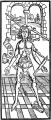
Fig. 5. A Wound Man. Title page of the Book of Cirurgia by Hieronymus Brunschwig (Strassburg, 1497)
- McMurrich1930 fig06.jpg
Fig. 6. Situs figure from Peyligk’s Philosophise Naturalis Compendium (Leipzig, 1499). After K. Sudhoff, Studien zur Geschichte der Medizin, Heft 8, pi. 7, 1909
- McMurrich1930 fig07.jpg
Fig. 7. Situs figure from the Antropologium, de hominis dignitate of Magnus Hundt (Leipzig, 1501). After Choulant
- McMurrich1930 fig08.jpg
Fig. 8. The brain and sense organs from the Antropologium of Magnus Hundt (Leipzig, 1501). After Sudhoff, Studien, Heft 8, pi.
- McMurrich1930 fig09.jpg
Fig. 9. Situs figure from the Spiegel der Artzny of Laurentius Phryesen (Strassburg, 1518)


Chapter VII Leonardo’s Anatomical Methods
- McMurrich1930 fig10.jpg
Fig. 10. Transverse sections of the leg. (QV, 20.)
- McMurrich1930 fig11.jpg
Fig. 11. Figures in which the muscles of the leg are represented by cords or wires. (QV, 4.)
- McMurrich1930 fig12.jpg
Fig. 12. Figures of the surface anatomy of the leg with a comparison of the hip muscles of a man and a horse, the muscles being represented by cords or wires. (QV, 22.)
Chapter IX Leonardo’s Canon of Proportions
- McMurrich1930 fig13.jpg
Fig. 13. The figure of a man inscribed in a circle and in a square. A drawing in the Royal Academy, Venice (Anderson)
- McMurrich1930 fig14.jpg
Fig. 14. Figure illustrating the proportions of the head. (QVI, 1.)
- McMurrich1930 fig15.jpg
Fig. 15. Figure showing the lines of measurement used in determining the proportions of the leg. (QVI, llv.)
- McMurrich1930 fig16.jpg
Fig. 16. Figures illustrating the proportions of the face and eye. A drawing in the Royal Palace, Turin (Anderson)
- McMurrich1930 fig17.jpg
Fig. 17. Proportions of the human body in the standing, kneeling and sitting postures. (QVI, 8.)
- McMurrich1930 fig18.jpg
Fiq. 18. The Bone Man from the Priifling five-figure series (1158). From Sudhoff, Studien, Heft 1, pi. 13, 1907
Chapter X The Skeleton
- McMurrich1930 fig19.jpg
Fig. 19. Skeleton from a Provengal manuscript in the University Library, Basel, Codex D II, 11 (End of thirteenth century). From Sudhoff, Studien, Heft 4, pi. 1, 190S
- McMurrich1930 fig20.jpg
Fig. 20. Skeleton from the Dresden Codex No. 301 (1323). From Sudhoff, Studien, Heft 4, pi. 6, 1908
- McMurrich1930 fig21.jpg
Fig. 21. Skeleton from the De arte phisicali of John Arderne (circa 1412). From Sudhoff, Studien, Heft 8, pi. 3, 1915
- McMurrich1930 fig22.jpg
Fig. 22. Skeleton by Richard Helain (1493). From Sudhoff, Archiv, vol. 1, p. 57, 1907
- McMurrich1930 fig23.jpg
Fig. 23. Skeleton from the British Museum Additional Ms. No. 21618. From Sudhoff, Archiv, vol. 8, p. 140, 1915
- McMurrich1930 fig24.jpg
Fig. 24. The vertebral column by Leonardo. (AnA, 8v.)
- McMurrich1930 fig25.jpg
Fig. 25. The cervical vertebra. (AnA, 8v.)
- McMurrich1930 fig26.jpg
Fig. 26. The skull cut to show the frontal and maxillary sinuses. (AnB, 41v.)
- McMurrich1930 fig27.jpg
Fig. 27. The bones of the arm in supination and pronation, together with the scapula and biceps muscle. (AnA, lv.)
- McMurrich1930 fig28.jpg
Fig. 28. The bones of the hand, with a dissection of the tendons and ligaments of the fingers. (AnA, lOv.) 124
- McMurrich1930 fig29.jpg
Fig. 29. Various figures of the bones of the foot with a sketch of the bones of the shoulder. (AnA, 12.) 125
Chapter XI The Muscles
Fig. 30. The Muscle Man from the Raudnitz five-figure series (1399). From Sudhoff, Archiv, vol. 3, pi. 12, 1910
Fig. 31. The abdominal muscles from Pietro di Abano’s Conciliator differentiarum (1496). From Sudhoff, Archiv, vol. 3, pi. 2, 1910
Fig. 32. The muscles of the neck and shoulder. (AnA, 3v.)
Fig. 33. Two representations of the muscles of the back and shoulder. (AnA, 16.) 137
Fig. 34. A cord diagram of the muscles supposed to stabilize the cervical vertebra in movements of the head. Also a sketch showing the insertions of muscles into the spine of a vertebra. (QII, 5v.)
Fig. 35. Diagrammatic representation of the superior serratus posterior and the serratus anterior. (Q0, 8.)
Fig. 36. The muscles of the shoulder, trunk and leg. (AnA, 15v.)
Fig. 37. Figures showing the form of the diaphragm. (QI, 5.)
Fig. 38. The abdominal muscles. (QI, 5.)
Fig. 39. The scapular and brachial muscles. (AnA, 2.)
Fig. 40. The muscles of the arm and forearm. (AnA, 9v.)
Fig. 41. Dissections of the muscles, tendons and ligaments of the hand and fingers. (AnA, 19.)
Fig. 42. The muscles and tendons of the sole of the foot. (AnA, 11.)
Fig. 43. Diagram of the structure of the heart in Ioannes Adelphus’ edition of Mondino’s Anathomia (Strassburg, 1513). After C. Singer, Fasciculo di medicina, vol. 1, fig. 59, 1925
Chapter XII The Heart
Fig. 44. Two figures of the heart. (QII, 3v.)
Fig. 45. Dissection of the heart showing papillary muscles and a moderator band. (QII, 14.)
Fig. 46. The thoracic and abdominal viscera, the heart dissected and showing several moderator bands in each ventricle. (QIV, 7.)
Fig. 47. Sketches of the base of the heart and of the papillary muscles and chordae tendineae of the left ventricle. (QIV, 14.)
Fig. 48. The tricuspid valve from above and from below, showing the attachments of the chordae tendineae. (QII, 8v.)
Fig. 49. Studies of the vortices in the pockets of the semilunar valves. (QIV, 11.)
Fig. 50. Figures illustrating the comparison of the heart and bloodvessels with a sprouting nut with its plumule and radicle. In the figure to the right the azygos vein is well shown. (AnB, 11.)
Chapter XIII The Blood-Vessels
Fig. 51. The superficial veins of the arm and a sketch comparing the arteries of a centenarian with those of a child. (AnB, 10.)
Fig. 52. Early study of the heart and blood-vessels. (QV, 1.)
Fig. 53. Dissections of the heart, lungs, abdominal viscera and bloodvessels. (QIII, lOv.)
Fig. 54. The great vessels of a centenarian. (AnB, 33.)
Fig. 55. The superficial pectoral and epigastric veins. (AnA, 6.)
Fig. 56. Figures of the hepatic artery and portal vein. (AnB, 34v.)
Fig. 57. The iliac vein and its branches. (AnB, 6v.)
Fig. 58. The hypogastric vessels and the umbilical vein. Above is a frontal section through the cervical vertebrae showing the costotransverse foramina. (AnB, 4.)
Fig. 59. An early sketch of the digestive tract and longitudinal and transverse sections of the penis. (QIII, 3v.)
Fig. 60. Above a supposed arrangement of the intestine; below the stomach, liver and spleen with splenic vein; to the right the caecum and appendix. (AnB, 14v.)
Fig. 61. A second arrangement of the intestines. To the right suggestion as to the mode of entrance of the ureter into the bladder. (AnB, 14.)
Fig. 62. The lungs, diaphragm, liver, stomach and spleen of an animal. (AnB, 37 v.)
Fig. 63. The mesentery. (AnB, 3.)
Fig. 64. The great omentum with the hypogastric vessels and the umbilical vein. To the left the deep epigastric veins. (AnB, 22v.)
Fig. 65. Dissection of the neck, in which an animal’s larynx is represented as human. (QV, 16.)
Chapter XV The Organs of Respiration
Fig. 66. Various figures of the larynx and trachea. The surface modeling of the leg. (AnA, 3.)
Fig. 67. The heart and bronchi after maceration away of the lung parenchyma. To the right representations of the bronchi. (QII, 1.)
Fig. 68. Sketch of the lungs and heart, showing the pleural cavities. (QIV. 3.)
Chapter XVI The Excretory and Reproductive Organs
- McMurrich1930 fig69.jpg
Fig. 69. The male organs of reproduction. (QIII, 4.) 199
- McMurrich1930 fig70.jpg
Fig. 70. The female organs of reproduction. (QI, 12.) 200
Chapter XVII The Nervous System
- McMurrich1930 fig71.jpg
Fig. 71. A section through the skull and brain showing the brain membranes. (QV, Ov.)
- McMurrich1930 fig72.jpg
Fig. 72. The ventricles of the brain and the cranial nerves. (QV, 8.)
- McMurrich1930 fig73.jpg
Fig. 73. The ventricles of the brain and a view of its base. (QV, 7.)
- McMurrich1930 fig74.jpg
Fig. 74. Cerebral localization. From G. Reisch: Margarita philosophise (Strassburg, 1504). After C. Singer: Fasciculo di medicina, part 1, fig. 69, 1925
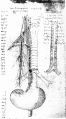
Fig. 75. Figure showing the course and distribution of the reversive (vagus) nerve. To the right a longitudinal section of the trachea. (AnB, 33v.)
- McMurrich1930 fig76.jpg
Fig. 76. Figures showing the arrangement of the brachial plexus. (AnB, 23 v.)
- McMurrich1930 fig77.jpg
Fig. 77. Another figure of the brachial plexus. (AnB, 3v.)
- McMurrich1930 fig78.jpg
Fig. 78. The lumbo-sacral plexus. (AnB, 6.)
- McMurrich1930 fig79.jpg
Fig. 79. Figure showing the course of the long saphenous nerve. (QV, 20 v.)
- McMurrich1930 fig80.jpg
Fig. 80. The branching of the common iliac vessels and the sciatic nerve. (QIV, 9.)
Fig. 81. The cervical portion of the spinal cord, showing the origins of the spinal nerves and what may be a suggestion of the ganglion ated cord. (AnB, 23.)


Chapter XVIII The Sense Organ
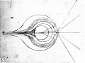
Fig. 82. Diagram of the structure of the eye. (CA, 337 II., A.)
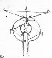
Fig. 83. Diagram showing two possibilities of refraction within the eye. (D, 10.)


Chapter XIX Embryology
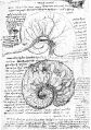
Fig. 84. Two figures of the membranes and circulation of the fetal calf. (AnB, 28.)
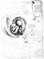
Fig. 85. Representations of the human fetus at term and of the ungulate placenta. (QIII, 8.)
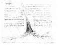
Fig. 86. Diagram of the umbilical and hypogastric vessels. (AnB, 29 v.)
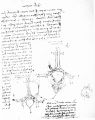
Fig. 87. Diagram of the human fetal circulation. (QI, 1.)


Chapter XX Comparative Anatomy
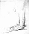
Fig. 88. Dissection of the foot of a bear. (QV, 11.)
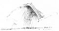
Fig. 89. Dissection of a bird’s wing. (QIV, 1.)


| Historic Disclaimer - information about historic embryology pages |
|---|
|
Reference: McMurrich JP. Leonardo da Vinci - the anatomist. (1930) Carnegie institution of Washington, Williams & Wilkins Company, Baltimore.
Cite this page: Hill, M.A. (2024, April 18) Embryology Leonardo da Vinci - the anatomist (1930) Illustrations. Retrieved from https://embryology.med.unsw.edu.au/embryology/index.php/Leonardo_da_Vinci_-_the_anatomist_(1930)_Illustrations
- © Dr Mark Hill 2024, UNSW Embryology ISBN: 978 0 7334 2609 4 - UNSW CRICOS Provider Code No. 00098G