Lecture - Gastrointestinal Development: Difference between revisions
mNo edit summary |
mNo edit summary |
||
| (46 intermediate revisions by the same user not shown) | |||
| Line 1: | Line 1: | ||
{{Header}} | |||
=Endoderm Development= | =Endoderm Development= | ||
== Introduction == | == Introduction == | ||
[[File:Gray0982a.jpg|thumb|The early developing gastrointestinal tract]]This lecture will cover the early development of the endoderm layer of the trilaminar embryo as it contributes to the lining, glands and organs of the gastrointestinal tract ('''GIT'''). | [[File:Gray0982a.jpg|thumb|The early developing gastrointestinal tract]]This lecture will cover the early development of the endoderm layer of the trilaminar embryo as it contributes to the lining, glands and organs of the gastrointestinal tract ('''GIT'''). The endoderm contribution to the respiratory system will be covered in a [[Lecture - Respiratory Development|separate lecture]]. | ||
Note that we will be returning in the laboratory and later (endocrine, neural crest) to discuss the gastrointestinal tract, associated organs and physical growth changes. | |||
Gastrulation, or gut formation, was historically the easiest observable feature of frog development. In human development, during the 4th week the 3 distinct portions (fore-, mid- and hind-gut) extend the length of the embryo and will contribute different structures. | |||
The oral cavity (mouth) is formed following breakdown of the [[B#buccopharyngeal membrane|buccopharyngeal membrane]] (= oropharyngeal or oral) and the opening means that it contains amniotic fluid, which is also swallowed later in development. | |||
The large mid-gut is generated by lateral embryonic folding which "pinches off" a pocket of the yolk sac, the 2 compartments continue to communicate through the vitelline duct. | |||
The hindgut (cloaca) will later be divided into separate urogenital and rectal regions that end at the cloacal membrane. | |||
Note that we will be returning in the laboratory and later (head, endocrine, neural crest) to discuss the gastrointestinal tract, associated organs and physical growth changes. | |||
[[Media:ANAT2341Lecture 2018 - Endoderm.pdf|2018 Lecture - Print PDF]] | |||
{| class="wikitable mw-collapsible mw-collapsed" | |||
! Some Recent Articles | |||
|- | |||
| '''Tongue''' | |||
{{#pmid:29945863}} | |||
:"Adult tongue epithelium is continuously renewed from epithelial progenitor cells, a process that requires hedgehog (HH) signaling. In summary, we show that SOX2 functions downstream of HH signaling to regulate lingual epithelium homeostasis." {{SOX}} | |||
{{#pmid:29784581}} | |||
:"Although the gross embryological contributions to tongue formation have been known for many years, it is only relatively recently that the molecular pathways regulating these processes have begun to be discovered. In particular, there is now evidence that the Hedgehog, {{TGF-beta}}, {{WNT}} and {{Notch}} signaling pathways all play an important role in mediating appropriate signaling interactions between the epithelial, cranial neural crest and mesodermal cell populations that are required to form the tongue." | |||
|- | |||
| '''Midgut Rotation''' | |||
[[File:Human embryo midgut loop 01.jpg|600px]] | |||
[[Carnegie stage 14]] (CS14) orientation of the midgut loop and its mesentery during the 5th week follows the helical body axis. | |||
* '''A''' - Dorsal view of the reconstruction of a CS14 embryo (s5029). Note the left-sided juxtaposition of the head relative to the caudal end of the body, reflecting the helical body axis. The successive parts of the midgut are shown in a rainbow color gradient (see legend color codes). Note that the vitelline artery (3) and the right vitelline vein (7) traverse the vitelline duct (8) at the apex of the midgut loop. The arrows indicate the changes occurring during straightening of the body axis in CS15 and CS16 embryos. | |||
* '''B''' - shows the position of the developing midgut mesentery (10) between both limbs of the midgut. Note the limited craniocaudal extension of the mesentery at this stage (10). The beige area identifies the region where the intestinal mesenchyme is attached to the dorsal body wall. | |||
* '''C''' - Histological section of embryo s5029 with right vitelline vein (7), vitelline artery (3), cecum (Ce) and developing dorsal midgut mesentery (10). Due to the helical body axis, the caudal end of the body is cut near transversely (with left (L) and right (R) sides), whereas more cranially, the body is cut almost sagittally (V: ventral; D: dorsal). Note that the midgut mesentery (10) is ~4-fold thinner than the mesenchymal mass surrounding the intestine. | |||
[http://www.biomedcentral.com/1471-213X/15/31 BMC Developmental Biology 2015, 15:31] | |||
|} | |||
==Lecture Objectives== | ==Lecture Objectives== | ||
| Line 13: | Line 54: | ||
* Brief understanding of mechanical changes (rotations) during GIT development | * Brief understanding of mechanical changes (rotations) during GIT development | ||
* Brief understanding of gastrointestinal abnormalities | * Brief understanding of gastrointestinal abnormalities | ||
==Lecture Resources== | ==Lecture Resources== | ||
{| class="wikitable mw-collapsible mw-collapsed" | {| class="wikitable mw-collapsible mw-collapsed" | ||
! Movies | ! Movies | ||
|- | |- | ||
| colspan=2 valign="bottom"|{{GIT_cartoons}} | | colspan=2 valign="bottom"|{{GIT_cartoons}} | ||
| Line 27: | Line 67: | ||
|} | |} | ||
{| class="wikitable mw-collapsible mw-collapsed" | {| class="wikitable mw-collapsible mw-collapsed" | ||
! References | ! References | ||
|- | |- | ||
| {{Embryo logocitation}} | | {{Embryo logocitation}} | ||
| | | | ||
{{Gastrointestinal Tract Links}} | {{Gastrointestinal Tract Links}} | ||
* Lecture Archive: | * Lecture Archive: [https://embryology.med.unsw.edu.au/embryology/index.php?title=Lecture_-_Gastrointestinal_Development&oldid=301545 2017] | [[Media:2015ANAT2341_Lecture_9_-_Gastrointestinal Development.pdf|2015 PDF]] | ||
[https://embryology.med.unsw.edu.au/embryology/index.php?title=Lecture_-_Gastrointestinal_Development&oldid=195582 2014] | [[Media:ANAT2341_Lecture_9_-_2014_Gastrointestinal_Development.pdf|2014 PDF]] | |||
|- | |- | ||
| {{MPT2011cover_citation}} | | {{MPT2011cover_citation}} | ||
| The following chapter links only work with a UNSW connection. | | The following chapter links only work with a UNSW connection. | ||
* [ | * Chapter 11 [https://ebookcentral-proquest-com.wwwproxy1.library.unsw.edu.au/lib/unsw/reader.action?docID=2074364&ppg=286 Alimentary System] | ||
|- | |- | ||
| {{SBBF2009cover_citation}} | | {{SBBF2009cover_citation}} | ||
| The following chapter links only work with a UNSW connection. | | The following chapter links only work with a UNSW connection. | ||
Chapter 14 [https://ebookcentral-proquest-com.wwwproxy1.library.unsw.edu.au/lib/unsw/reader.action?docID=2074524&ppg=359 Development of the Gastrointestinal Tract] | |||
|} | |} | ||
{| class="wikitable mw-collapsible mw-collapsed" | {| class="wikitable mw-collapsible mw-collapsed" | ||
! | ! colspan=2|2015 Lecture - Video and Audio | ||
|- | |- | ||
| | | '''Lecture Video''' | ||
<html5media>File:ANAT2341-2015Lecture-GIT.mp4</html5media> | |||
[[Media:ANAT2341-2015Lecture-GIT.mp4|Video (in new window)]] | |||
'''Lecture Audio''' | |||
<html5media>File:ANAT2341-2015Lecture-GIT.mp3</html5media> | |||
[[Media:ANAT2341-2015Lecture-GIT.mp3|Audio (in new window)]] | |||
| See also | |||
<br> | |||
{{OneMinuteClock}}<br> | |||
<br> | |||
[[One_Minute_Embryology#Endoderm_Development|'''1 Minute Embryology''']] | |||
[https://thebox.unsw.edu.au/video/1-minute-embryology-endoderm-development UNSW theBox] | |||
|} | |} | ||
== Germ Layer Contributions == | |||
* '''{{endoderm}}''' - epithelium and associated glands. | |||
* ''' | * '''{{mesoderm}}''' (splanchnic) - mesentry, connective tissues, smooth muscle, blood vessels. | ||
* ''' | * '''{{ectoderm}}''' (neural crest) - enteric nervous system. | ||
Both endoderm and mesoderm will also have major contributions to associated organs. | |||
Folding of the embryonic disc occurs ventrally around the notochord, which forms a rod-like region running rostro-caudally in the midline. | Folding of the embryonic disc occurs ventrally around the notochord, which forms a rod-like region running rostro-caudally in the midline. | ||
| Line 83: | Line 134: | ||
|} | |} | ||
===Coelomic Cavity=== | |||
* The mesoderm initially undergoes segmentation to form paraxial, intermediate mesoderm and '''lateral plate mesoderm'''. | |||
* Paraxial mesoderm segments into somites and lateral plate mesoderm divides into somatic and '''splanchnic mesoderm'''. | |||
* The space forming between them is the '''coelomic cavity''', that will form the 3 major body cavities (pericardial, pleural, '''peritoneal''') | |||
* Most of the gastrointestinal tract will eventually lie within the peritoneal cavity. | |||
<gallery mode="packed-hover" caption="Mesoderm and Ectoderm Cartoons"> | |||
File:Mesoderm-cartoon1.jpg|Trilaminar Embryo | |||
File:Mesoderm-cartoon2.jpg|Paraxial and Lateral Plate | |||
File:Mesoderm-cartoon3.jpg|Somites | |||
File:Mesoderm-cartoon4.jpg|Somatic and Splanchnic | |||
</gallery> | |||
(Note only the righhand side is shown, lefthand side would be identical.) | |||
==Week 4== | |||
(Gestational age {{GA}} 6 weeks) Carnegie stage 11 | |||
{| | {| | ||
| [[File:Stage11_sem100.jpg| | | [[File:Stage11 bf3.jpg|200px]] | ||
| [[File:Stage_11_historic-Atwell1930-1.jpg|240px]] | |||
| [[File:Stage11_sem100.jpg|300px]] | |||
|- | |||
| Embryo (stage 11 ventral view) | |||
| Embryo (midline section) | |||
| Embryo (EM section) endoderm, splanchnic mesoderm, Intraembryonic coelom | |||
|- | |||
| [[File:Stage11_bf9.jpg|300px]] | |||
| [[File:Stage11_sem4.jpg|300px]] | |||
| | |||
|- | |||
| Stomodeum | |||
| Buccopharyngeal membrane | |||
| | |||
|} | |||
<gallery> | <gallery> | ||
File:Stage7_cloacal-oral-membranes.jpg|Stage 7 Membranes | File:Stage7_cloacal-oral-membranes.jpg|Stage 7 Membranes | ||
| Line 104: | Line 179: | ||
</gallery> | </gallery> | ||
== Week 5 | ===Liver Development=== | ||
{| | |||
| [[File:Gray0982a.jpg|200px]] | |||
| [[File:Stage_13_image_073.jpg|200px|Liver and Stomach]] | |||
| [[File:Stage13 bf10.jpg|200px|Stage 13 Embryo]] | |||
|} | |||
{{Liver}} contributions from endoderm and splanchnic mesoderm at the level of the transverse septum (week 4) | |||
* Stage 11 - hepatic diverticulum development | |||
* Stage 12 - cell differentiation, septum transversum forming liver stroma, hepatic diverticulum forming hepatic trabeculae | |||
* Stage 13 - epithelial cord proliferation enmeshing stromal capillaries | |||
The {{liver}} initially occupies the entire anterior body. All blood vessels enter the liver (placental, vitelline) and leave to enter the heart. | |||
===Stomach=== | |||
{| | |||
| [[File:Gray0982a.jpg|200px]] | |||
| [[File:Stage14_stomach.jpg|200px]] | |||
| {{Stomach_rotation_movie}} | |||
|} | |||
* During week 4 at the level where the stomach will form the tube begins to dilate, forming an '''enlarged lumen'''. | |||
* The dorsal border grows more rapidly than ventral first rotation (of 90 degrees), which establishes the greater curvature of the stomach. | |||
* A second rotation (of 90 degrees) occurs on the longitudinal axis establishing the adult orientation of the stomach. | |||
==Week 5== | |||
({{GA}} 7 weeks) | |||
=== Canalization === | |||
{| border='0px' | {| border='0px' | ||
|- | |- | ||
| | | {{GIT_growth_movie}} | ||
| | | | ||
* Beginning at week 5 endoderm in the GIT wall proliferates | * Beginning at week 5 endoderm in the GIT wall proliferates | ||
* | * By week 6 totally blocking (occluding) | ||
* | * over the next two weeks this tissue degenerates reforming a hollow gut tube. | ||
* By the end of week 8 the GIT endoderm tube is a tube once more. | * By the end of week 8 the GIT endoderm tube is a tube once more. | ||
* The process is called recanalization (hollow, then solid, then hollow again) | |||
* Abnormalities in this process can lead to abnormalities such as atresia, stenosis or duplications. | |||
* The | |||
* | |||
|} | |} | ||
=== | ===Mesentery Development=== | ||
[[File:Greater-omentum.jpg|thumb|Greater Omentum]] | |||
{| | {| | ||
| | | {{Greater_omentum_movie}} {{Lesser sac movie}} | ||
* | | | ||
* | * Ventral mesentery lost except at level of stomach and liver. | ||
* | ** contributing the lesser omentum and falciform ligament. | ||
* | * Dorsal mesentery forms the adult structure along the length of the tract and allows blood vessel, lymph and neural connection. | ||
* | * At the level of the stomach the dorsal mesogastrium extends as a fold forming the greater omentum | ||
** continues to grow and extend down into the peritoneal cavity and eventually lies anterior to the small intestines. | |||
** This fold of mesentery will also fuse to form a single sheet. | |||
'''Spleen''' | |||
* Mesoderm within the dorsal mesogastrium (week 5) form a long strip of cells adjacent to the forming stomach above the developing pancreas. | |||
* Vascular and immune organ, no direct GIT function. | |||
|} | |||
| [[File: | ==Week 8 - 10== | ||
({{GA}} 10-12 weeks) | |||
===Intestine Herniation=== | |||
[[File:Stage_22_image_088.jpg|thumb|Week 8 herniated midgut]] | |||
[[File:Human- fetal week 10 sagittal plane D.jpg|thumb|Week 10]] | |||
{| border='0px' | |||
|- | |||
| {{Gastrointestinal stage 22 movie}} | |||
| | |||
* '''neural crest''' migration into the wall forms enteric nervous system (peristalsis, secretion) | |||
* midgut grows in length as a loop extending ventrally, returning as hindgut | |||
* connected by dorsal mesentery | |||
* rotates to form adult anatomical position (abnormalities of rotation) | |||
* continued body growth "engulfs" the intestine by about week 11. | |||
|} | |} | ||
=== | ===Intestine Rotation=== | ||
[[File:Normal intestinal rotation cartoon.jpg|500px]] | |||
Normal intestinal rotation (note these are gestational age {{GA}} weeks){{#pmid:20549505|PMID20549505}} | |||
|} | |||
===Hindgut=== | ===Hindgut=== | ||
* | [[File:Stage12_sem9_cloacal_membrane.jpg|thumb|Cloacal membrane (Week 4, Stage 12)]] | ||
* | {| | ||
| {{Urogenital septum movie}} | |||
| | |||
* Initially the '''cloaca''' forms a common urinary, genital, GIT space | |||
* This is divided by formation of a '''septum''' into anterior urinary and dorsal rectal (superior Tourneux fold; lateral Rathke folds) | |||
* hindgut - distal third transverse colon, descending and sigmoid colon, rectum. | |||
* anal pit - distal third of anorectal canal (ectodermal) | |||
|} | |||
==Gastrointestinal Tract Divisions== | |||
{| | |||
| During the 4th week the 3 distinct portions (fore-, mid- and hind-gut) extend the length of the embryo and will contribute different components of the GIT. These 3 divisions are also later defined by the vascular (artery) supply to each of theses divisions. | |||
[[File: | # '''Foregut''' - celiac artery (Adult: pharynx, esophagus, stomach, upper duodenum, respiratory tract, liver, gallbladder pancreas) | ||
# '''Midgut''' - superior mesenteric artery (Adult: lower duodenum, jejunum, ileum, cecum, appendix, ascending colon, half transverse colon) | |||
# '''Hindgut''' - inferior mesenteric artery (Adult: half transverse colon, descending colon, rectum, superior part anal canal) | |||
| [[File:GIT_blood_supply.jpg|400px]] | |||
Gastrointestinal Tract Blood Supply | |||
|} | |||
==Fetal== | |||
{| | |||
| [[File:Fetal small Intestine length growth graph.jpg|300px]] | |||
| [[File:Fetal_liver_weight_growth_graph.jpg|300px]] | |||
|- | |- | ||
| | | Small Intestine length (mm) | ||
| | | Liver Growth (weight grams) | ||
|- | |- | ||
| | | | ||
| | | 1 to 124 grams (birth) | ||
|- | |- | ||
|} | |} | ||
=== | ==Liver== | ||
* Differentiates to form the hepatic diverticulum and hepatic primordium, generates the gall bladder then divides into right and left hepatic (liver) buds. | |||
* Hepatic Buds - form hepatocytes, produce bile from week 13 (forms meconium of newborn) | |||
** Left Hepatic Bud - left lobe, quadrate, caudate (both q and c anatomically Left) caudate lobe of human liver consists of 3 anatomical parts: Spiegel's lobe, caudate process, and paracaval portion. | |||
* | ** Right Hepatic Bud - right lobe | ||
* Bile duct - 3 connecting stalks (cystic duct, hepatic ducts) which fuse. | |||
* Early liver also involved in blood formation, after the yolk sac and blood islands acting as a primary site. | |||
* | |||
* | |||
* | |||
* | |||
* | |||
[[ | [[Gastrointestinal_Tract_-_Liver_Development|Liver Development]] | ||
:''' | ==Pancreas== | ||
[[File:Stage22_pancreas_a.jpg|thumb|Pancreas (week 8)]] | |||
* Pancreatic buds - endoderm, covered in splanchnic mesoderm | |||
* Pancreatic bud formation – duodenal level endoderm, splanchnic mesoderm forms dorsal and ventral mesentery, '''dorsal bud''' (larger, first), '''ventral bud''' (smaller, later) | |||
* Duodenum growth/rotation – brings ventral and dorsal buds together, fusion of buds, exocrine function (postnatal function) | |||
* Pancreatic duct – ventral bud duct and distal part of dorsal bud | |||
* Pancreatic islets - endocrine function ('''week 10''' onwards) | |||
[[File:Pancreas_rotation.jpg|Pancreas rotation cartoon]] | |||
[[File: | |||
[[Gastrointestinal_Tract_-_Pancreas_Development|Pancreas Development]] | |||
==Spleen== | |||
{| | |||
| | |||
* Mesoderm within the dorsal mesogastrium form a long strip of cells adjacent to the forming stomach above the developing pancreas. | * Mesoderm within the dorsal mesogastrium form a long strip of cells adjacent to the forming stomach above the developing pancreas. | ||
* The spleen is located on the left side of the abdomen and has a role initially in blood and then immune system development. | * The spleen is located on the left side of the abdomen and has a role initially in blood and then immune system development. | ||
* The spleen's haematopoietic function (blood cell formation) is lost with embryo development and lymphoid precursor cells migrate into the developing organ. | * The spleen's haematopoietic function (blood cell formation) is lost with embryo development and lymphoid precursor cells migrate into the developing organ. | ||
* Vascularization of the spleen arises initially by branches from the dorsal aorta. | * Vascularization of the spleen arises initially by branches from the dorsal aorta. | ||
| [[File:Stage 22 image 087.jpg|thumb|300px|Spleen week 8 stage 22 embryo]] | |||
| [[File: | |||
|} | |} | ||
==Enteric Nervous System== | |||
Topic covered in the neural crest lecture. Table below summarises the two major neural plexuses. | |||
{{GIT plexus table}} | |||
== Gastrointestinal Tract Abnormalities == | |||
[[File:Australian_abnormalities_81-92_git.jpg|thumb|Australian Statistics [[Gastrointestinal Tract - Abnormalities]]]] | |||
[[File: | {| class="wikitable mw-collapsible mw-collapsed" | ||
! USA Statistics | |||
|- | |||
| | |||
{{USA_Selected_defect_table_2006}} | |||
|} | |||
===Lumen Abnormalities=== | ===Lumen Abnormalities=== | ||
There are several types of abnormalities that impact upon the continuity of the gastrointestinal tract lumen. | {| | ||
| valign=top|There are several types of abnormalities that impact upon the continuity of the gastrointestinal tract lumen. | |||
====Atresia==== | |||
* Interuption of the lumen (esophageal atresia, duodenal atresia, extrahepatic biliary atresia, anorectal atresia) | |||
====Stenosis==== | |||
* | * Narrowing of the lumen (duodenal stenosis, pyloric stenosis) | ||
[[File: | ====Duplication==== | ||
* Incomplete recanalization resulting in parallel lumens, this is really a specialized form of stenosis. | |||
| [[File:Gastrointestinal tract duplication sites.jpg|300px|Gastrointestinal tract duplication sites based upon 78 clinical studies.{{#pmid:718292|PMID718292]] | |||
|} | |||
===Meckel's Diverticulum=== | ===Meckel's Diverticulum=== | ||
{| | |||
| valign=top| | |||
* This abnormality is a very common (incidence of 1–2% in the general population) and results from improper closure and absorption of the vitelline duct during early development. | |||
** vitelline duct (omphalomesenteric duct, yolk stalk) is a transient developmental duct that connects the yolk to the primitive GIT. | |||
| [[File:Meckel%27s_diverticulum_01.jpg|150px]] | |||
Meckel's Diverticulum | |||
|} | |||
===Intestinal Malrotation=== | ===Intestinal Malrotation=== | ||
{| | |||
Presents clinically in symptomatic malrotation as: | | Presents clinically in symptomatic malrotation as: | ||
* Neonates - bilious vomiting and bloody stools. | * Neonates - bilious vomiting and bloody stools. | ||
* Newborn - bilious vomiting and failure to thrive. | * Newborn - bilious vomiting and failure to thrive. | ||
* Infants - recurrent abdominal pain, intestinal obstruction, malabsorption/diarrhea, peritonitis/septic shock, solid food intolerance, common bile duct obstruction, abdominal distention, and failure to thrive. | * Infants - recurrent abdominal pain, intestinal obstruction, malabsorption/diarrhea, peritonitis/septic shock, solid food intolerance, common bile duct obstruction, abdominal distention, and failure to thrive. | ||
'''Ladd's Bands''' - are a series of bands crossing the duodenum which can cause duodenal obstruction. | '''Ladd's Bands''' - are a series of bands crossing the duodenum which can cause duodenal obstruction. | ||
: | |||
:Links: [[Gastrointestinal_Tract_-_Abnormalities#Intestinal_Malrotation|Intestinal Malrotation]] | |||
| [[File:Intestinal_malrotation.jpg|200px]] | |||
Intestinal malrotation | |||
|} | |||
===Intestinal Aganglionosis=== | ===Intestinal Aganglionosis=== | ||
{| | |||
| (intestinal aganglionosis, Hirschsprung's disease, aganglionic colon, megacolon, congenital aganglionic megacolon, congenital megacolon) | |||
* A condition caused by the lack of enteric nervous system (neural ganglia) in the intestinal tract responsible for gastric motility (peristalsis). | * A condition caused by the lack of enteric nervous system (neural ganglia) in the intestinal tract responsible for gastric motility (peristalsis). | ||
* Neural crest cells | |||
** migrate initially into the cranial end of the GIT. | |||
** migrate during embryonic development caudally down the GIT. | |||
* Aganglionosis typically at the anal end of GIT. | |||
** increased severity as it extends cranially. | |||
| [[File:Megacolon_surgery_01.jpg|200px]] | |||
|} | |||
=== Gastroschisis === | === Gastroschisis === | ||
{| | {| | ||
| | | Gastroschisis (omphalocele, paraomphalocele, laparoschisis, abdominoschisis, abdominal hernia) is a congenital abdominal wall defect which results in herniation of fetal abdominal viscera (intestines and/or organs) into the amniotic cavity. | ||
Incidence of gastroschisis has been reported at 1.66/10,000, occuring more frequently in young mothers (less than 20 years old). | |||
By definition, it is a body wall defect, not a gastrointestinal tract defect, which in turn impacts upon GIT development. | |||
This indirect developmental effect (one system impacting upon another) occurs in several other systems. | |||
* '''Omphalocele''' - appears similar to gastroschisis, herniation of the bowel, liver and other organs into the intact umbilical cord, the tissues being '''covered by membranes''' unless the latter are ruptured. | |||
| {{Ultrasound Gastroschisis}} | |||
|- | |||
|} | |||
===Final Thoughts- After Birth=== | |||
Remember that the GIT does not function until after birth consider: | |||
* [[:File:Guthrie_card.jpg|metabolic disorders]] discovered by [[Neonatal_Diagnosis|neonatal diagnosis]] | |||
* Neonatal feeding difficulties due to cleft lip and cleft palate. | |||
File: | |||
'''Links:''' [[Gastrointestinal Tract - Abnormalities]] | |||
==References== | |||
<references/> | |||
== Terms == | == Terms == | ||
Expand the table below to see GIT related terms. | |||
{{GIT terms}} | |||
{{Glossary}} | |||
{{ | {{2018ANAT2341}} | ||
{{Footer}} | {{Footer}} | ||
[[Category:Gastrointestinal Tract]] | |||
Latest revision as of 13:12, 6 August 2018
| Embryology - 18 Apr 2024 |
|---|
| Google Translate - select your language from the list shown below (this will open a new external page) |
|
العربية | català | 中文 | 中國傳統的 | français | Deutsche | עִברִית | हिंदी | bahasa Indonesia | italiano | 日本語 | 한국어 | မြန်မာ | Pilipino | Polskie | português | ਪੰਜਾਬੀ ਦੇ | Română | русский | Español | Swahili | Svensk | ไทย | Türkçe | اردو | ייִדיש | Tiếng Việt These external translations are automated and may not be accurate. (More? About Translations) |
Endoderm Development
Introduction
This lecture will cover the early development of the endoderm layer of the trilaminar embryo as it contributes to the lining, glands and organs of the gastrointestinal tract (GIT). The endoderm contribution to the respiratory system will be covered in a separate lecture.
Gastrulation, or gut formation, was historically the easiest observable feature of frog development. In human development, during the 4th week the 3 distinct portions (fore-, mid- and hind-gut) extend the length of the embryo and will contribute different structures.
The oral cavity (mouth) is formed following breakdown of the buccopharyngeal membrane (= oropharyngeal or oral) and the opening means that it contains amniotic fluid, which is also swallowed later in development.
The large mid-gut is generated by lateral embryonic folding which "pinches off" a pocket of the yolk sac, the 2 compartments continue to communicate through the vitelline duct.
The hindgut (cloaca) will later be divided into separate urogenital and rectal regions that end at the cloacal membrane.
Note that we will be returning in the laboratory and later (head, endocrine, neural crest) to discuss the gastrointestinal tract, associated organs and physical growth changes.
| Some Recent Articles |
|---|
| Tongue
Castillo-Azofeifa D, Seidel K, Gross L, Golden EJ, Jacquez B, Klein OD & Barlow LA. (2018). SOX2 regulation by hedgehog signaling controls adult lingual epithelium homeostasis. Development , 145, . PMID: 29945863 DOI.
Cobourne MT, Iseki S, Birjandi AA, Adel Al-Lami H, Thauvin-Robinet C, Xavier GM & Liu KJ. (2018). How to make a tongue: Cellular and molecular regulation of muscle and connective tissue formation during mammalian tongue development. Semin. Cell Dev. Biol. , , . PMID: 29784581 DOI.
|
| Midgut Rotation
Carnegie stage 14 (CS14) orientation of the midgut loop and its mesentery during the 5th week follows the helical body axis.
|

Lecture Objectives

- Understanding of germ layer contributions to the early gastrointestinal tract (GIT)
- Understanding of the folding of the GIT
- Understanding of three main GIT embryonic divisions
- Understanding of associated organ development (liver, pancreas, spleen)
- Brief understanding of mechanical changes (rotations) during GIT development
- Brief understanding of gastrointestinal abnormalities
Lecture Resources
| Movies | ||||||||||||||||||||||||||||||||||||||||||||||||||||||||||||||||||||||
|---|---|---|---|---|---|---|---|---|---|---|---|---|---|---|---|---|---|---|---|---|---|---|---|---|---|---|---|---|---|---|---|---|---|---|---|---|---|---|---|---|---|---|---|---|---|---|---|---|---|---|---|---|---|---|---|---|---|---|---|---|---|---|---|---|---|---|---|---|---|---|
| ||||||||||||||||||||||||||||||||||||||||||||||||||||||||||||||||||||||
Week 4-5 Stage 13 |
Week 8 Stage 22 | |||||||||||||||||||||||||||||||||||||||||||||||||||||||||||||||||||||


| 2015 Lecture - Video and Audio | |
|---|---|
| Lecture Video
<html5media>File:ANAT2341-2015Lecture-GIT.mp4</html5media> Lecture Audio <html5media>File:ANAT2341-2015Lecture-GIT.mp3</html5media> |
See also
 |
Germ Layer Contributions
- endoderm - epithelium and associated glands.
- mesoderm (splanchnic) - mesentry, connective tissues, smooth muscle, blood vessels.
- ectoderm (neural crest) - enteric nervous system.
Both endoderm and mesoderm will also have major contributions to associated organs.
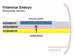
Folding of the embryonic disc occurs ventrally around the notochord, which forms a rod-like region running rostro-caudally in the midline.
In relation to the notochord:
- Laterally (either side of the notochord) lies mesoderm.
- Rostrally (above the notochord end) lies the buccopharyngeal membrane, above this again is the mesoderm region forming the heart.
- Caudally (below the notochord end) lies the primitive streak (where gastrulation occurred), below this again is the cloacal membrane.
- Dorsally (above the notochord) lies the neural tube then ectoderm.
- Ventrally (beneath the notochord) lies the mesoderm then endoderm.
The ventral endoderm (shown yellow) has grown to line a space called the yolk sac. Folding of the embryonic disc "pinches off" part of this yolk sac forming the first primative GIT.
| <html5media height="340" width="300">File:Endoderm 003.mp4</html5media> | 
|
Coelomic Cavity
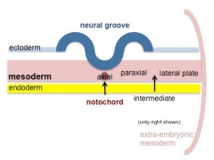
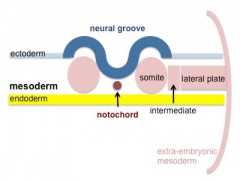
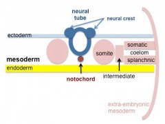
- The mesoderm initially undergoes segmentation to form paraxial, intermediate mesoderm and lateral plate mesoderm.
- Paraxial mesoderm segments into somites and lateral plate mesoderm divides into somatic and splanchnic mesoderm.
- The space forming between them is the coelomic cavity, that will form the 3 major body cavities (pericardial, pleural, peritoneal)
- Most of the gastrointestinal tract will eventually lie within the peritoneal cavity.
- Mesoderm and Ectoderm Cartoons
Trilaminar Embryo
Paraxial and Lateral Plate
Somites
Somatic and Splanchnic


(Note only the righhand side is shown, lefthand side would be identical.)
Week 4
(Gestational age GA 6 weeks) Carnegie stage 11

|

|

|
| Embryo (stage 11 ventral view) | Embryo (midline section) | Embryo (EM section) endoderm, splanchnic mesoderm, Intraembryonic coelom |

|

|
|
| Stomodeum | Buccopharyngeal membrane |
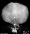
Stage 7 Membranes

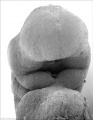
Stage 11 25 days, Low power ventral view of the Buccopharyngeal Membrane

Higher power ventrolateral view of the Buccopharyngeal Membrane

Close up view of the degenerating Buccopharyngeal Membrane
Stage 12 Week 4, 26 days
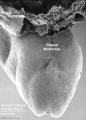
Stage 12 Cloacal membrane


Liver Development

|

|

|
liver contributions from endoderm and splanchnic mesoderm at the level of the transverse septum (week 4)
- Stage 11 - hepatic diverticulum development
- Stage 12 - cell differentiation, septum transversum forming liver stroma, hepatic diverticulum forming hepatic trabeculae
- Stage 13 - epithelial cord proliferation enmeshing stromal capillaries
The liver initially occupies the entire anterior body. All blood vessels enter the liver (placental, vitelline) and leave to enter the heart.
Stomach
|
|

|
|
- During week 4 at the level where the stomach will form the tube begins to dilate, forming an enlarged lumen.
- The dorsal border grows more rapidly than ventral first rotation (of 90 degrees), which establishes the greater curvature of the stomach.
- A second rotation (of 90 degrees) occurs on the longitudinal axis establishing the adult orientation of the stomach.
Week 5
(GA 7 weeks)
Canalization
|
|
Mesentery Development

|
Spleen
|
Week 8 - 10
(GA 10-12 weeks)
Intestine Herniation


|
|
Intestine Rotation

Normal intestinal rotation (note these are gestational age GA weeks)[1]
Hindgut
|
|
Gastrointestinal Tract Divisions
| During the 4th week the 3 distinct portions (fore-, mid- and hind-gut) extend the length of the embryo and will contribute different components of the GIT. These 3 divisions are also later defined by the vascular (artery) supply to each of theses divisions.
|

Gastrointestinal Tract Blood Supply |
Fetal

|

|
| Small Intestine length (mm) | Liver Growth (weight grams) |
| 1 to 124 grams (birth) |
Liver
- Differentiates to form the hepatic diverticulum and hepatic primordium, generates the gall bladder then divides into right and left hepatic (liver) buds.
- Hepatic Buds - form hepatocytes, produce bile from week 13 (forms meconium of newborn)
- Left Hepatic Bud - left lobe, quadrate, caudate (both q and c anatomically Left) caudate lobe of human liver consists of 3 anatomical parts: Spiegel's lobe, caudate process, and paracaval portion.
- Right Hepatic Bud - right lobe
- Bile duct - 3 connecting stalks (cystic duct, hepatic ducts) which fuse.
- Early liver also involved in blood formation, after the yolk sac and blood islands acting as a primary site.
Pancreas

- Pancreatic buds - endoderm, covered in splanchnic mesoderm
- Pancreatic bud formation – duodenal level endoderm, splanchnic mesoderm forms dorsal and ventral mesentery, dorsal bud (larger, first), ventral bud (smaller, later)
- Duodenum growth/rotation – brings ventral and dorsal buds together, fusion of buds, exocrine function (postnatal function)
- Pancreatic duct – ventral bud duct and distal part of dorsal bud
- Pancreatic islets - endocrine function (week 10 onwards)

Spleen
|

Enteric Nervous System
Topic covered in the neural crest lecture. Table below summarises the two major neural plexuses.
| Myenteric plexus | Submucosal plexus |
|---|---|
| Auerbach's plexus | Meissner's plexus |
| Leopold Auerbach (1828–1897) a German anatomist and neuropathologist. | Georg Meissner (1829–1905) a German anatomist and physiologist. |
|
|
| Links: enteric nervous system | intestine | neural crest | PMID 25428846 |
Gastrointestinal Tract Abnormalities

| USA Statistics | ||||||||||||||||||||||||||||||||||||||||||||||||||||||||||||||||||||||||
|---|---|---|---|---|---|---|---|---|---|---|---|---|---|---|---|---|---|---|---|---|---|---|---|---|---|---|---|---|---|---|---|---|---|---|---|---|---|---|---|---|---|---|---|---|---|---|---|---|---|---|---|---|---|---|---|---|---|---|---|---|---|---|---|---|---|---|---|---|---|---|---|---|
| ||||||||||||||||||||||||||||||||||||||||||||||||||||||||||||||||||||||||
Lumen Abnormalities
There are several types of abnormalities that impact upon the continuity of the gastrointestinal tract lumen.
Atresia
Stenosis
Duplication
|

|
Meckel's Diverticulum
|

Meckel's Diverticulum |
Intestinal Malrotation
Presents clinically in symptomatic malrotation as:
|

Intestinal malrotation |
Intestinal Aganglionosis
(intestinal aganglionosis, Hirschsprung's disease, aganglionic colon, megacolon, congenital aganglionic megacolon, congenital megacolon)
|

|
Gastroschisis
| Gastroschisis (omphalocele, paraomphalocele, laparoschisis, abdominoschisis, abdominal hernia) is a congenital abdominal wall defect which results in herniation of fetal abdominal viscera (intestines and/or organs) into the amniotic cavity.
Incidence of gastroschisis has been reported at 1.66/10,000, occuring more frequently in young mothers (less than 20 years old). By definition, it is a body wall defect, not a gastrointestinal tract defect, which in turn impacts upon GIT development. This indirect developmental effect (one system impacting upon another) occurs in several other systems.
|
|
Final Thoughts- After Birth
Remember that the GIT does not function until after birth consider:
- metabolic disorders discovered by neonatal diagnosis
- Neonatal feeding difficulties due to cleft lip and cleft palate.
Links: Gastrointestinal Tract - Abnormalities
References
Terms
Expand the table below to see GIT related terms.
| Gastrointestinal Tract Terms | ||
|---|---|---|
| ||
|
Glossary Links
- Glossary: A | B | C | D | E | F | G | H | I | J | K | L | M | N | O | P | Q | R | S | T | U | V | W | X | Y | Z | Numbers | Symbols | Term Link
| 2018 ANAT2341 - Timetable | Course Outline | Moodle | Tutorial 1 | Tutorial 2 | Tutorial 3 |
Labs: 1 Preimplantation and Implantation | 2 Reproductive Technology Revolution | 3 Group Projects | 4 GM manipulation mouse embryos | 5 Early chicken eggs | 6 Female reproductive tract | 7 Skin regeneration | 8 Vertebral development | 9 Organogenesis Lab | 10 Cardiac development | 11 Group projects | 12 Stem Cell Journal Club |
|
Lectures: 1 Introduction | 2 Fertilization | 3 Week 1/2 | 4 Week 3 | 5 Ectoderm | 6 Placenta | 7 Mesoderm | 8 Endoderm | 9 Research Technology | 10 Cardiovascular | 11 Respiratory | 12 Neural crest | 13 Head | 14 Musculoskeletal | 15 Limb | 16 Renal | 17 Genital | 18 Endocrine | 19 Sensory | 20 Fetal | 21 Integumentary | 22 Birth | 23 Stem cells | 24 Revision |
| Student Projects: Group Projects Information Project 1 | Project 3 | Project 4 | Project 5 | 2018 Test Student | Copyright |
Cite this page: Hill, M.A. (2024, April 18) Embryology Lecture - Gastrointestinal Development. Retrieved from https://embryology.med.unsw.edu.au/embryology/index.php/Lecture_-_Gastrointestinal_Development
- © Dr Mark Hill 2024, UNSW Embryology ISBN: 978 0 7334 2609 4 - UNSW CRICOS Provider Code No. 00098G