Lecture - 2016 Course Introduction
| Embryology - 25 Apr 2024 |
|---|
| Google Translate - select your language from the list shown below (this will open a new external page) |
|
العربية | català | 中文 | 中國傳統的 | français | Deutsche | עִברִית | हिंदी | bahasa Indonesia | italiano | 日本語 | 한국어 | မြန်မာ | Pilipino | Polskie | português | ਪੰਜਾਬੀ ਦੇ | Română | русский | Español | Swahili | Svensk | ไทย | Türkçe | اردو | ייִדיש | Tiếng Việt These external translations are automated and may not be accurate. (More? About Translations) |
Course Introduction

Course coordinator |
This first lecture will be a general introduction to the course and the subject of Embryology.
|
Lecture Objectives

- Understand the course objectives and assessment.
- Brief understanding of the historic background of embryology.
- Brief understanding of Australian data.
- Broad overview of human development.
<html5media height="384" width="352">File:Human development 001.mp4</html5media>
Here is the whole course in One Minute.
| Introduction Movies | |||||||||||
|---|---|---|---|---|---|---|---|---|---|---|---|
|
|
| |||||||||
| Fertilization | Embryonic Development (week 1 - 8) | Birth (week 37) |
History
History - Embryologists | Embryology History | Human Embryo Collections

17-18C Braune - The Position of the Uterus and Fetus at Term (1872)
| Human Embryo Collections | ||
|---|---|---|
Wilhelm His (1831-1904) His's Normentafel (Normal Table) |

|

|
Franz Keibel (1861 - 1929) Franz Keibel and Curt Elze (1908) Normal Plates of the Development of the Human Embryo |

|

|

Franklin Mall (1862-1917) |

| |
| Begun by Dr. Hideo Nishimura (1912–1995)
Developed by Kohei Shiota and currently curated by Shigehito Yamada.
|

| |


| Animal Models | |
|---|---|
| |
| |
 |
mouse
|
| Fly Development - The fruitfly (drosophila) was and is the traditional geneticist's tool. It has been transformed to an magnificent embryologist's tool, with developmental mechanisms being uncovered in this system combined with homolgy gene searches in other species. The fly genome was one of the first to be been completely sequenced. In early development nurse cells sacrifice their cytoplasmic contents to allow egg growth and early pattern formation is through the localization of maternal messenger RNAs (mRNAs). | |
 |
Worm Development - Early embryological studies of the worm Caenorhabditis elegans (C.Elegans, so called because of its "elegant" curving movement) characterized the fate of each and every cell in the worm through all stages of development. This worm has recently had its entire genome sequenced. |
| Zebrafish Development - Zebrafish are seen as the latest and greatest "model' for embryological development studies. They can be easily genetically altered and develop as practically "see through" embryos, all internal development can be clearly observed from the outside in the living embryo. |
| In Vitro Fertilization (1978) | Stem Cells (1981) | Induced Stem Cells (2006) | Molecular Development |

|

|

|

|
Australian Data
1 August 2014 at 03:53:30 PM (Canberra time), the resident population of Australia is projected to be: 23,550,233.

| Australian Statistics | |
|---|---|
|

|
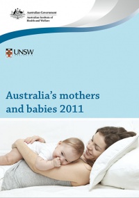
| Australia’s mothers and babies (2011) | Assisted reproductive technology in Australia and New Zealand (2010) |
| Average maternal age in 2011 was 30.0 years, the same as 2009 but still more than the earlier years (2000, 29.0 years; 2002, 29.4 years). | Assisted Reproductive Technology (ART) was used by 3.8% (2009, 3.6%) of women who gave birth. |
| Victoria - 10 most reported birth anomalies | ||||||||||||||||||||
|---|---|---|---|---|---|---|---|---|---|---|---|---|---|---|---|---|---|---|---|---|
| Based upon statistics from the Victorian Perinatal Data Collection Unit in Victoria between 2003-2004. | ||||||||||||||||||||
|


Human Development

Glossary Links
- Glossary: A | B | C | D | E | F | G | H | I | J | K | L | M | N | O | P | Q | R | S | T | U | V | W | X | Y | Z | Numbers | Symbols | Term Link
Cite this page: Hill, M.A. (2024, April 25) Embryology Lecture - 2016 Course Introduction. Retrieved from https://embryology.med.unsw.edu.au/embryology/index.php/Lecture_-_2016_Course_Introduction
- © Dr Mark Hill 2024, UNSW Embryology ISBN: 978 0 7334 2609 4 - UNSW CRICOS Provider Code No. 00098G