K12 Professional Development 2014: Difference between revisions
mNo edit summary |
mNo edit summary |
||
| (15 intermediate revisions by the same user not shown) | |||
| Line 9: | Line 9: | ||
The section entitled [[#Additional Useful Resources|Additional Useful Resources]] links directly to resources and currently there is no | The section entitled [[#Additional Useful Resources|Additional Useful Resources]] links directly to resources and currently there is no content designed for K12 student use, but content and resources on these pages may be useful in designing your own specific content around these topics. | ||
Note that content on K12 pages has been simplified in both terminology and descriptions from that found on other embryology pages designed for university level students. That does not mean that other embryology pages are not useful for students, but may be to difficult to place in context given the advanced terminology and descriptions used on non-K12 pages. | Note that content on K12 pages has been simplified in both terminology and descriptions from that found on other embryology pages designed for university level students. That does not mean that other embryology pages are not useful for students, but may be to difficult to place in context given the advanced terminology and descriptions used on non-K12 pages. | ||
Note that clicking the "Expand" text on pages will open collapsible tables with more information or resources. | |||
{| class="wikitable mw-collapsible mw-collapsed" | {| class="wikitable mw-collapsible mw-collapsed" | ||
| Line 22: | Line 24: | ||
Student exercise sheets have been designed to be printed and used in the classroom. | Student exercise sheets have been designed to be printed and used in the classroom. | ||
|} | |} | ||
[[Media:K12_Embryology_Professional_Development_2014.pdf|PDF of this Page]] | |||
Below are a list of the current K12 designed resources. | Below are a list of the current K12 designed resources. | ||
| Line 33: | Line 37: | ||
{| | {| | ||
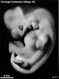
| [[File:Stage16_bf8.jpg|120px|link=K12_Comparative_Embryology]] | | [[File:Stage16_bf8.jpg|120px|link=K12_Comparative_Embryology]] | ||
| This page has been designed as an introduction to Comparative Embryology.The page has 4 student exercises contained within collapsible tables. These exercises have worksheets that can be printed out to be completed by students. Also look at | | This page has been designed as an introduction to Comparative Embryology. The page has 4 student exercises contained within collapsible tables. These exercises have worksheets that can be printed out to be completed by students. Also look at the series of student designed projects on animal development. | ||
:'''Links:''' [[K12_Comparative_Embryology|Comparative Embryology]] | :'''K12 Links:''' [[K12_Comparative_Embryology|Comparative Embryology]] | ||
:Student Projects: [[2009_Group_Project_1|Rabbit]] | [[2009_Group_Project_2|Fly]] | [[2009_Group_Project_3|Zebrafish]] | [[2009_Group_Project_4|Mouse]] | [[2009_Group_Project_5|Frog]] | [[Animal_Development|Related page - Animal Development]] | :Student Projects: [[2009_Group_Project_1|Rabbit]] | [[2009_Group_Project_2|Fly]] | [[2009_Group_Project_3|Zebrafish]] | [[2009_Group_Project_4|Mouse]] | [[2009_Group_Project_5|Frog]] | [[Animal_Development|Related page - Animal Development]] | ||
| Line 46: | Line 50: | ||
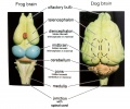
| This page was designed originally as a presentation for brain awareness week. Students should understand that the description of early brain development is the same for most species. A more specific page on K12 Comparative Brain Anatomy is under development. | | This page was designed originally as a presentation for brain awareness week. Students should understand that the description of early brain development is the same for most species. A more specific page on K12 Comparative Brain Anatomy is under development. | ||
:'''Links:''' [[K12_Brain_Awareness_Week|Comparative Brain Anatomy]] | :'''K12 Links:''' [[K12_Brain_Awareness_Week|Comparative Brain Anatomy]] | ||
|} | |} | ||
==Additional Useful Resources== | ==Additional Useful Resources== | ||
| Line 57: | Line 61: | ||
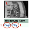
| The movies page has many different animations showing developmental processes. Some of these movies are simplified cartoons, while others are derived from developmental research articles. | | The movies page has many different animations showing developmental processes. Some of these movies are simplified cartoons, while others are derived from developmental research articles. | ||
The moves can be | The moves can be: | ||
# Opened on a new page with a description and links to additional content, resources and references. | |||
# Played as a movie by itself, movie can be expanded to full screen, as well as stopped and started to allow discussion. | |||
:'''Links:''' [[Movies]] | :'''Links:''' [[Movies]] | ||
| Line 72: | Line 78: | ||
| Thalidomide is a drug that was introduced on to the market on October 1, 1957 in West Germany. Thalidomide soon became a drug prescribed to pregnant women to combat symptoms associated with morning sickness. | | Thalidomide is a drug that was introduced on to the market on October 1, 1957 in West Germany. Thalidomide soon became a drug prescribed to pregnant women to combat symptoms associated with morning sickness. | ||
When taken during the first trimester of pregnancy, thalidomide prevented the proper growth of the fetus resulting in horrific birth defects in thousands of children around the world. | When taken during the first trimester of pregnancy, thalidomide prevented the proper growth of the fetus resulting in horrific birth defects in thousands of children around the world. This is an example for students of inadequate drug testing and a lack of understanding of environmental effects on human development. This is often cited today as a reason to have significant testing of drugs before release and classification of drugs based upon their affects on development. | ||
:'''Links:''' [[Abnormal_Development_-_Thalidomide|Thalidomide]] | :'''Links:''' [[Abnormal_Development_-_Thalidomide|Thalidomide]] | [[Australian Drug Categories]] | ||
|} | |} | ||
===Cell Division=== | ===Cell Division=== | ||
| Line 100: | Line 106: | ||
[[File:Chromatin_Structure.png|800px]] | [[File:Chromatin_Structure.png|800px]] | ||
{| | |||
| [[File:Nucleus structure cartoon 01.jpg|200px]] | |||
| This cartoon shows the general structure of the nucleus when the cell is transcriptionally active and not dividing. Chromosomes are unravelled and spread throughout the nucleus in "chromosome territories". | |||
|} | |||
{| | |||
| width=120px|[[File:Autosomal_dominant_inheritance.jpg|120px]] | |||
| There are also a set of cartoon diagrams illustrating [[Molecular_Development_-_Genetics#Inheritance_Genetics|genetic mechanisms of inheritance]]. There is also a downloadable student worksheet exercise on inheritance patterns (see below). | |||
{| class="wikitable mw-collapsible mw-collapsed" | |||
! Teacher Information - Genetic Inheritance Exercise | |||
|- | |||
| [[Media:K12 Genetic Inheritance Exercise 1.pdf|'''Exercise 1''']] - Print out the document linked from [[Media:K12 Genetic Inheritance Exercise 1.pdf|Exercise 1]] (4 pages) of cartoons with and without the labelling. The first 2 sheets are for teacher use, page 3 and 4 are the unlabelled student worksheets. | |||
The links below are a search of an online genetics database (OMIM) that will list some known genetic disorders of these patterns. Firstly, note how many disorders have appeared in the results. Secondly, clicking any entry on the list will open that disorder with more information about clinical features, gene location, historic and current research. | |||
* [http://www.ncbi.nlm.nih.gov/omim/?term=autosomal+recessive '''autosomal recessive'''] | |||
* [http://www.ncbi.nlm.nih.gov/omim/?term=autosomal+dominant '''autosomal dominant'''] | |||
* [http://www.ncbi.nlm.nih.gov/omim/?term=X+linked+dominant '''X linked dominant'''] | |||
* [http://www.ncbi.nlm.nih.gov/omim/?term=X+linked+recessive '''X linked recessive'''] | |||
Here is an example entry for [http://omim.org/entry/310200 Duchenne Muscular Dystrophy]. An X linked recessive disorder that leads to muscular degeneration in boys due to a mutation in the dystrophin gene located on the X chromosome. (More? [[Musculoskeletal_System_-_Abnormalities#Duchenne_Muscular_Dystrophy|brief description]]) | |||
|} | |||
|} | |||
:'''Links:''' [[Molecular Development - Genetics|Genetics]] | [[Molecular_Development_-_Genetics#Inheritance_Genetics|Inheritance Genetics]] | |||
===Current Reproductive Technologies=== | ===Current Reproductive Technologies=== | ||
{| | {| | ||
Latest revision as of 14:30, 17 October 2014
| Embryology - 23 Apr 2024 |
|---|
| Google Translate - select your language from the list shown below (this will open a new external page) |
|
العربية | català | 中文 | 中國傳統的 | français | Deutsche | עִברִית | हिंदी | bahasa Indonesia | italiano | 日本語 | 한국어 | မြန်မာ | Pilipino | Polskie | português | ਪੰਜਾਬੀ ਦੇ | Română | русский | Español | Swahili | Svensk | ไทย | Türkçe | اردو | ייִדיש | Tiếng Việt These external translations are automated and may not be accurate. (More? About Translations) |
Introduction

This page introduces K12 teaching resources related to embryology and development. The current page is designed to help teachers find useful curated online resources for use in biology classes related to embryology and development. The content and links on this current page are under ongoing development, I am happy to receive feedback and requests for design of specific K12 content and exercises for your classes.
The initial resources I will be presenting relate to comparative embryology and comparative brain anatomy.
The section entitled Additional Useful Resources links directly to resources and currently there is no content designed for K12 student use, but content and resources on these pages may be useful in designing your own specific content around these topics.
Note that content on K12 pages has been simplified in both terminology and descriptions from that found on other embryology pages designed for university level students. That does not mean that other embryology pages are not useful for students, but may be to difficult to place in context given the advanced terminology and descriptions used on non-K12 pages.
Note that clicking the "Expand" text on pages will open collapsible tables with more information or resources.
| Content Reuse |
|---|
| Please note content on this site has been derived from a number of sources and under a range of different copyright conditions. I am happy for educational reuse in the classroom without republication. I specifically do not allow republication of content on the internet. This is to prevent misuse of content out of context and also to prevent multiple search results for the same content. In general, opening an image will show the specific copyright restrictions associated with the image.
Please contact me if you require information on educational reuse outside of your classrooms. Student exercise sheets have been designed to be printed and used in the classroom. |
Below are a list of the current K12 designed resources.
- Links: K12 Professional Development 2014 - Embryology for K12 Students | Museum of Human Disease - Professional Development Days
Comparative Embryology
|
This page has been designed as an introduction to Comparative Embryology. The page has 4 student exercises contained within collapsible tables. These exercises have worksheets that can be printed out to be completed by students. Also look at the series of student designed projects on animal development.
|
Comparative Brain Anatomy
|
This page was designed originally as a presentation for brain awareness week. Students should understand that the description of early brain development is the same for most species. A more specific page on K12 Comparative Brain Anatomy is under development.
|
Additional Useful Resources
The following pages are not designed for K12 students but may provide background information and resources for teachers in other Biology areas.
Movies
|
The movies page has many different animations showing developmental processes. Some of these movies are simplified cartoons, while others are derived from developmental research articles.
The moves can be:
|
This is the first MRI recording of childbirth. |
Thalidomide

Thalidomide molecular structure |
Thalidomide is a drug that was introduced on to the market on October 1, 1957 in West Germany. Thalidomide soon became a drug prescribed to pregnant women to combat symptoms associated with morning sickness.
When taken during the first trimester of pregnancy, thalidomide prevented the proper growth of the fetus resulting in horrific birth defects in thousands of children around the world. This is an example for students of inadequate drug testing and a lack of understanding of environmental effects on human development. This is often cited today as a reason to have significant testing of drugs before release and classification of drugs based upon their affects on development.
|
Cell Division
These pages are designed for university level students but also contain images and movies that can be used with students.
The page link opens a movie page with additional descriptions about what is being shown in the movie.
|
This movie shows chromosomes being segregated during mitosis, the last image is a normal microscopic view of the 2 daughter cells following cell division. Chromosomes have been labelled (white) and align at metaphase, then separate into the two daughter cells. Note the chromosomes unfold at the end of mitosis.
Exercise - Allow the students to see the unlabelled movie and then identify specific stages of mitosis by time. The labelled movie can be shown after the student have attempted the exercise.
| |||
|
This movie shows how in the oocyte (egg) during meiosis 1 excess DNA is segregated into a specialised structure, the polar body. The polar body contains DNA in an exclusion body that does not contribute to the embryo. DNA is labelled blue and the same oocyte is shown in both bright field (left) and fluorescence (right). Note the remaining oocyte chromosomes arrest at metaphase 2.
|
Chromosomal Structure
This generally available cartoon image shows the major structures in DNA compaction from duplex to chromosome. Note that this process of compaction occurs only during preparation for cell division and that cells carrying out their normal function have uncompacted DNA to allow gene transcription.


|
This cartoon shows the general structure of the nucleus when the cell is transcriptionally active and not dividing. Chromosomes are unravelled and spread throughout the nucleus in "chromosome territories". |

|
There are also a set of cartoon diagrams illustrating genetic mechanisms of inheritance. There is also a downloadable student worksheet exercise on inheritance patterns (see below).
|
- Links: Genetics | Inheritance Genetics
Current Reproductive Technologies

Louise Brown, the first IVF baby as an adult. |
The general public more commonly recognise the term In Vitro Fertilisation (IVF).
Note the Nobel Prize in Physiology or Medicine 2010 was awarded to Robert G. Edwards "for the development of in vitro fertilization" who battled societal and establishment resistance to his development of the in vitro fertilization procedure, which has so far led to the birth of around 4 million people. (More? Assisted Reproductive Technology | Nobel Prize 2010) 10 April 2013 - Sir Robert Edwards has died aged 87. BBC News Exercise - Students should identify the many different ways in which pregnancy can be now be achieved using ART. |

|
Statistical data on assisted reproductive technology is released regularly in this publication. Student can download the full report and there is an overview executive summary available. |
Anatomy and function of the Human Ear

Immune System

|
This is a university lecture on the immune system, but also contains easily understood simplified cartoons and electron micrographs of B and T cells. |
HSC NSW
| Biology Syllabus |
|---|
Blueprint of Life
|
External Links
External Links Notice - The dynamic nature of the internet may mean that some of these listed links may no longer function. If the link no longer works search the web with the link text or name. Links to any external commercial sites are provided for information purposes only and should never be considered an endorsement. UNSW Embryology is provided as an educational resource with no clinical information or commercial affiliation.
Glossary Links
- Glossary: A | B | C | D | E | F | G | H | I | J | K | L | M | N | O | P | Q | R | S | T | U | V | W | X | Y | Z | Numbers | Symbols | Term Link
Cite this page: Hill, M.A. (2024, April 23) Embryology K12 Professional Development 2014. Retrieved from https://embryology.med.unsw.edu.au/embryology/index.php/K12_Professional_Development_2014
- © Dr Mark Hill 2024, UNSW Embryology ISBN: 978 0 7334 2609 4 - UNSW CRICOS Provider Code No. 00098G