K12 Comparative Embryology: Difference between revisions

From Embryology
mNo edit summary
mNo edit summary
Line 25: Line 25:
|}
|}


| <html5media height="505" width="400">File:Embryo stages 003.mp4</html5media>
| [[File:Embryo stages 01.gif]]


[[Media:Embryo_stages_003.mp4|'''Click Here''' to play on mobile device]]
[[Media:Embryo_stages_003.mp4|'''Click Here''' to play on mobile device]]


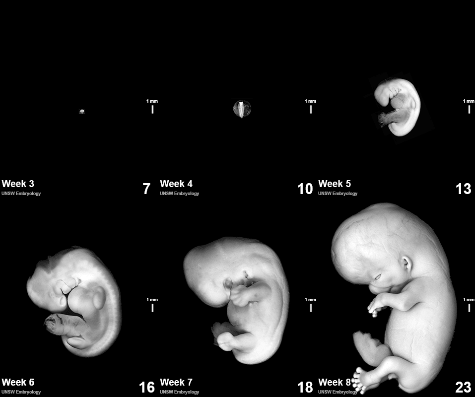
This movie shows human embryo development between week 4 to 8 after fertilisation.
This movie shows human embryo development between week 3 to 8 after fertilisation.
|}
|}


Revision as of 11:03, 5 September 2016

Embryology - 19 Apr 2024    Facebook link Pinterest link Twitter link  Expand to Translate  
Google Translate - select your language from the list shown below (this will open a new external page)

العربية | català | 中文 | 中國傳統的 | français | Deutsche | עִברִית | हिंदी | bahasa Indonesia | italiano | 日本語 | 한국어 | မြန်မာ | Pilipino | Polskie | português | ਪੰਜਾਬੀ ਦੇ | Română | русский | Español | Swahili | Svensk | ไทย | Türkçe | اردو | ייִדיש | Tiếng Việt    These external translations are automated and may not be accurate. (More? About Translations)

K12 Professional Development 2016

Introduction

All human and animal embryos go through very similar stages of early development. See also Humans and Animal Embryology.

What are the key things in development that we share?

This page introduces a few of the concepts of development shared with all animals.


K12 Links: Start Here | Week 1 | Week 2 | Week 3 | Week 4 | Week 5 to 8 | Arms and Legs | Heart | Fetus | Brain Growth | Eyes and Ears | Animal Development Times | Humans and Animal Embryology | Comparative Embryology | Thalidomide


Teacher Note 
Mark Hill.jpg This is currently only a draft designed to help K12 students understand comparative embryology.

I am currently looking to simplify concepts and include images on this page. I am happy to receive feedback as too what you may like to be included here. I have also begun to add some simple exercises that can be used in class to help understand concepts in embryonic development and comparison. Note some of the links on this page leave the K12 notes section and may be beyond the level of your students, bookmark this page to easily return here. This page can be printed using the lefthand menu "Toolbox" or Printable version.

K12 Professional Development 2014

Embryo stages 01.gif

Click Here to play on mobile device

This movie shows human embryo development between week 3 to 8 after fertilisation.

Human Carnegie Stages

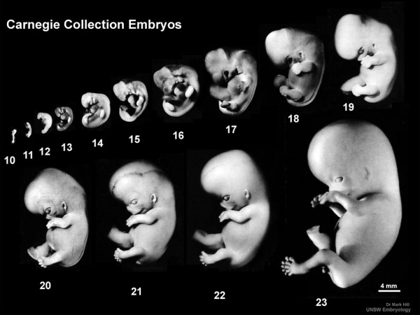
Carnegie stages are named after the famous USA institute which began collecting and classifying embryos in the early 1900's. Human stages are based on the external and/or internal features of the embryo, and are not directly dependent on either age or size.


The human embryonic period proper is divided into 23 Carnegie stages covering the first 8 weeks after fertilisation (post-ovulation). This period is most of the first trimester and the second and third trimester is called Fetal Development and is mainly about growth.


Other features used in this classification include: ranges of age in days, number of segments (somites) present, and embryonic size (CRL, crown rump length).


Similar types of measurements are also used to stage how animal embryos develop. You can therefore compare other animal embryos each other and to human development by using these staging criteria.

Human Carnegie stage 10-23.jpg
The Carnegie Embryos

Developmental Misconceptions

Many of these are truely historic, and while essentially wrong, science works through testing these alternate theories, and is some cases some can even be partially correct.

Preformationist Theory
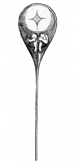
N. Hartsoeker (1694) image of how he imagined a sperm would look if it contained a preformed individual.


Note that Hartsoeker later rejected this preformationist view, carrying out calculations of size that showed that if all of the animals of any species had been enclosed in the first male or female, those animals that now inhabit the earth would have to be infinitely and incomprehensively small.


This was replaced by the "epigenesis theory", the organism form arises gradually, changing shape and acquiring its adult parts over time.

Hilfer1990 Fig01.jpg
Recapitulation Theory
Ernst Haeckel (1834 – 1919) "ontogeny recapitulates phylogeny" claimed that an individual organism's biological development (ontogeny), parallels and summarises its species evolutionary development (phylogeny). First a single-celled organism, then evolve into a fish, then an amphibian, then a reptile, then a bird, and finally reach a mammal.

Current developmental biology shows that animals follow similar developmental programs, but do not go through a "species change" during development.

Ovary Germinal Epithelium
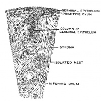
The epithelium surrounding the ovary was originally thought to provide the source of follicles and oocytes and therefore called the "germinal epithelium".


Current developmental biology shows that this is not the source of primordial germ cells or follicle components, but is a cuboidal epithelium overlying a dense connective tissue layer (tunica albuginea).

Keith1921 fig006.jpg
Genetic Theory
Genetics - "genes determine our physiology and chemical behaviour".

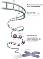
While genetics is essentially correct, we now know that inheritance mechanisms exist outside the DNA sequence of our genes and include DNA methylation, histone modification, and those of the microRNA machinery.


Molecular Development - Epigenetics

Epigenetics cartoon.jpg


Cite this page: Hill, M.A. (2024, April 19) Embryology K12 Comparative Embryology. Retrieved from https://embryology.med.unsw.edu.au/embryology/index.php/K12_Comparative_Embryology

What Links Here?
© Dr Mark Hill 2024, UNSW Embryology ISBN: 978 0 7334 2609 4 - UNSW CRICOS Provider Code No. 00098G