K12 - Communication: Difference between revisions
| Line 29: | Line 29: | ||
| valign="top" width="200px"|'''Retinal Cell Organization''' | | valign="top" width="200px"|'''Retinal Cell Organization''' | ||
These are the names of the cells and neurones required to convert light into electrical signals. | These are the names of the cells and neurones required to convert light into electrical signals. | ||
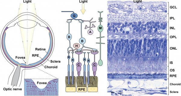
* '''G''' - retinal ganglion cells | * '''G''' - retinal ganglion cells (form the ganglion cell layer) | ||
* '''M''' - Müller cell | * '''M''' - Müller cell | ||
* '''A''' - amacrine cell | * '''A''' - amacrine cell | ||
* '''B''' - bipolar cell | * '''B''' - bipolar cell | ||
* '''H''' - horizontal cell | * '''H''' - horizontal cell | ||
* '''R''' - rod | * '''R''' - rod (detect light) | ||
* '''C''' - cone | * '''C''' - cone (detect light) | ||
| valign="top"|'''Human Retina Histology''' | | valign="top"|'''Human Retina Histology''' | ||
Revision as of 14:07, 14 March 2012
Introduction
Biological communication occurs at many levels, from one cell communication with its neighbour in a tissue (paracrine, to signals released into the blood from one cell to signal to another cell or tissue (endocrine or hormone signaling). The signalling that occurs in the brain, spinal cord and other nervous tissues involves electrical (action potentials) signaling.
This page will introduce development of signaling in our special sensory nervous systems: the eyes for vision and the ears for hearing. Both systems convert signals in one medium (light or sound) into an electrical signal that our brain can understand.
- This content is only as a brief introduction, designed for K12 students. More detailed information can be found on the listed linked pages.
Hearing - Sound
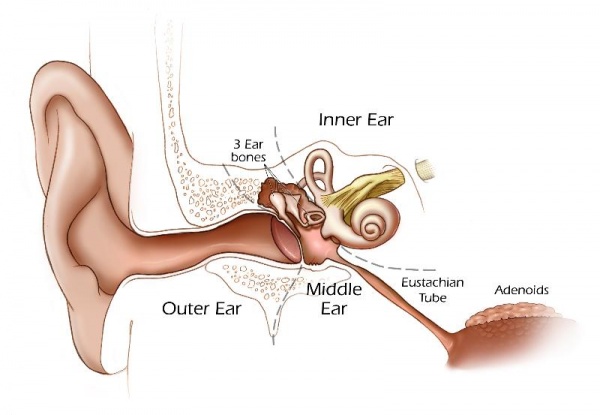
This cartoon shows the adult "ear" with the 3 main divisions (outer, middle, inner).
Vision - Light
This cartoon[1] shows the eyeball (left), a cartoon of the retina cells (middle) and an actual slice of the human retina.
The Eye
|
Retinal Cell Organization
These are the names of the cells and neurones required to convert light into electrical signals.
|
Human Retina Histology
This is a thin section of the retina that has been stained with dyes so that you can see the structure shown in the cartoon. |
Glossary Links
- Glossary: A | B | C | D | E | F | G | H | I | J | K | L | M | N | O | P | Q | R | S | T | U | V | W | X | Y | Z | Numbers | Symbols | Term Link
Cite this page: Hill, M.A. (2024, April 19) Embryology K12 - Communication. Retrieved from https://embryology.med.unsw.edu.au/embryology/index.php/K12_-_Communication
- © Dr Mark Hill 2024, UNSW Embryology ISBN: 978 0 7334 2609 4 - UNSW CRICOS Provider Code No. 00098G
- ↑ <pubmed>20855501</pubmed>| PMC3101587 | JCB

